

Search Count: 114
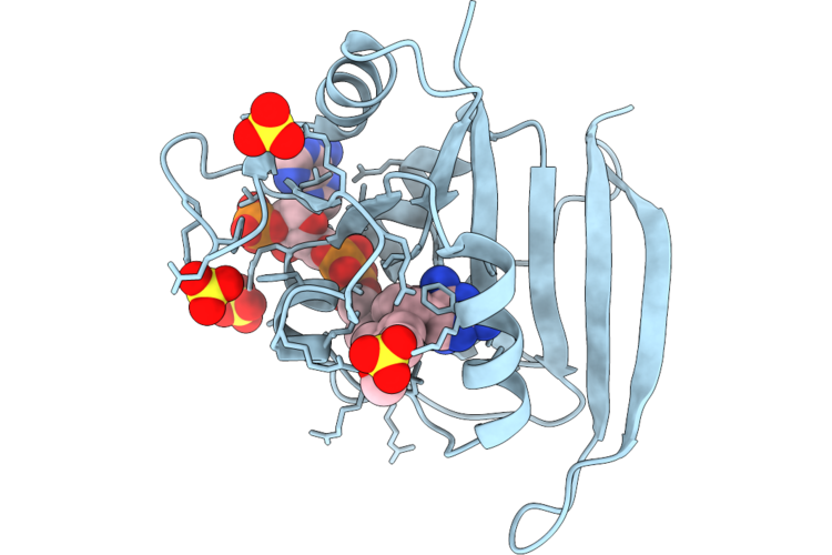 |
Structure Of Dihydrofolate Reductase From Burkholderia Thailandensis Complexed With Trimethoprim And Dihydrofolate
Organism: Burkholderia thailandensis
Method: X-RAY DIFFRACTION Resolution:1.84 Å Release Date: 2026-08-26 Classification: OXIDOREDUCTASE Ligands: TOP, NDP, SO4 |
Organism: Burkholderia thailandensis
Method: X-RAY DIFFRACTION
Release Date: 2026-08-26
Ligands: TOP, NDP, SO4
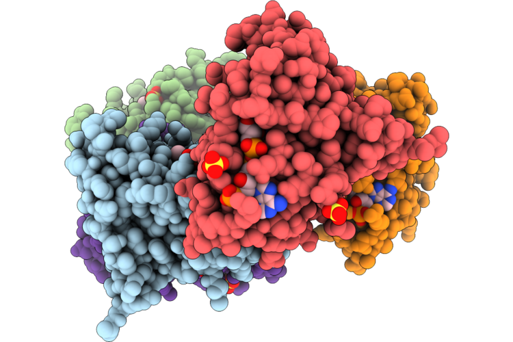 |
Structure Of Dihydrofolate Reductase From Burkholderia Thailandensis (I99L Variant) Complexed With Trimethoprim And Dihydrofolate
Organism: Burkholderia thailandensis
Method: X-RAY DIFFRACTION Resolution:2.19 Å Release Date: 2026-08-26 Classification: OXIDOREDUCTASE Ligands: TOP, NDP, SO4 |
Organism: Burkholderia thailandensis
Method: X-RAY DIFFRACTION
Release Date: 2026-08-26
Ligands: TOP, NDP, SO4
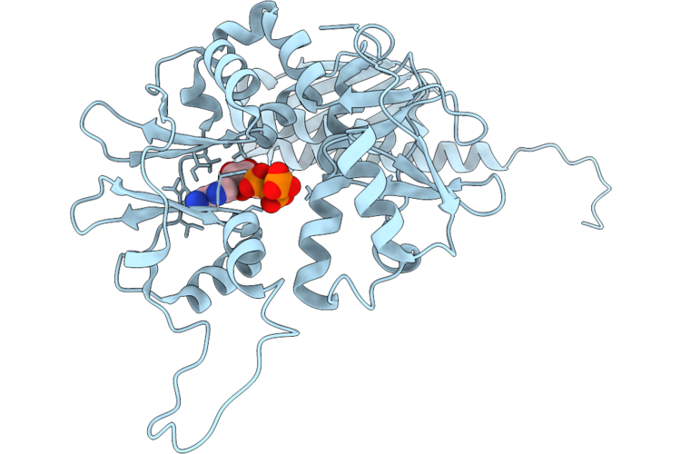 |
P. Abyssi Hibernation Factor Hib Bound To Atp (Hib-Ptc Conformation)
Organism: Pyrococcus abyssi ge5
Method: ELECTRON MICROSCOPY Release Date: 2026-08-12 Classification: RIBOSOME Ligands: ATP |
Organism: Pyrococcus abyssi ge5
Method: ELECTRON MICROSCOPY
Release Date: 2026-08-12
Ligands: ATP
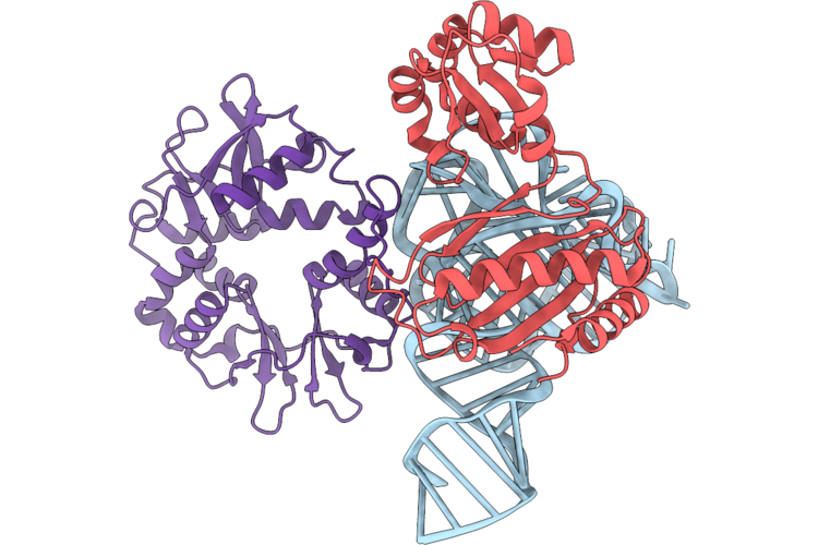 |
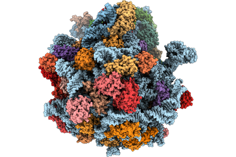
Cryo-Em Structure Of The N-Terminal Domain Of Hib Bound To The L1 Stalk Of Pyrococcus Abyssi 70S
Organism: Pyrococcus abyssi ge5
Method: ELECTRON MICROSCOPY Release Date: 2026-08-12 Classification: RIBOSOME |
Organism: Pyrococcus abyssi ge5
Method: ELECTRON MICROSCOPY
Release Date: 2026-08-12
 |
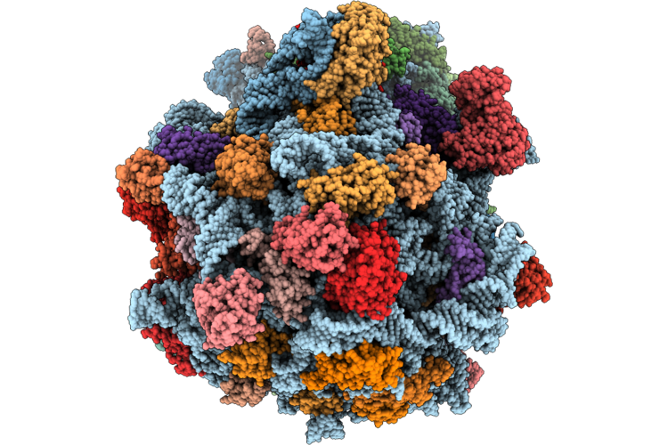
Cryo-Em Structure Of P. Abyssi Hiba:Ribosome With An Sd:Antisd Duplex
Organism: Pyrococcus abyssi ge5
Method: ELECTRON MICROSCOPY Resolution:2.20 Å Release Date: 2026-07-22 Classification: RIBOSOME Ligands: MG, ZN |
Organism: Pyrococcus abyssi ge5
Method: ELECTRON MICROSCOPY
Release Date: 2026-07-22
Ligands: MG, ZN
 |
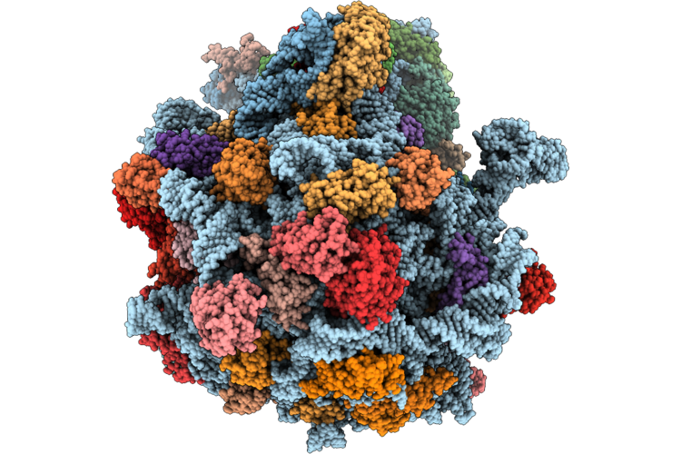
Cryo-Em Structure Of P. Abyssi 70S Ribosome In Complex With Hibernation Factor Hiba And Sbds
Organism: Pyrococcus abyssi ge5
Method: ELECTRON MICROSCOPY Resolution:2.30 Å Release Date: 2026-07-22 Classification: RIBOSOME Ligands: MG, ZN |
Organism: Pyrococcus abyssi ge5
Method: ELECTRON MICROSCOPY
Release Date: 2026-07-22
Ligands: MG, ZN
 |
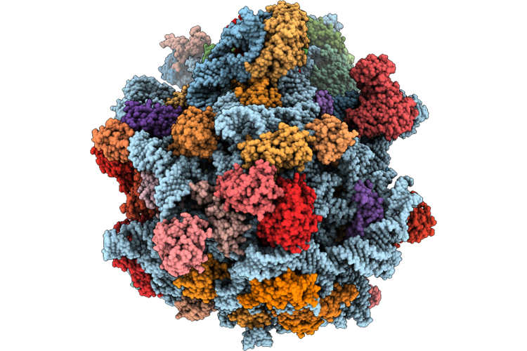
Cryo-Em Structure Of P. Abyssi 70S Ribosome In Complex With Hibernation Factor Hiba In Ptc Conformation
Organism: Pyrococcus abyssi ge5
Method: ELECTRON MICROSCOPY Resolution:2.10 Å Release Date: 2026-07-22 Classification: RIBOSOME Ligands: MG, ZN |
Organism: Pyrococcus abyssi ge5
Method: ELECTRON MICROSCOPY
Release Date: 2026-07-22
Ligands: MG, ZN
 |
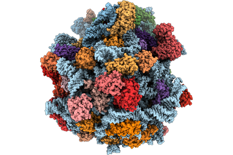
Cryo-Em Structure Of P. Abyssi 70S Ribosome In Complex With Hibernation Factor Hiba (Ptc Conformation With E-Site Trna)
Organism: Pyrococcus abyssi ge5
Method: ELECTRON MICROSCOPY Resolution:2.11 Å Release Date: 2026-07-22 Classification: RIBOSOME Ligands: MG, ZN |
Organism: Pyrococcus abyssi ge5
Method: ELECTRON MICROSCOPY
Release Date: 2026-07-22
Ligands: MG, ZN
 |
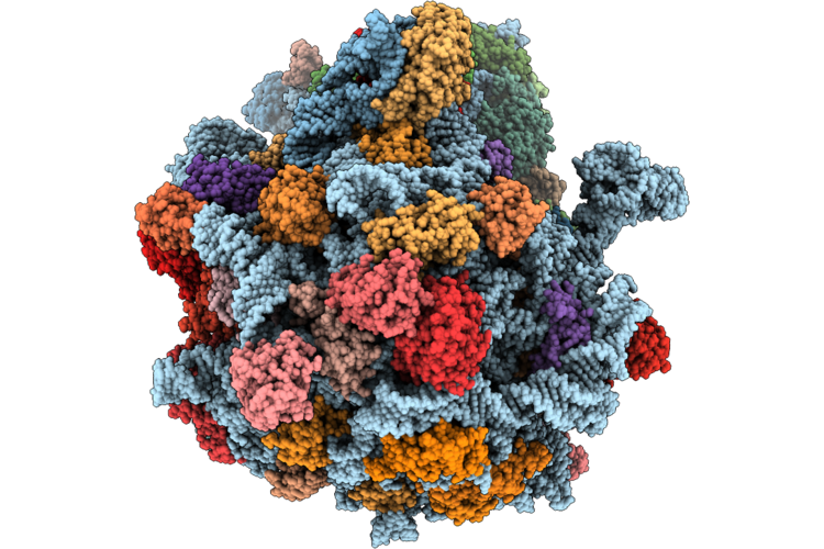
Cryo-Em Structure Of P. Abyssi 70S Ribosome In Complex With Hibernation Factor Hiba (L1 Stalk Conformation)
Organism: Pyrococcus abyssi ge5
Method: ELECTRON MICROSCOPY Resolution:2.10 Å Release Date: 2026-07-22 Classification: RIBOSOME Ligands: MG, ZN |
Organism: Pyrococcus abyssi ge5
Method: ELECTRON MICROSCOPY
Release Date: 2026-07-22
Ligands: MG, ZN
 |
Cryo-Em Structure Of P. Abyssi 70S Ribosome In Complex With Hibernation Factor Hiba (Hiba-Ul5 Conformation)
Organism: Pyrococcus abyssi ge5
Method: ELECTRON MICROSCOPY Resolution:2.10 Å Release Date: 2026-05-13 Classification: RIBOSOME Ligands: MG, ZN |
Organism: Pyrococcus abyssi ge5
Method: ELECTRON MICROSCOPY
Release Date: 2026-05-13
Ligands: MG, ZN
 |
High-Resolution Cryo-Em Structure Of Saccharolobus Solfataricus 30S Ribosomal Subunit Bound To Mrna And Initiator Trna
Method: ELECTRON MICROSCOPY
Release Date: 2025-01-15 Classification: TRANSLATION Ligands: MG, SPM, ZN |
Method: ELECTRON MICROSCOPY
Release Date: 2025-01-15
Ligands: MG, SPM, ZN
 |
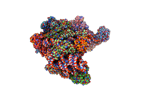
Cryo-Em Structure Of Saccharolobus Solfataricus 30S Initiation Complex Bound To Ss-Map Leaderless Mrna
Organism: Escherichia coli, Saccharolobus solfataricus p2
Method: ELECTRON MICROSCOPY Release Date: 2025-01-15 Classification: TRANSLATION Ligands: MG, SPM, ZN |
Organism: Escherichia coli, Saccharolobus solfataricus p2
Method: ELECTRON MICROSCOPY
Release Date: 2025-01-15
Ligands: MG, SPM, ZN
 |
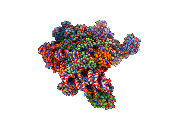
Cryo-Em Structure Of Saccharolobus Solfataricus 30S Initiation Complex Bound To Sd Mrna
Organism: Escherichia coli, Saccharolobus solfataricus p2
Method: ELECTRON MICROSCOPY Release Date: 2025-01-15 Classification: TRANSLATION Ligands: MG, SPM, ZN |
Organism: Escherichia coli, Saccharolobus solfataricus p2
Method: ELECTRON MICROSCOPY
Release Date: 2025-01-15
Ligands: MG, SPM, ZN
 |
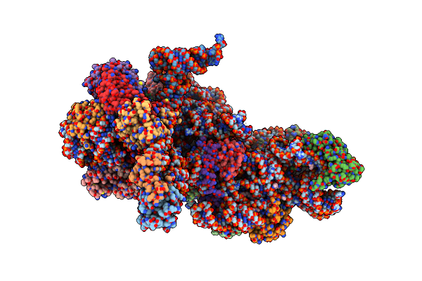
Cryo-Em Structure Of Saccharolobus Solfataricus 30S Initiation Complex Bound To Sd Mrna With H44 In Up Position
Organism: Escherichia coli, Pyrococcus abyssi, Saccharolobus solfataricus p2
Method: ELECTRON MICROSCOPY Release Date: 2025-01-15 Classification: TRANSLATION Ligands: MG, SPM, ZN |
Organism: Escherichia coli, Pyrococcus abyssi, Saccharolobus solfataricus p2
Method: ELECTRON MICROSCOPY
Release Date: 2025-01-15
Ligands: MG, SPM, ZN
 |
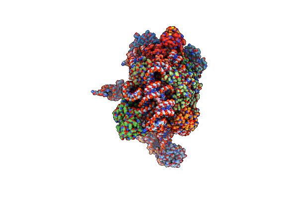
Cryo-Em Structure Of Saccharolobus Solfataricus 30S Initiation Complex Bound To Ss-Aif2Beta Leaderless Mrna With H44 In Up Position
Organism: Escherichia coli, Saccharolobus solfataricus p2
Method: ELECTRON MICROSCOPY Release Date: 2025-01-15 Classification: TRANSLATION Ligands: SPM, MG, ZN, GTP |
Organism: Escherichia coli, Saccharolobus solfataricus p2
Method: ELECTRON MICROSCOPY
Release Date: 2025-01-15
Ligands: SPM, MG, ZN, GTP
 |
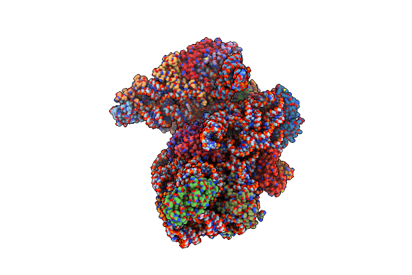
Cryo-Em Structure Of Saccharolobus Solfataricus 30S Initiation Complex Bound To Ss-Aef1A-Like Mrna
Organism: Escherichia coli, Saccharolobus solfataricus p2
Method: ELECTRON MICROSCOPY Release Date: 2025-01-15 Classification: TRANSLATION Ligands: MG, SPM, ZN |
Organism: Escherichia coli, Saccharolobus solfataricus p2
Method: ELECTRON MICROSCOPY
Release Date: 2025-01-15
Ligands: MG, SPM, ZN
 |
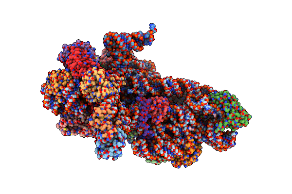
Cryo-Em Structure Of Saccharolobus Solfataricus 30S Initiation Complex Bound To Ss-Map Leaderless Mrna With H44 In Up Position
Organism: Escherichia coli, Saccharolobus solfataricus p2
Method: ELECTRON MICROSCOPY Release Date: 2025-01-15 Classification: TRANSLATION Ligands: SPM, MG, ZN, ATP |
Organism: Escherichia coli, Saccharolobus solfataricus p2
Method: ELECTRON MICROSCOPY
Release Date: 2025-01-15
Ligands: SPM, MG, ZN, ATP
 |
Cryo-Em Structure Of Saccharolobus Solfataricus 30S Initiation Complex Bound To Ss-Aif2Beta Leaderless Mrna
Organism: Escherichia coli, Saccharolobus solfataricus p2
Method: ELECTRON MICROSCOPY Release Date: 2025-01-15 Classification: TRANSLATION Ligands: SPM, MG, ZN, GTP |
Organism: Escherichia coli, Saccharolobus solfataricus p2
Method: ELECTRON MICROSCOPY
Release Date: 2025-01-15
Ligands: SPM, MG, ZN, GTP
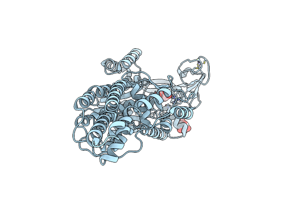 |
Escherichia Coli Methionyl-Trna Synthetase Mutant L13M,I297C
Organism: Escherichia coli
Method: X-RAY DIFFRACTION Resolution:1.55 Å Release Date: 2023-08-16 Classification: TRANSLATION Ligands: ZN, CIT |
Organism: Escherichia coli
Method: X-RAY DIFFRACTION
Release Date: 2023-08-16
Ligands: ZN, CIT
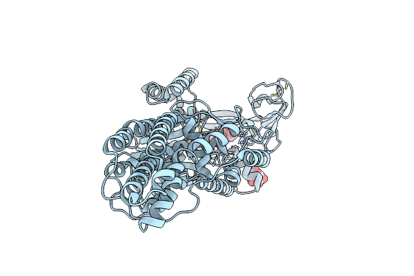 |
Escherichia Coli Methionyl-Trna Synthetase Mutant L13M,I297C Complexed With Beta3-Methionine.
Organism: Escherichia coli
Method: X-RAY DIFFRACTION Resolution:1.53 Å Release Date: 2023-08-16 Classification: TRANSLATION Ligands: ZN, CIT, B3M |
Organism: Escherichia coli
Method: X-RAY DIFFRACTION
Release Date: 2023-08-16
Ligands: ZN, CIT, B3M