
Search Count: 375
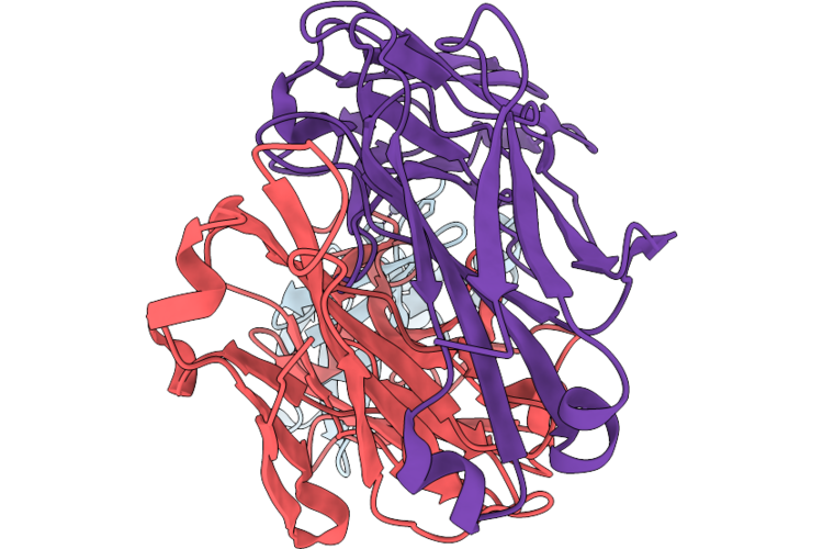 |
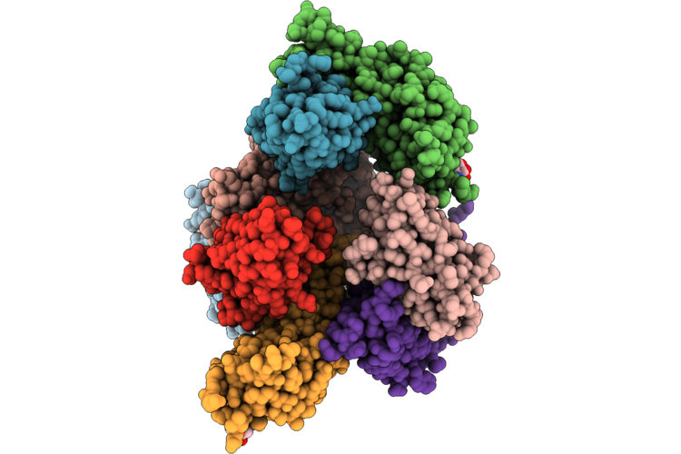
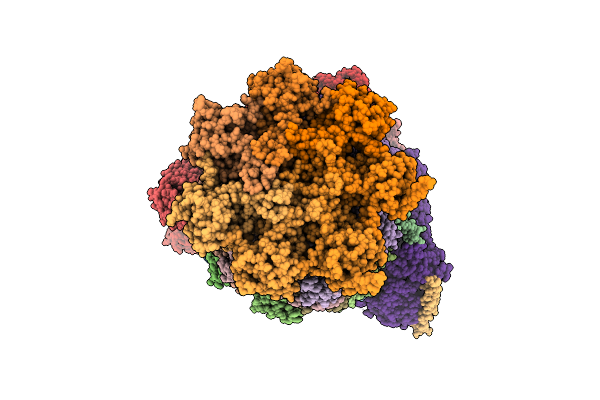
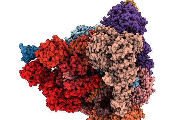
Structure Of Mab Phtd3 In Complex With Phtd
Organism: Streptococcus pneumoniae, Homo sapiens
Method: ELECTRON MICROSCOPY Release Date: 2026-06-17 Classification: ANTIMICROBIAL PROTEIN Ligands: ZN |
Organism: Streptococcus pneumoniae, Homo sapiens
Method: ELECTRON MICROSCOPY
Release Date: 2026-06-17
Ligands: ZN
 |
Crystal Structure Of A Flavin Dependent Baeyer Villiger Monooxygenase From Micromonospora Lupini Nbc_00409 In Complex With Fad
Organism: Micromonospora lupini
Method: X-RAY DIFFRACTION Resolution:3.20 Å Release Date: 2026-05-27 Classification: BIOSYNTHETIC PROTEIN Ligands: FAD |
Organism: Micromonospora lupini
Method: X-RAY DIFFRACTION
Release Date: 2026-05-27
Ligands: FAD
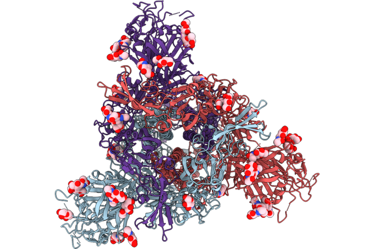 |
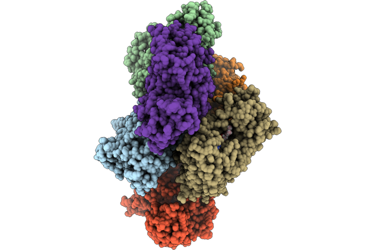
Prefusion Structure Of Hku4 Spike Glycoprotein
Organism: Bat coronavirus hku4
Method: ELECTRON MICROSCOPY Release Date: 2026-05-27 Classification: VIRAL PROTEIN Ligands: NAG |
Organism: Bat coronavirus hku4
Method: ELECTRON MICROSCOPY
Release Date: 2026-05-27
Ligands: NAG
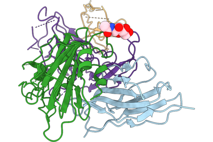 |
Crystal Structure Of Nanobody Tnb438 With Mers-Cov Rbd
Organism: Middle east respiratory syndrome-related coronavirus, Homo sapiens
Method: X-RAY DIFFRACTION Resolution:4.37 Å Release Date: 2026-05-27 Classification: VIRAL PROTEIN/IMMUNE SYSTEM Ligands: NAG |
Organism: Middle east respiratory syndrome-related coronavirus, Homo sapiens
Method: X-RAY DIFFRACTION
Release Date: 2026-05-27
Ligands: NAG
 |
Crystal Structure Of Nanobody Tnb494 With Mers-Cov Rbd
Organism: Homo sapiens, Middle east respiratory syndrome-related coronavirus
Method: X-RAY DIFFRACTION Resolution:2.70 Å Release Date: 2026-05-27 Classification: VIRAL PROTEIN/IMMUNE SYSTEM Ligands: NAG |
Organism: Homo sapiens, Middle east respiratory syndrome-related coronavirus
Method: X-RAY DIFFRACTION
Release Date: 2026-05-27
Ligands: NAG
 |
Crystal Structure Of Nanobody Tnb165 With Mers-Cov Rbd
Organism: Middle east respiratory syndrome-related coronavirus, Homo sapiens
Method: X-RAY DIFFRACTION Resolution:2.50 Å Release Date: 2026-04-29 Classification: VIRAL PROTEIN/IMMUNE SYSTEM Ligands: NAG |
Organism: Middle east respiratory syndrome-related coronavirus, Homo sapiens
Method: X-RAY DIFFRACTION
Release Date: 2026-04-29
Ligands: NAG
 |
Crystal Structure Of Nanobody Tnb150 With Mers-Cov Rbd
Organism: Middle east respiratory syndrome-related coronavirus, Homo sapiens
Method: X-RAY DIFFRACTION Resolution:3.24 Å Release Date: 2026-04-29 Classification: VIRAL PROTEIN/IMMUNE SYSTEM Ligands: NAG |
Organism: Middle east respiratory syndrome-related coronavirus, Homo sapiens
Method: X-RAY DIFFRACTION
Release Date: 2026-04-29
Ligands: NAG
 |
Crystal Structure Of Nanobody Tnb316 With Nanobody B9 And Mers-Cov Rbd
Organism: Homo sapiens, Middle east respiratory syndrome-related coronavirus
Method: X-RAY DIFFRACTION Resolution:1.96 Å Release Date: 2026-04-29 Classification: VIRAL PROTEIN/IMMUNE SYSTEM |
 |
Q23.Md39 In Complex With Fabs From Antibodies Ch01 And 35O22
Organism: Homo sapiens, Human immunodeficiency virus 1
Method: ELECTRON MICROSCOPY Release Date: 2026-04-22 Classification: VIRAL PROTEIN/IMMUNE SYSTEM Ligands: NAG |
Organism: Homo sapiens, Human immunodeficiency virus 1
Method: ELECTRON MICROSCOPY
Release Date: 2026-04-22
Ligands: NAG
 |
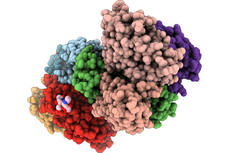
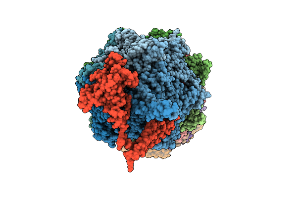
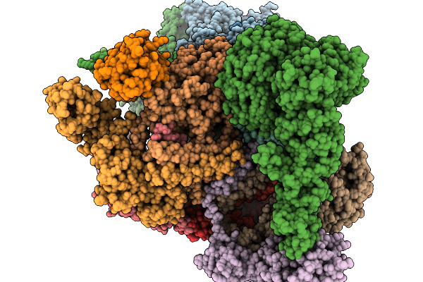
Structure Of Human 26S Proteasome Complexed With Midnolin(1-111+337-468)
Organism: Homo sapiens, Escherichia coli str. k-12 substr. mg1655, Synthetic construct
Method: ELECTRON MICROSCOPY Resolution:3.42 Å Release Date: 2026-04-01 Classification: HYDROLASE Ligands: ZN, ATP, MG, ADP, LDZ |
Organism: Homo sapiens, Escherichia coli str. k-12 substr. mg1655, Synthetic construct
Method: ELECTRON MICROSCOPY
Release Date: 2026-04-01
Ligands: ZN, ATP, MG, ADP, LDZ
 |
Substrate-Free Human 26S Proteasome Purified By Midnolin, 20S Proteasome, Rpts And Rpn11 Part
Organism: Homo sapiens
Method: ELECTRON MICROSCOPY Release Date: 2026-04-01 Classification: HYDROLASE Ligands: ZN, ATP, MG, ADP, LDZ |
Organism: Homo sapiens
Method: ELECTRON MICROSCOPY
Release Date: 2026-04-01
Ligands: ZN, ATP, MG, ADP, LDZ
 |
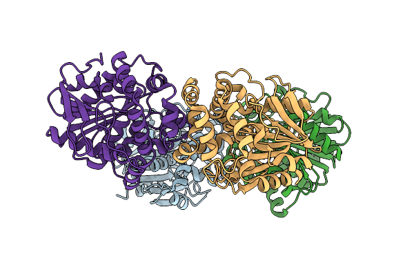
Crystal Structure Of The Fluoroacetate Dehalogenase Rpa1163 - Trp185Tyr/His280Ala With 2-Fluoro-3-Phenylpropanoic Acid
Organism: Rhodopseudomonas palustris cga009
Method: X-RAY DIFFRACTION Resolution:1.47 Å Release Date: 2026-04-01 Classification: TRANSFERASE |
Organism: Rhodopseudomonas palustris cga009
Method: X-RAY DIFFRACTION
Release Date: 2026-04-01
 |
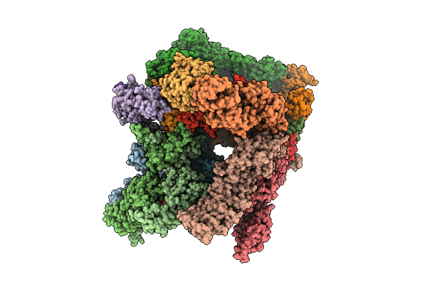
Structure Of Human 26S Proteasome Complexed With Midnolin, 19S Proteasome With Ubl Bound
Organism: Escherichia coli k-12, Homo sapiens, Purpureocillium lilacinum
Method: ELECTRON MICROSCOPY Resolution:3.65 Å Release Date: 2026-04-01 Classification: HYDROLASE Ligands: ADP, ATP, ZN, MG |
Organism: Escherichia coli k-12, Homo sapiens, Purpureocillium lilacinum
Method: ELECTRON MICROSCOPY
Release Date: 2026-04-01
Ligands: ADP, ATP, ZN, MG
 |
Structure Of Human 26S Proteasome Complexed With Midnolin, 19S Proteasome With Ubl And Catch Domain Resolved
Organism: Escherichia coli k-12, Homo sapiens, Pseudotamlana agarivorans
Method: ELECTRON MICROSCOPY Release Date: 2026-04-01 Classification: HYDROLASE Ligands: ADP, ATP, ZN, MG |
Organism: Escherichia coli k-12, Homo sapiens, Pseudotamlana agarivorans
Method: ELECTRON MICROSCOPY
Release Date: 2026-04-01
Ligands: ADP, ATP, ZN, MG
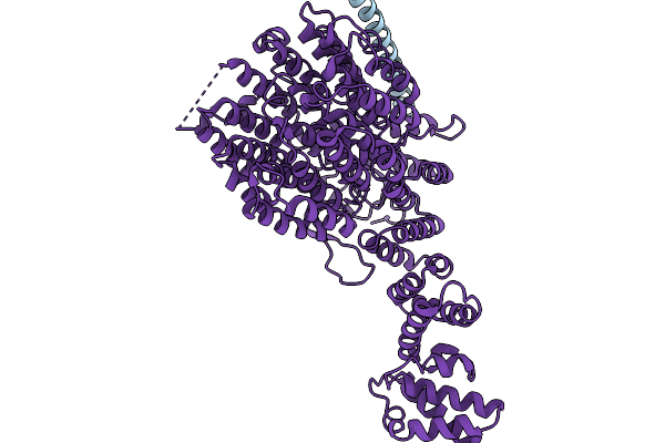 |
Focused Refinement Of Rpn1 And The C-Terminal Helix Of Midnolin In The Substrate-Engaged Human 26S Proteasome
Organism: Homo sapiens
Method: ELECTRON MICROSCOPY Release Date: 2026-03-25 Classification: HYDROLASE |
Organism: Homo sapiens
Method: ELECTRON MICROSCOPY
Release Date: 2026-03-25
 |
Substrate-Engaged Human 26S Proteasome Bound To Midnolin With Rpt1 At Top Of Spiral Staircase
Organism: Homo sapiens
Method: ELECTRON MICROSCOPY Release Date: 2026-03-25 Classification: HYDROLASE Ligands: ZN, ATP, MG, ADP, LDZ |
Organism: Homo sapiens
Method: ELECTRON MICROSCOPY
Release Date: 2026-03-25
Ligands: ZN, ATP, MG, ADP, LDZ
 |
Substrate-Engaged Human 26S Proteasome Bound To Midnolin With Rpt5 At Top Of Spiral Staircase
Organism: Homo sapiens
Method: ELECTRON MICROSCOPY Release Date: 2026-03-25 Classification: HYDROLASE Ligands: ZN, ATP, MG, ADP, LDZ |
Organism: Homo sapiens
Method: ELECTRON MICROSCOPY
Release Date: 2026-03-25
Ligands: ZN, ATP, MG, ADP, LDZ
 |
Substrate-Engaged Human 26S Proteasome Bound To Midnolin With Rpt2 At Top Of Spiral Staircase
Organism: Homo sapiens
Method: ELECTRON MICROSCOPY Release Date: 2026-03-25 Classification: HYDROLASE Ligands: ADP, ATP, MG, LDZ, ZN |
Organism: Homo sapiens
Method: ELECTRON MICROSCOPY
Release Date: 2026-03-25
Ligands: ADP, ATP, MG, LDZ, ZN
 |
Focused Refinement Of 19S In The Substrate-Engaged Human 26S Proteasome Bound To Midnolin With Rpt6 At Top Of Spiral Staircase
Organism: Homo sapiens
Method: ELECTRON MICROSCOPY Release Date: 2026-03-25 Classification: HYDROLASE Ligands: ADP, ZN, ATP, MG |
Organism: Homo sapiens
Method: ELECTRON MICROSCOPY
Release Date: 2026-03-25
Ligands: ADP, ZN, ATP, MG
 |
Crystal Structure Of The Fluoroacetate Dehalogenase Rpa1163 - Lys181Met/Trp185Tyr/His280Ala With 2-Fluoro-3-Phenylpropanoic Acid
Organism: Rhodopseudomonas palustris cga009
Method: X-RAY DIFFRACTION Resolution:2.08 Å Release Date: 2026-03-25 Classification: TRANSFERASE |
Organism: Rhodopseudomonas palustris cga009
Method: X-RAY DIFFRACTION
Release Date: 2026-03-25