

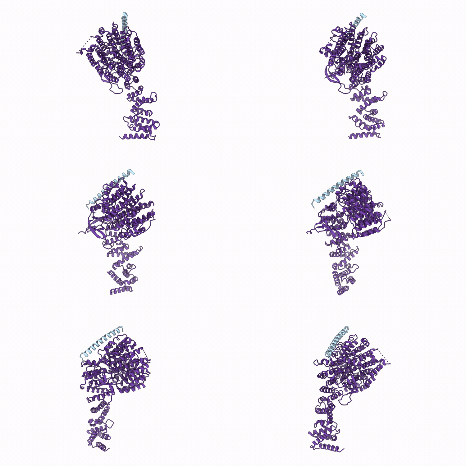
9MBO image
Deposition Date 2025-03-17
Release Date 2026-03-25
Last Version Date 2026-04-08
Entry Detail
PDB ID:
9MBO
Keywords:
Title:
Focused refinement of RPN1 and the C-terminal helix of midnolin in the substrate-engaged human 26S proteasome
Biological Source:
Source Organism(s):
Homo sapiens (Taxon ID: 9606)
Expression System(s):
Method Details:
Experimental Method:
Resolution:
2.83 Å
Aggregation State:
PARTICLE
Reconstruction Method:
SINGLE PARTICLE
Macromolecular Entities
Structures with similar UniProt ID
Protein Blast
Polymer Type:polypeptide(L)
Molecule:Midnolin
Gene (Uniprot):MIDN
Chain IDs:A
Chain Length:468
Number of Molecules:1
Biological Source:Homo sapiens
Structures with similar UniProt ID
Protein Blast
Polymer Type:polypeptide(L)
Molecule:26S proteasome non-ATPase reg
Gene (Uniprot):PSMD2
Chain IDs:B
Chain Length:908
Number of Molecules:1
Biological Source:Homo sapiens
Ligand Molecules
Primary Citation
Structural dynamics of the midnolin-proteasome during ubiquitin-independent substrate turnover.
Nat Commun 17 ? ? (2026)
PMID: 41896529 DOI: 10.1038/s41467-026-71002-0

Abstact

The 26S proteasome typically degrades proteins marked by ubiquitin chains. However, a distinct, ubiquitin-independent degradation pathway for nuclear proteins exists, mediated by the adaptor protein midnolin, yet its molecular mechanism remains poorly understood. Here, we present nine cryo-electron microscopy structures of the human 26S proteasome in complex with midnolin, which collectively delineate a near-complete catalytic cycle. Our structures reveal that midnolin binds to the proteasome via the RPN1 subunit by its C-terminal helix. Unexpectedly, its ubiquitin-like domain interacts with the RPN11 deubiquitinase in a non-catalytic role. This interaction positions the adjacent Catch domain, which is responsible for substrate binding, directly above the proteasomal entrance, potentially facilitating substrate entry into the proteasome. Furthermore, we observe four consecutive spiral staircase conformations of the AAA+ ATPase hexamer during substrate translocation. These findings provide insights into the mechanisms underlying ubiquitin-independent nuclear protein degradation and may help develop strategies for targeting nuclear proteins via direct proteasomal degradation.

Legend

Protein

Chemical

Disease

Primary Citation of related structures
Feedback Form
Name
Email
Institute
Feedback