
Search Count: 1,667
All
Selected
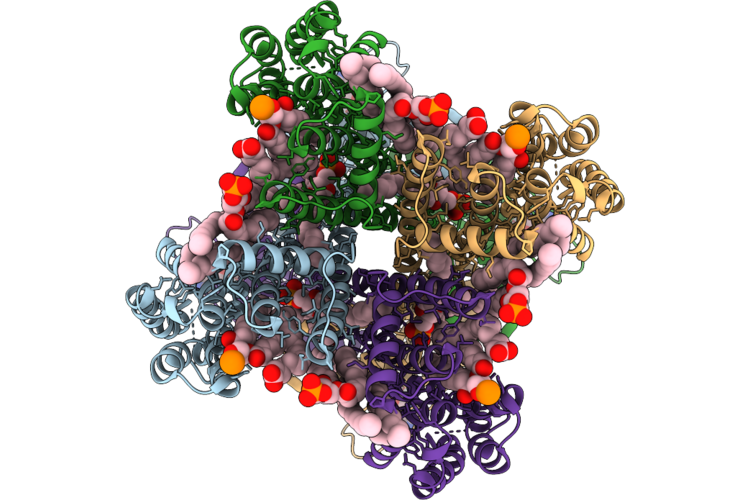 |
Organism: Winmispira thermophila
Method: ELECTRON MICROSCOPY Resolution:2.51 Å Release Date: 2026-04-29 Classification: TRANSPORT PROTEIN Ligands: CMP, PCW |
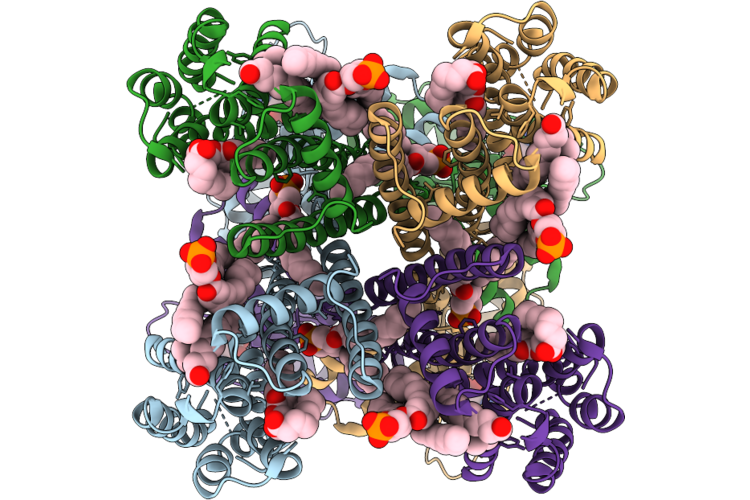 |
Organism: Spirochaeta thermophila
Method: ELECTRON MICROSCOPY Release Date: 2026-04-22 Classification: TRANSPORT PROTEIN Ligands: CMP, PEE |
 |
Sthk Intermediate State At Low Temperature, Camp-Bound In The Presence Of Dope
Organism: Spirochaeta thermophila
Method: ELECTRON MICROSCOPY Release Date: 2026-04-22 Classification: TRANSPORT PROTEIN Ligands: CMP, PEE |
 |
Crystal Structure Of Human Pkmyt1 Protein Kinase Domain With Naphthyridinone Inhibitor Compound 11
Organism: Homo sapiens
Method: X-RAY DIFFRACTION Resolution:1.74 Å Release Date: 2026-03-25 Classification: TRANSFERASE Ligands: A1E5A, SO4 |
 |
Crystal Structure Of Human Pkmyt1 Protein Kinase Domain With Naphthyridinone Inhibitor Compound 16
Organism: Homo sapiens
Method: X-RAY DIFFRACTION Resolution:1.59 Å Release Date: 2026-03-25 Classification: TRANSFERASE Ligands: A1E5J, DMS, EDO, SO4 |
 |
Phosphorylation Dependent Recognition Of Ripk1 By Phosphatidylinositol 3,4,5-Trisphosphate 5-Phosphatase 1
|
 |
Organism: Homo sapiens, Synthetic construct
Method: ELECTRON MICROSCOPY Release Date: 2026-03-04 Classification: SIGNALING PROTEIN/IMMUNE SYSTEM Ligands: ODN |
 |
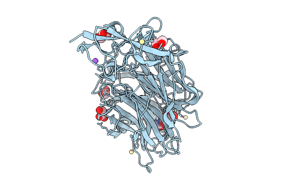
Organism: Treponema denticola
Method: X-RAY DIFFRACTION Resolution:1.63 Å Release Date: 2026-02-25 Classification: HYDROLASE Ligands: CD, NA, B3P, CIT, EDO, PGE |
 |
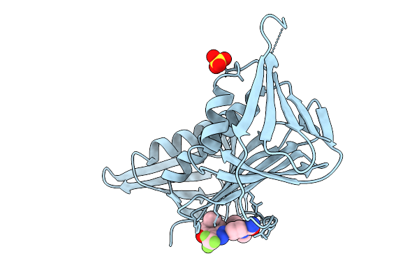
Crystal Structure Of Treponema Denticola Sialidase (Tde_0471) Bound To Neu5Ac2En (Dana)
Organism: Treponema denticola
Method: X-RAY DIFFRACTION Resolution:1.55 Å Release Date: 2026-02-25 Classification: HYDROLASE Ligands: DAN, EDO, PEG, CD |
 |
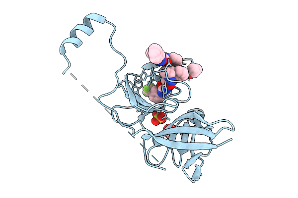
Crystal Structure Of Treponema Denticola Sialidase (Tde_0471) Bound To Neu5Ac (Nana)
Organism: Treponema denticola
Method: X-RAY DIFFRACTION Resolution:1.56 Å Release Date: 2026-02-25 Classification: HYDROLASE Ligands: CD, SLB, EDO, PEG, PGE |
 |
Crystal Structure Of Treponema Denticola Sialidase (Tde_0471) D165A Mutant Bound To 3'-Sialyllactose (Only Neu5Ac Visible)
Organism: Treponema denticola
Method: X-RAY DIFFRACTION Resolution:1.81 Å Release Date: 2026-02-25 Classification: HYDROLASE Ligands: SIA, GOL, PEG, CD, NA |
 |
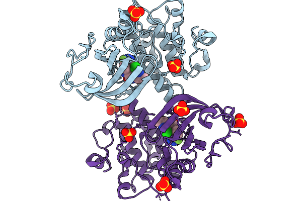
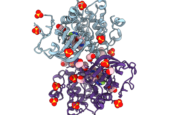
Organism: Homo sapiens
Method: ELECTRON MICROSCOPY Resolution:3.26 Å Release Date: 2026-02-25 Classification: TRANSPORT PROTEIN Ligands: GDS |
 |
Organism: Homo sapiens
Method: X-RAY DIFFRACTION Resolution:1.90 Å Release Date: 2026-02-25 Classification: DNA BINDING PROTEIN Ligands: E0G, CL, SO4 |
 |
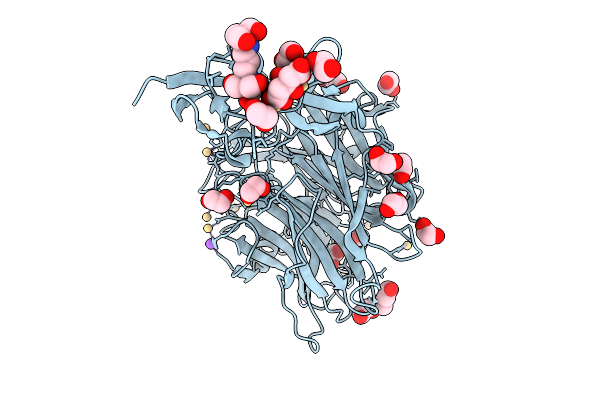
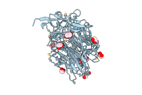
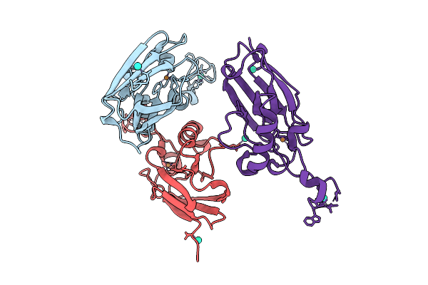
Crystal Structure Of Zika Virus Ns2B-Ns3 Protease In Complex With Compound 1
Organism: Zika virus
Method: X-RAY DIFFRACTION Resolution:2.48 Å Release Date: 2026-01-28 Classification: VIRAL PROTEIN Ligands: A1H2Z, SO4, IPA |
 |
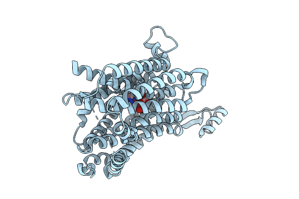
Organism: Pseudomonas aeruginosa (strain atcc 15692 / dsm 22644 / cip 104116 / jcm 14847 / lmg 12228 / 1c / prs 101 / pao1)
Method: X-RAY DIFFRACTION Resolution:2.38 Å Release Date: 2025-12-24 Classification: ELECTRON TRANSPORT Ligands: CU, TB |
 |
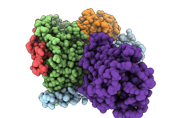
Cryo-Em Structure Of The Vaccinia Virus Entry/Fusion Complex (Efc) Lacking The F9 Subunit
Organism: Orthopoxvirus vaccinia
Method: ELECTRON MICROSCOPY Release Date: 2025-12-17 Classification: VIRAL PROTEIN |
 |
Cryo-Em Structure Of The Vaccinia Virus Entry/Fusion Complex (Efc) Including The F9 Subunit
Organism: Orthopoxvirus vaccinia
Method: ELECTRON MICROSCOPY Release Date: 2025-12-17 Classification: VIRAL PROTEIN |
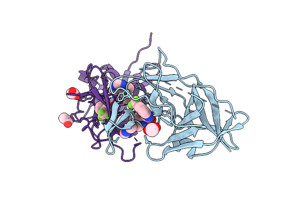 |
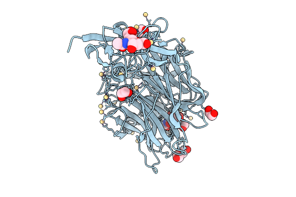
Crystal Structure Of Zika Virus Ns2B-Ns3 Protease In Complex With Compound 2
Organism: Zika virus
Method: X-RAY DIFFRACTION Resolution:2.29 Å Release Date: 2025-12-03 Classification: HYDROLASE Ligands: A1I16, ACY |
 |
Organism: Marinobacter sp. dsm 11874
Method: X-RAY DIFFRACTION Resolution:1.68 Å Release Date: 2025-11-26 Classification: TRANSPORT PROTEIN Ligands: 1GP |
 |
Organism: Marinobacter sp. dsm 11874
Method: X-RAY DIFFRACTION Resolution:2.00 Å Release Date: 2025-11-26 Classification: TRANSPORT PROTEIN Ligands: G3P |