

Search Count: 837
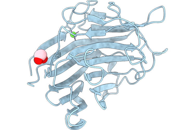 |
Engineered Endo-1,3-Beta-Glucanase From Cellulosimicrobium Cellulans Crystallized At Ph 6.2, Ccgluecdmh1-E120Q
Organism: Cellulosimicrobium cellulans
Method: X-RAY DIFFRACTION Resolution:1.76 Å Release Date: 2026-07-08 Classification: HYDROLASE Ligands: EDO, CA |
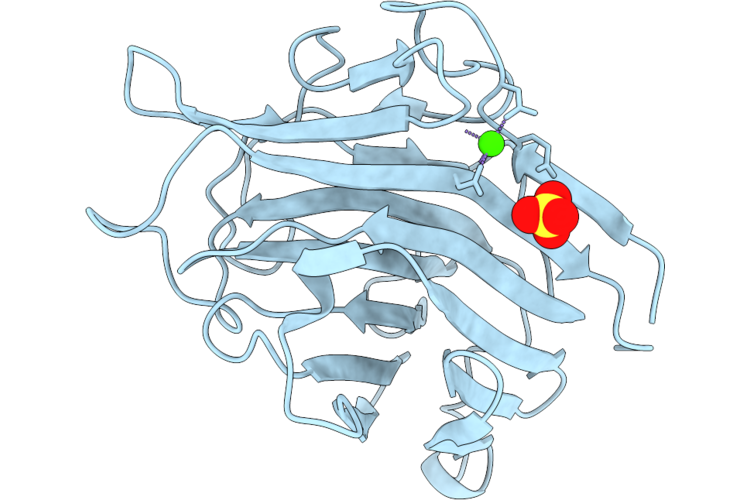 |
Engineered Endo-1,3-Beta-Glucanase From Cellulosimicrobium Cellulans Crystallized At Ph6.2,Ccgluecdmh1
Organism: Cellulosimicrobium cellulans
Method: X-RAY DIFFRACTION Resolution:1.28 Å Release Date: 2026-07-08 Classification: HYDROLASE Ligands: CA, SO4 |
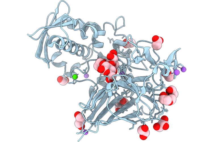 |
Gh64 Family Beta-1,3-Glucanase From Massilia Violaceinigra In Complex With A Glucose Disaccharide
Organism: Massilia violaceinigra
Method: X-RAY DIFFRACTION Resolution:1.99 Å Release Date: 2026-07-08 Classification: HYDROLASE Ligands: EDO, ACT, GOL, PGE, CA, CL, NA |
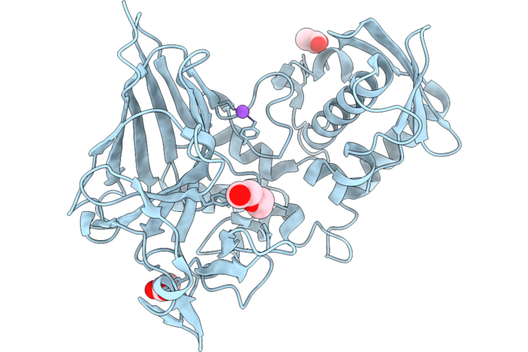 |
Gh64 Family Beta-1,3-Glucanase From Massilia Violaceinigra
Organism: Massilia violaceinigra
Method: X-RAY DIFFRACTION Resolution:2.30 Å Release Date: 2026-07-08 Classification: HYDROLASE |
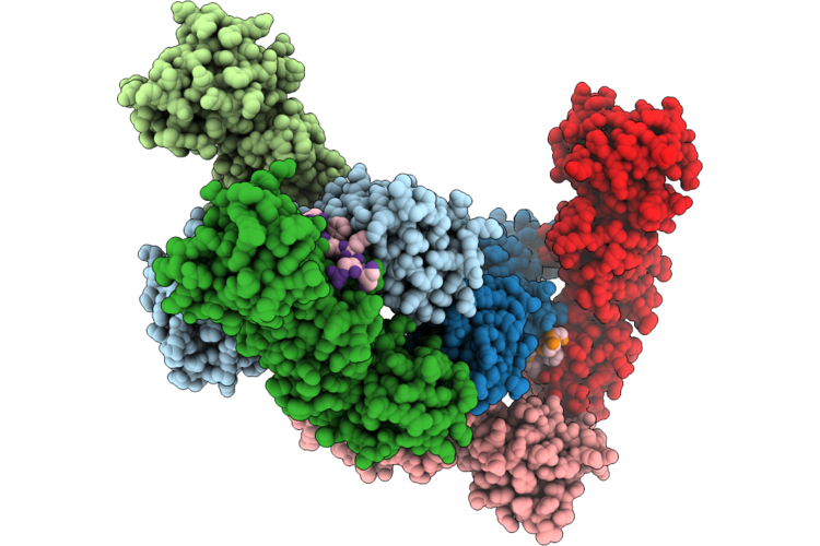 |
Topbp1 Brct0-2 In Complex With Phosphorylated Htatsf1
Organism: Homo sapiens, Mus musculus
Method: X-RAY DIFFRACTION Resolution:2.99 Å Release Date: 2026-07-01 Classification: PEPTIDE BINDING PROTEIN |
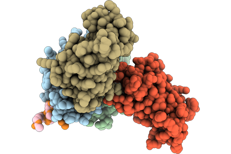 |
The Crystal Structure Of Brd4-Bds In Complex With H2A Peptide
Organism: Homo sapiens
Method: X-RAY DIFFRACTION Resolution:2.61 Å Release Date: 2026-07-01 Classification: PROTEIN BINDING |
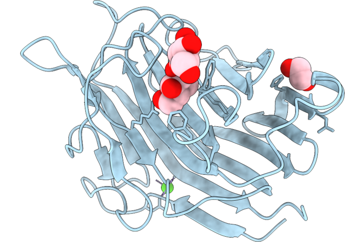 |
Engineered Endo-1,3-Beta-Glucanase From Cellulosimicrobium Cellulans In Complex With A Glucose Disaccharide At Ph4.6,Ccgluecdmh1+Dp2
Organism: Cellulosimicrobium cellulans
Method: X-RAY DIFFRACTION Resolution:1.33 Å Release Date: 2026-07-01 Classification: HYDROLASE Ligands: CA, EDO |
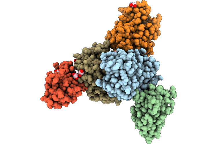 |
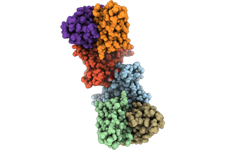
Crystal Structure Of The Sb32-Tgp Complex
Organism: Galaxea fascicularis, Synthetic construct
Method: X-RAY DIFFRACTION Resolution:1.82 Å Release Date: 2026-06-24 Classification: PROTEIN BINDING Ligands: GOL |
 |
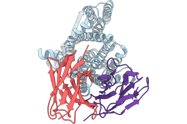
Cryo-Em Structure Of The Consensus Inward-Facing Apo Nhaa Dimer At Ph 7.5
Organism: Escherichia coli, Mus musculus
Method: ELECTRON MICROSCOPY Resolution:2.70 Å Release Date: 2026-06-17 Classification: TRANSPORT PROTEIN Ligands: CDL, PGT |
 |
Cryo-Em Structure Of The Inward-Facing Apo Nhaa In The Unplugged State At Ph 7.5
Organism: Escherichia coli, Mus musculus
Method: ELECTRON MICROSCOPY Release Date: 2026-06-17 Classification: TRANSPORT PROTEIN |
 |
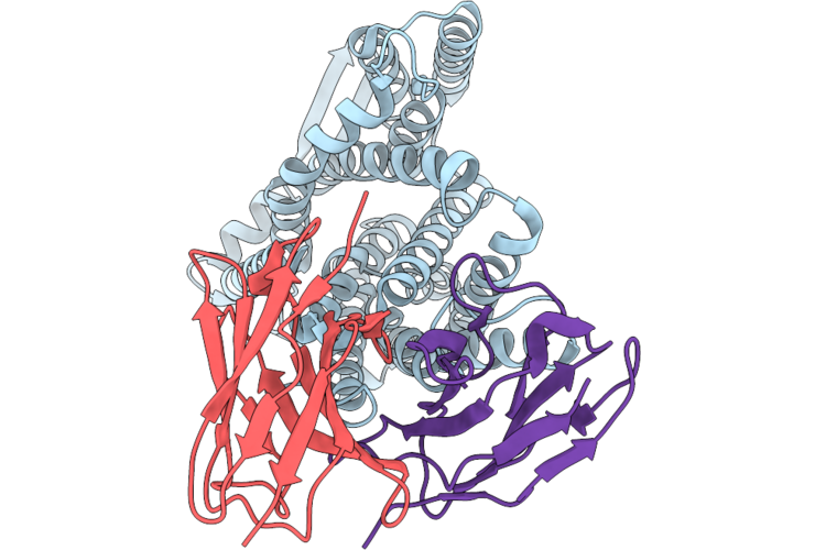
Cryo-Em Structure Of The Inward-Facing Apo Nhaa In The Plugged State At Ph 7.5
Organism: Escherichia coli, Mus musculus
Method: ELECTRON MICROSCOPY Release Date: 2026-06-17 Classification: TRANSPORT PROTEIN |
 |
Cryo-Em Structure Of The Inward-Facing Apo Nhaa With Flexible N-Terminus At Ph 7.5
Organism: Escherichia coli, Mus musculus
Method: ELECTRON MICROSCOPY Release Date: 2026-06-17 Classification: TRANSPORT PROTEIN |
 |
Cryo-Em Structure Of The Inward-Facing Apo Nhaa In The Unplugged State At Ph 6.3
Organism: Escherichia coli, Mus musculus
Method: ELECTRON MICROSCOPY Release Date: 2026-06-17 Classification: TRANSPORT PROTEIN |
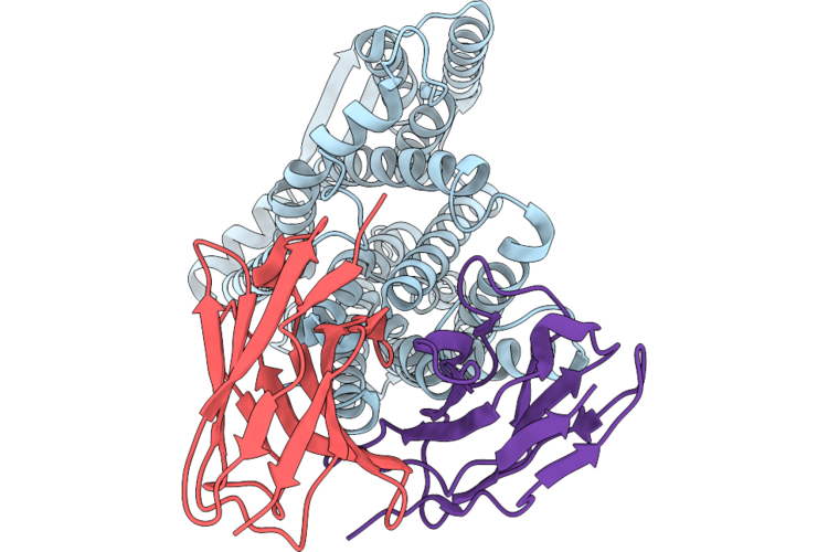 |
Cryo-Em Structure Of The Inward-Facing Apo Nhaa In The Plugged State At Ph 6.3
Organism: Escherichia coli, Mus musculus
Method: ELECTRON MICROSCOPY Release Date: 2026-06-17 Classification: TRANSPORT PROTEIN |
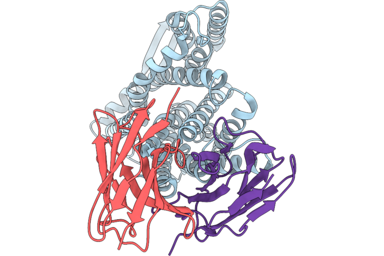 |
Cryo-Em Structure Of The Inward-Facing Apo Nhaa With Flexible N-Terminus At Ph 6.3
Organism: Escherichia coli, Mus musculus
Method: ELECTRON MICROSCOPY Release Date: 2026-06-17 Classification: TRANSPORT PROTEIN |
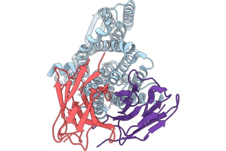 |
Cryo-Em Structure Of The Inward-Facing Apo Nhaa In The Unplugged State At Ph 5.5
Organism: Escherichia coli, Mus musculus
Method: ELECTRON MICROSCOPY Release Date: 2026-06-17 Classification: TRANSPORT PROTEIN |
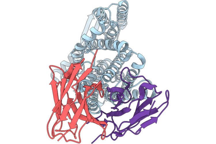 |
Cryo-Em Structure Of The Inward-Facing Apo Nhaa In The Plugged State At Ph 5.5
Organism: Escherichia coli, Mus musculus
Method: ELECTRON MICROSCOPY Release Date: 2026-06-17 Classification: TRANSPORT PROTEIN |
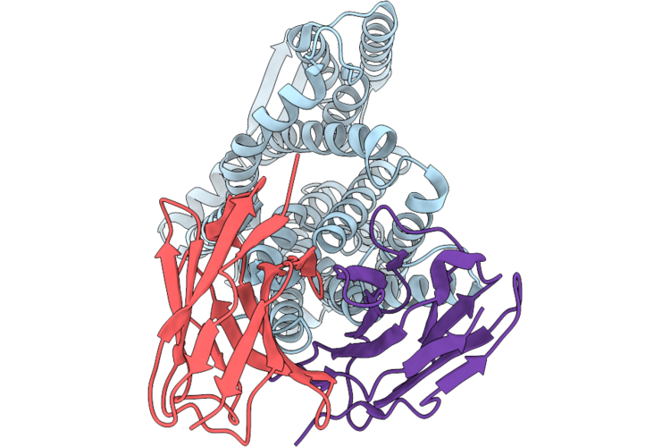 |
Cryo-Em Structure Of The Inward-Facing Apo Nhaa With Flexible N-Terminus At Ph 5.5
Organism: Escherichia coli, Mus musculus
Method: ELECTRON MICROSCOPY Release Date: 2026-06-17 Classification: TRANSPORT PROTEIN |
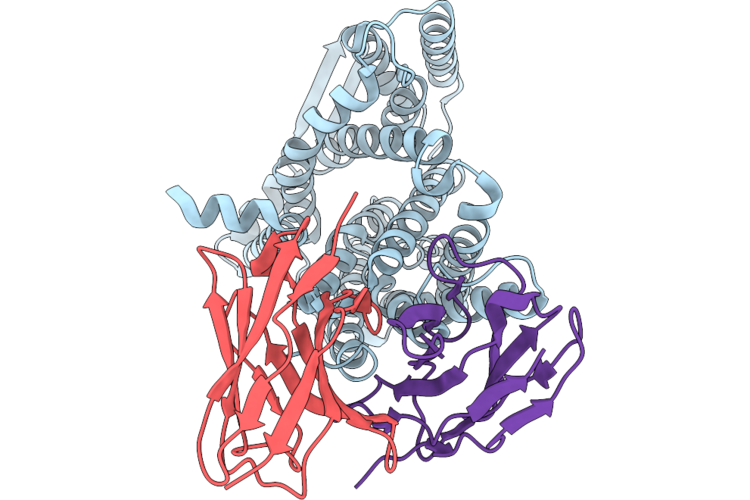 |
Cryo-Em Structure Of The Inward-Facing Apo Nhaa In The Open-Funnel State At Ph 8.5
Organism: Escherichia coli, Mus musculus
Method: ELECTRON MICROSCOPY Release Date: 2026-06-17 Classification: TRANSPORT PROTEIN Ligands: HOH |
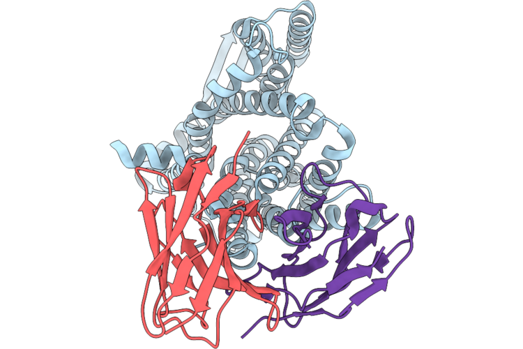 |
Cryo-Em Structure Of The Inward-Facing Apo Nhaa In The Unplugged State At Ph 8.5
Organism: Escherichia coli, Mus musculus
Method: ELECTRON MICROSCOPY Release Date: 2026-06-17 Classification: TRANSPORT PROTEIN |