

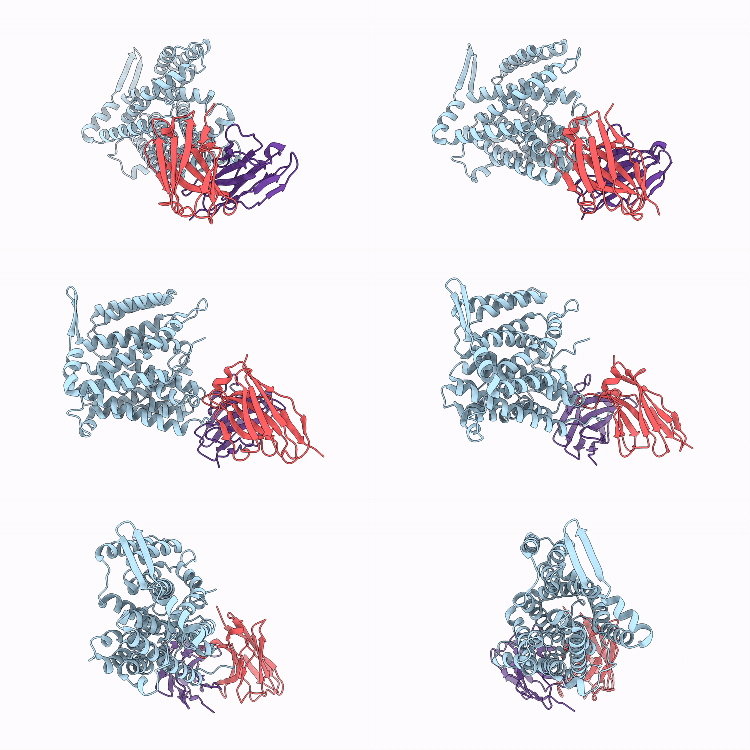
9RH6 image
Deposition Date 2025-06-08
Release Date 2026-06-17
Last Version Date 2026-08-05
Entry Detail
PDB ID:
9RH6
Title:
Cryo-EM structure of the inward-facing apo NhaA in the plugged state at pH 6.3
Biological Source:
Source Organism(s):
Escherichia coli (Taxon ID: 562)
Mus musculus (Taxon ID: 10090)
Expression System(s):
Method Details:
Experimental Method:
Resolution:
3.40 Å
Aggregation State:
PARTICLE
Reconstruction Method:
SINGLE PARTICLE
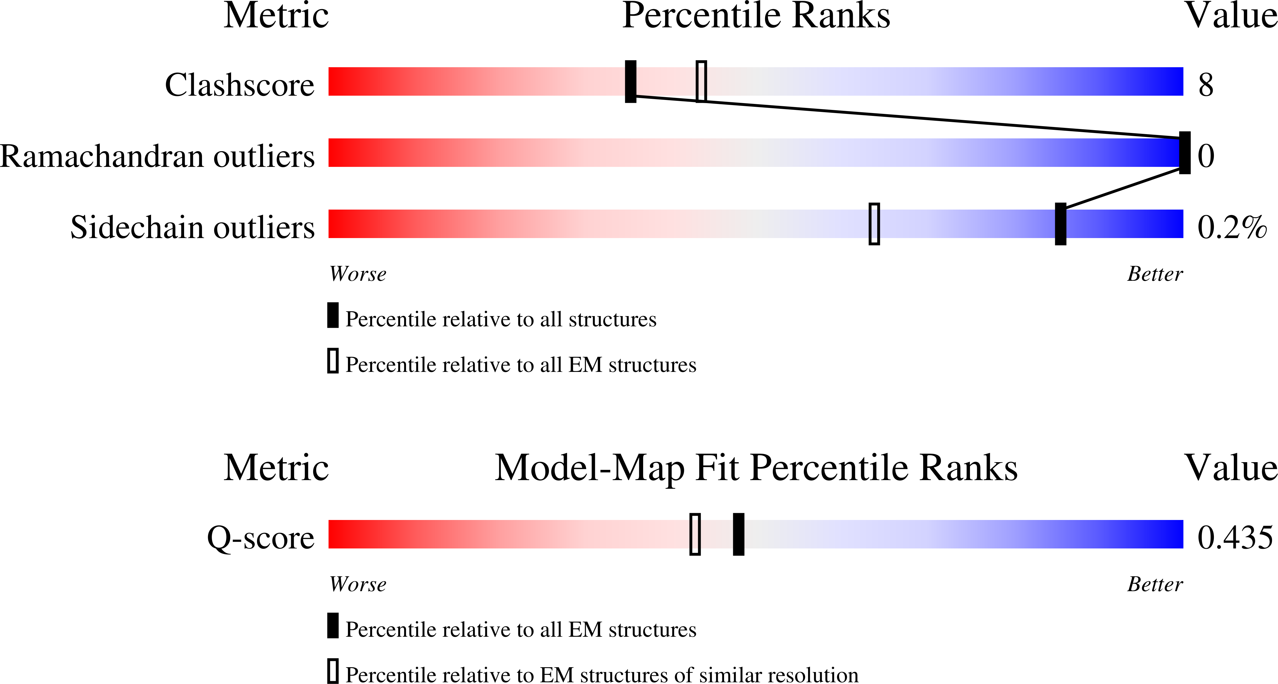
Macromolecular Entities
Polymer Type:polypeptide(L)
Molecule:Na(+)/H(+) antiporter NhaA
Gene (Uniprot):nhaA
Chain IDs:A
Chain Length:407
Number of Molecules:1
Biological Source:Escherichia coli
Structural Superimposition Protein Blast
Polymer Type:polypeptide(L)
Molecule:Fv6F9 heavy chain
Chain IDs:B
Chain Length:127
Number of Molecules:1
Biological Source:Mus musculus
Structural Superimposition Protein Blast
Polymer Type:polypeptide(L)
Molecule:Fv6F9 light chain
Chain IDs:C
Chain Length:119
Number of Molecules:1
Biological Source:Mus musculus
Ligand Molecules
Primary Citation
pH-dependent activation of the Na + /H + antiporter NhaA and conformational dynamics of its N-terminus.
Nat Commun 17 ? ? (2026)
PMID: 42285937 DOI: 10.1038/s41467-026-73424-2

Abstact

Na(+)/H(+) antiporters are vital for regulating intracellular pH and sodium ion levels across all domains of life. In Escherichia coli, NhaA is the principal Na(+)/H(+) antiporter, exhibiting strong pH sensitivity and rapid turnover, yet the structural transitions underlying its activation and substrate recognition have remained obscure. Here, we use single-particle cryo-electron microscopy to determine the conformational ensemble of NhaA across a physiological pH range and in the presence of Na(+), complemented by constant-pH molecular dynamics simulations. High-resolution structures of apo and Na(+)-bound NhaA reconstituted in lipid nanodiscs reveal progressive opening of the cytoplasmic funnel with increasing pH. We also visualize the previously unresolved N-terminal tail, which forms a dynamic plug at the cytoplasmic entrance under low-pH conditions and disengages at alkaline pH, coinciding with activation. The Na(+)-bound structure captures Na(+) coordination at the ion-binding site, and simulations suggest potential roles for the conserved charged residues. Together, these findings illuminate how pH sensing, N-terminal gating, and substrate binding are structurally coordinated in NhaA, providing a framework for understanding Na(+)/H(+) antiporter activation and regulation, and the basis for targeting clinical important antiporters.

Legend

Protein

Chemical

Disease

Primary Citation of related structures
Feedback Form
Name
Email
Institute
Feedback