
Search Count: 602
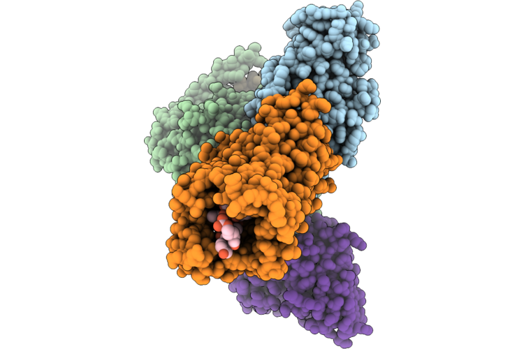 |
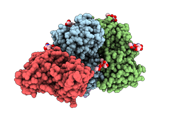
Organism: Homo sapiens, Escherichia coli, Oplophorus gracilirostris
Method: ELECTRON MICROSCOPY Resolution:2.80 Å Release Date: 2026-04-29 Classification: SIGNALING PROTEIN/IMMUNE SYSTEM Ligands: CLR |
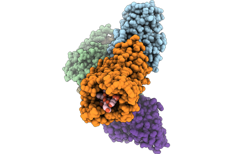 |
Organism: Homo sapiens, Escherichia coli, Oplophorus gracilirostris
Method: ELECTRON MICROSCOPY Resolution:2.70 Å Release Date: 2026-04-29 Classification: SIGNALING PROTEIN Ligands: CLR |
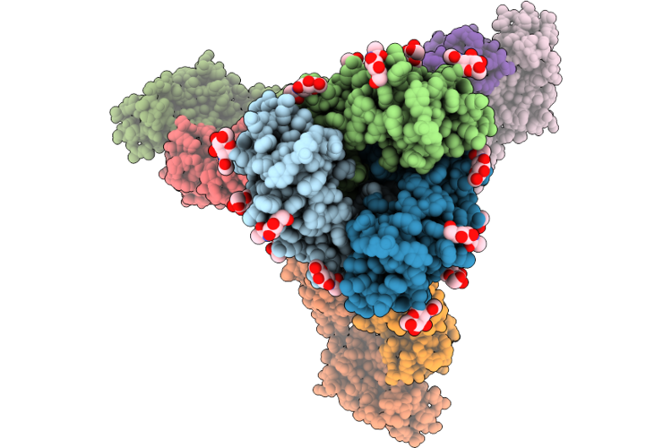 |
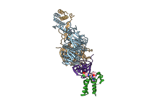
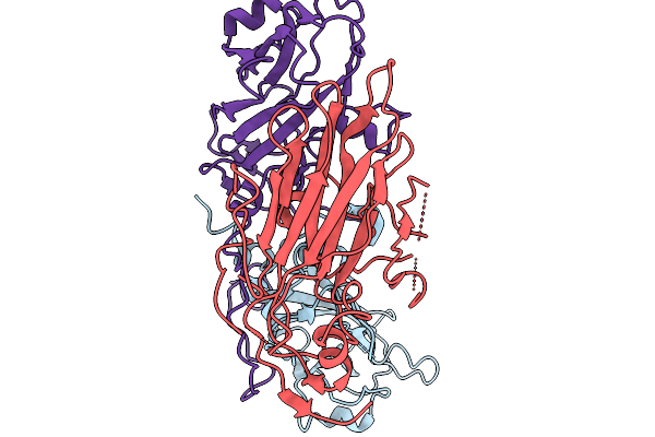
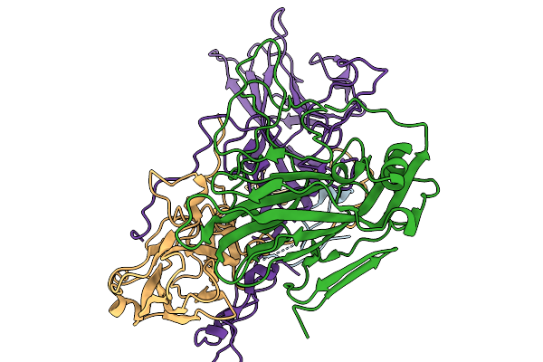
Cryo-Em Structure Of Sars-Cov-2 Wide-Type S Trimer In The Early Fusion Intermediate Conformation (E-Fic) Complexed With Ace2 And 76E1-Fab (Focused Refinement Of The S2-76E1)
Organism: Severe acute respiratory syndrome coronavirus 2, Homo sapiens
Method: ELECTRON MICROSCOPY Release Date: 2026-04-22 Classification: VIRAL PROTEIN/IMMUNE SYSTEM Ligands: NAG |
 |
Cryo-Em Structure Of Sars-Cov-2 Wide-Type S Trimer In The Early Fusion Intermediate Conformation (E-Fic) Complexed With Ace2 And 76E1-Fab
Organism: Severe acute respiratory syndrome coronavirus 2, Homo sapiens
Method: ELECTRON MICROSCOPY Release Date: 2026-04-22 Classification: VIRAL PROTEIN/HYDROLASE Ligands: NAG |
 |
Cryo-Em Structure Of Sars-Cov-2 Xbb.1.5 S Trimer In The Early Fusion Intermediate Conformation (E-Fic) Complexed With Ace2 And 76E1-Fab (Focused Refinement Of The S2-76E1)
Organism: Severe acute respiratory syndrome coronavirus 2, Homo sapiens
Method: ELECTRON MICROSCOPY Release Date: 2026-04-22 Classification: VIRAL PROTEIN/IMMUNE SYSTEM Ligands: NAG |
 |
Cryo-Em Structure Of Sars-Cov-2 Xbb.1.5 S Trimer In The Early Fusion Intermediate Conformation (E-Fic) Complexed With Ace2 And 76E1-Fab
Organism: Severe acute respiratory syndrome coronavirus 2, Homo sapiens
Method: ELECTRON MICROSCOPY Release Date: 2026-04-22 Classification: VIRAL PROTEIN/HYDROLASE Ligands: NAG |
 |
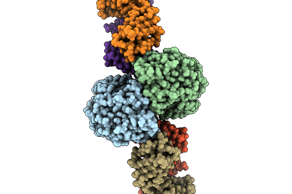
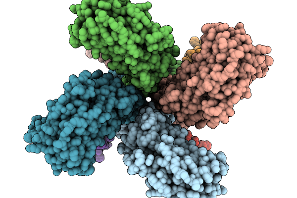
Ternary Complex Of An Improved Charged Molecular Glue Degrader Zz2-So2H, Brd4(Bd1) Neosubstrate, And The Ctlh E3 Ligase Receptor Module Ypel5-Wdr26
Organism: Homo sapiens
Method: ELECTRON MICROSCOPY Resolution:3.42 Å Release Date: 2026-04-08 Classification: LIGASE Ligands: ZN, A1IL8 |
 |
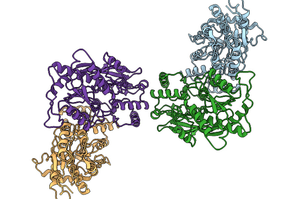
Ternary Complex Of A Charged Molecular Glue Degrader Zz1-So2H, Brd4(Bd1) Neosubstrate, And The Ctlh E3 Ligase Receptor Module Ypel5-Wdr26
Organism: Homo sapiens
Method: ELECTRON MICROSCOPY Resolution:3.38 Å Release Date: 2026-04-08 Classification: LIGASE Ligands: ZN, A1IL9 |
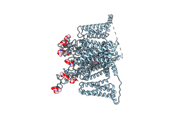 |
Organism: Homo sapiens
Method: ELECTRON MICROSCOPY Release Date: 2026-04-01 Classification: MEMBRANE PROTEIN Ligands: NAG, A1EQU, NA |
 |
Organism: Mus musculus
Method: ELECTRON MICROSCOPY Resolution:3.50 Å Release Date: 2026-03-25 Classification: MEMBRANE PROTEIN |
 |
Organism: Mus musculus
Method: ELECTRON MICROSCOPY Resolution:3.20 Å Release Date: 2026-03-25 Classification: MEMBRANE PROTEIN |
 |
Organism: Mus musculus
Method: ELECTRON MICROSCOPY Release Date: 2026-03-25 Classification: MEMBRANE PROTEIN |
 |
Organism: Mus musculus
Method: ELECTRON MICROSCOPY Release Date: 2026-03-25 Classification: MEMBRANE PROTEIN |
 |
Organism: Homo sapiens
Method: ELECTRON MICROSCOPY Resolution:3.05 Å Release Date: 2026-03-18 Classification: SIGNALING PROTEIN |
 |
Organism: Sus scrofa
Method: ELECTRON MICROSCOPY Release Date: 2026-03-04 Classification: MEMBRANE PROTEIN Ligands: DQC |
 |
Organism: Mus musculus, Sus scrofa
Method: ELECTRON MICROSCOPY Release Date: 2026-03-04 Classification: MEMBRANE PROTEIN Ligands: NAG |
 |
Organism: Sus scrofa
Method: ELECTRON MICROSCOPY Release Date: 2026-03-04 Classification: MEMBRANE PROTEIN Ligands: GGL, A1EA4 |
 |
Organism: Sus scrofa
Method: ELECTRON MICROSCOPY Release Date: 2026-03-04 Classification: MEMBRANE PROTEIN |
 |
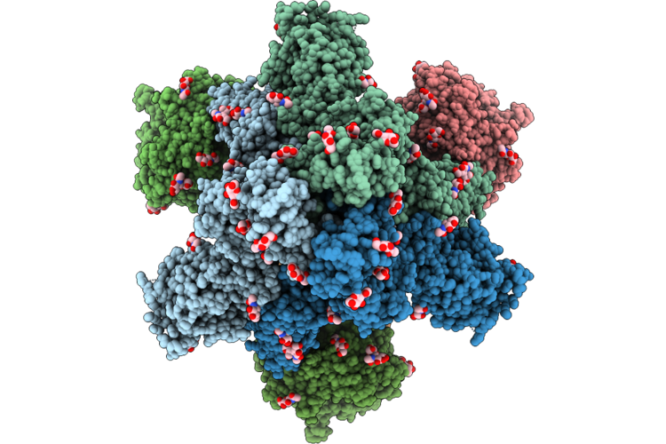
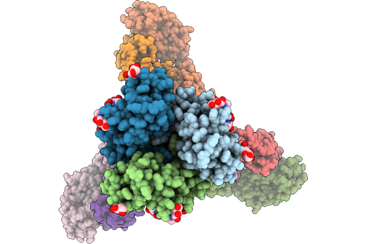
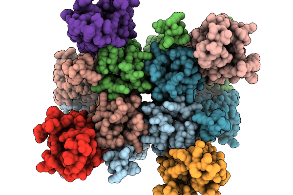
Organism: Poliovirus 1
Method: ELECTRON MICROSCOPY Resolution:2.95 Å Release Date: 2026-03-04 Classification: VIRUS LIKE PARTICLE |
 |
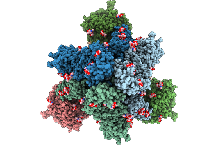
Organism: Poliovirus 1
Method: ELECTRON MICROSCOPY Release Date: 2026-03-04 Classification: VIRUS LIKE PARTICLE |