

Search Count: 2,589
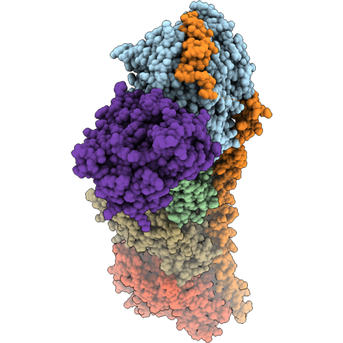 |
T2R-Ttl-Compound 40 Complex
Organism: Rattus norvegicus, Gallus gallus, Bos taurus
Method: X-RAY DIFFRACTION Resolution:2.64 Å Release Date: 2026-11-04 Classification: CELL CYCLE Ligands: GTP, MG, CA, GDP, MES, A1J5T, ACP |
Organism: Rattus norvegicus, Gallus gallus, Bos taurus
Method: X-RAY DIFFRACTION
Release Date: 2026-11-04
Ligands: GTP, MG, CA, GDP, MES, A1J5T, ACP
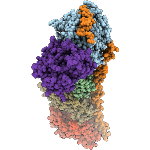 |
T2R-Ttl-Compound 63 Complex
Organism: Rattus norvegicus, Gallus gallus, Bos taurus
Method: X-RAY DIFFRACTION Resolution:2.61 Å Release Date: 2026-11-04 Classification: CELL CYCLE Ligands: GTP, MG, CA, IMD, GDP, MES, A1J5U, ACP |
Organism: Rattus norvegicus, Gallus gallus, Bos taurus
Method: X-RAY DIFFRACTION
Release Date: 2026-11-04
Ligands: GTP, MG, CA, IMD, GDP, MES, A1J5U, ACP
 |
Sb225002 With Tubulin
Organism: Sus scrofa, Mus musculus, Gallus gallus
Method: X-RAY DIFFRACTION Resolution:2.60 Å Release Date: 2026-10-28 Classification: PROTEIN BINDING Ligands: GTP, GOL, CA, MG, GDP, MES, A1E64, ACP |
Organism: Sus scrofa, Mus musculus, Gallus gallus
Method: X-RAY DIFFRACTION
Release Date: 2026-10-28
Ligands: GTP, GOL, CA, MG, GDP, MES, A1E64, ACP
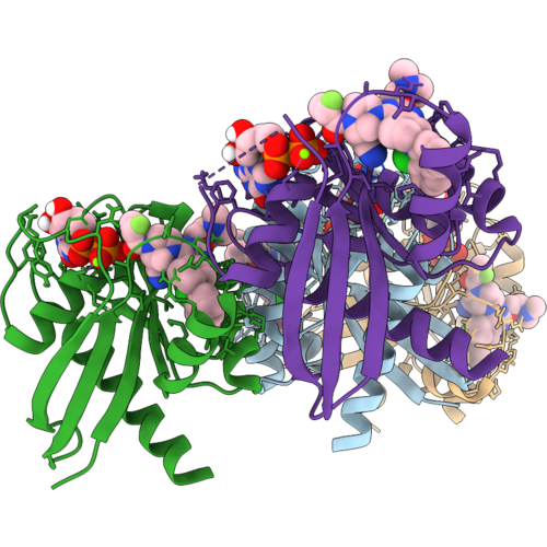 |
Crystal Structure Of Luci-Tag, Bound To Adagrasib
Organism: Synthetic construct
Method: X-RAY DIFFRACTION Resolution:2.94 Å Release Date: 2026-10-28 Classification: DE NOVO PROTEIN Ligands: M1X, GDP, MG |
Organism: Synthetic construct
Method: X-RAY DIFFRACTION
Release Date: 2026-10-28
Ligands: M1X, GDP, MG
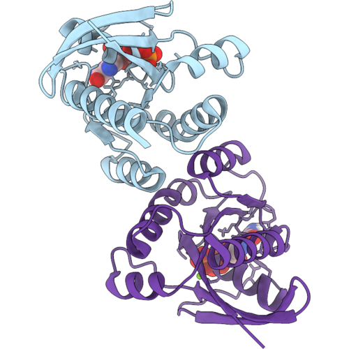 |
Crystal Structure Of Luci-Tag, Ligand-Free
Organism: Synthetic construct
Method: X-RAY DIFFRACTION Resolution:2.08 Å Release Date: 2026-10-28 Classification: DE NOVO PROTEIN Ligands: GDP, MG |
Organism: Synthetic construct
Method: X-RAY DIFFRACTION
Release Date: 2026-10-28
Ligands: GDP, MG
 |
Crystal Structure Of Luci-Tag, Bound To Ars-1323
Organism: Synthetic construct
Method: X-RAY DIFFRACTION Resolution:2.79 Å Release Date: 2026-10-28 Classification: DE NOVO PROTEIN Ligands: 91S, GDP, MG |
Organism: Synthetic construct
Method: X-RAY DIFFRACTION
Release Date: 2026-10-28
Ligands: 91S, GDP, MG
 |
Structure Of C. Tepidum Rel In An Open State In Complex With Apcpp And Gdp
Organism: Chlorobaculum tepidum
Method: X-RAY DIFFRACTION Resolution:2.65 Å Release Date: 2026-10-28 Classification: TRANSFERASE Ligands: APC, GDP, MG, MN, FMT |
Organism: Chlorobaculum tepidum
Method: X-RAY DIFFRACTION
Release Date: 2026-10-28
Ligands: APC, GDP, MG, MN, FMT
 |
Crystal Structure Of Cell Division Control Protein 42 Homolog
Organism: Homo sapiens
Method: X-RAY DIFFRACTION Resolution:2.90 Å Release Date: 2026-10-28 Classification: CELL CYCLE Ligands: SO4, MG, GDP |
Organism: Homo sapiens
Method: X-RAY DIFFRACTION
Release Date: 2026-10-28
Ligands: SO4, MG, GDP
 |
Crystal Structure Of Kras-G12D/Y96S Mutant In Complex With Mrtx-1133
Organism: Homo sapiens
Method: X-RAY DIFFRACTION Resolution:1.23 Å Release Date: 2026-10-28 Classification: HYDROLASE Ligands: MG, GDP, 6IC, GLY |
Organism: Homo sapiens
Method: X-RAY DIFFRACTION
Release Date: 2026-10-28
Ligands: MG, GDP, 6IC, GLY
 |
Crystal Structure Of P. Furiosus Srp54 Ng Domain G225E
Organism: Pyrococcus furiosus
Method: X-RAY DIFFRACTION Resolution:1.70 Å Release Date: 2026-10-28 Classification: RNA BINDING PROTEIN Ligands: GDP, MG |
Organism: Pyrococcus furiosus
Method: X-RAY DIFFRACTION
Release Date: 2026-10-28
Ligands: GDP, MG
 |
Tubulin Cofactors D,E,G Bound To Beta Tubulin
Organism: Homo sapiens, Trichoplusia ni
Method: ELECTRON MICROSCOPY Resolution:3.67 Å Release Date: 2026-10-21 Classification: CYTOSOLIC PROTEIN Ligands: GDP |
Organism: Homo sapiens, Trichoplusia ni
Method: ELECTRON MICROSCOPY
Release Date: 2026-10-21
Ligands: GDP
 |
Tubulin Cofactors D And E Bound To Beta Tubulin
Organism: Homo sapiens, Trichoplusia ni
Method: ELECTRON MICROSCOPY Resolution:4.78 Å Release Date: 2026-10-21 Classification: CYTOSOLIC PROTEIN Ligands: GDP |
Organism: Homo sapiens, Trichoplusia ni
Method: ELECTRON MICROSCOPY
Release Date: 2026-10-21
Ligands: GDP
 |
Dimer Of Tubulin Folding Cofactors D And E Bound To Beta Tubulin In The Back To Back Orientation
Organism: Homo sapiens, Trichoplusia ni
Method: ELECTRON MICROSCOPY Release Date: 2026-10-21 Classification: CYTOSOLIC PROTEIN Ligands: GDP |
Organism: Homo sapiens, Trichoplusia ni
Method: ELECTRON MICROSCOPY
Release Date: 2026-10-21
Ligands: GDP
 |
Dimer Of Tubulin Folding Cofactors D And E Bound To Beta Tubulin In A Side To Side Orientation
Organism: Homo sapiens, Trichoplusia ni
Method: ELECTRON MICROSCOPY Release Date: 2026-10-21 Classification: CYTOSOLIC PROTEIN Ligands: GDP |
Organism: Homo sapiens, Trichoplusia ni
Method: ELECTRON MICROSCOPY
Release Date: 2026-10-21
Ligands: GDP
 |
Dimer Of Tubulin Cofactors D,E, And Arl2 Bound To Beta Tubulin In A Back To Back Orientation
Organism: Homo sapiens, Trichoplusia ni
Method: ELECTRON MICROSCOPY Release Date: 2026-10-21 Classification: CYTOSOLIC PROTEIN Ligands: GDP |
Organism: Homo sapiens, Trichoplusia ni
Method: ELECTRON MICROSCOPY
Release Date: 2026-10-21
Ligands: GDP
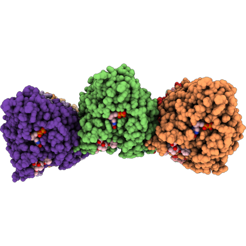 |
Plasmodium Falciparum 17-Pf Subpellicular Microtubule
Organism: Plasmodium falciparum
Method: ELECTRON MICROSCOPY Release Date: 2026-10-14 Classification: STRUCTURAL PROTEIN Ligands: GTP, MG, GDP, TA1 |
Organism: Plasmodium falciparum
Method: ELECTRON MICROSCOPY
Release Date: 2026-10-14
Ligands: GTP, MG, GDP, TA1
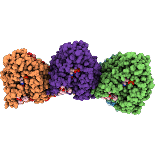 |
Plasmodium Falciparum 18-Pf Subpellicular Microtubule
Organism: Plasmodium falciparum
Method: ELECTRON MICROSCOPY Release Date: 2026-10-14 Classification: STRUCTURAL PROTEIN Ligands: GTP, MG, GDP, TA1 |
Organism: Plasmodium falciparum
Method: ELECTRON MICROSCOPY
Release Date: 2026-10-14
Ligands: GTP, MG, GDP, TA1
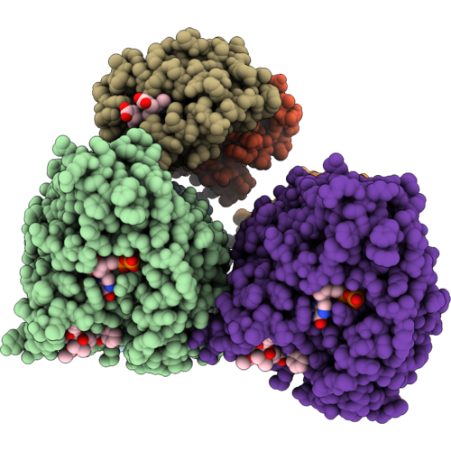 |
Plasmodium Falciparum 17-Pf Subpellicular Microtubule Associated With Human Hemoglobin (Composite Map)
Organism: Plasmodium falciparum, Homo sapiens
Method: ELECTRON MICROSCOPY Release Date: 2026-10-14 Classification: OXYGEN TRANSPORT/Structural Protein Ligands: GTP, MG, GDP, TA1, HEM |
Organism: Plasmodium falciparum, Homo sapiens
Method: ELECTRON MICROSCOPY
Release Date: 2026-10-14
Ligands: GTP, MG, GDP, TA1, HEM
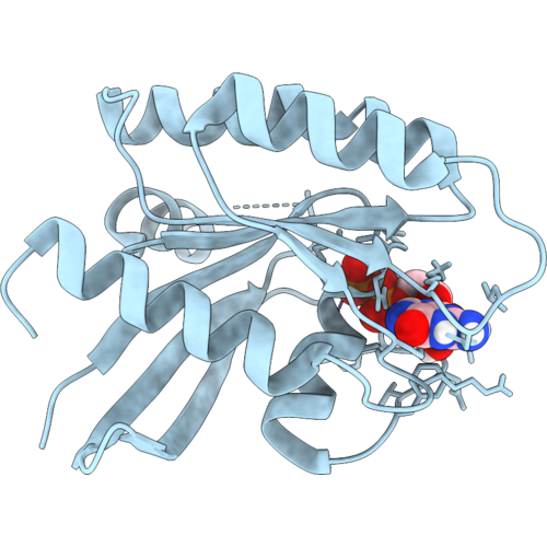 |
Structure Of Gdp-Bound Kras-G12C/H95Q, A Switch-Ii Pocket Inhibitor Resistance Mutant
Organism: Homo sapiens
Method: X-RAY DIFFRACTION Resolution:1.35 Å Release Date: 2026-10-07 Classification: SIGNALING PROTEIN Ligands: GDP, MG |
Organism: Homo sapiens
Method: X-RAY DIFFRACTION
Release Date: 2026-10-07
Ligands: GDP, MG
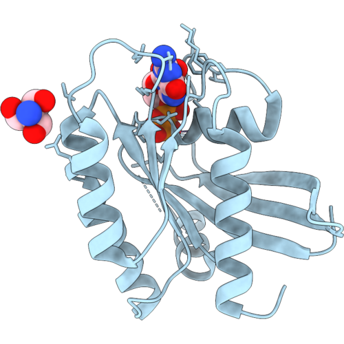 |
Structure Of Gdp-Bound Kras-G12C/Y96C, A Switch-Ii Pocket Inhibitor Resistance Mutant
Organism: Homo sapiens
Method: X-RAY DIFFRACTION Resolution:1.50 Å Release Date: 2026-10-07 Classification: SIGNALING PROTEIN Ligands: MG, GDP, TRS |
Organism: Homo sapiens
Method: X-RAY DIFFRACTION
Release Date: 2026-10-07
Ligands: MG, GDP, TRS