
Search Count: 1,734
 |
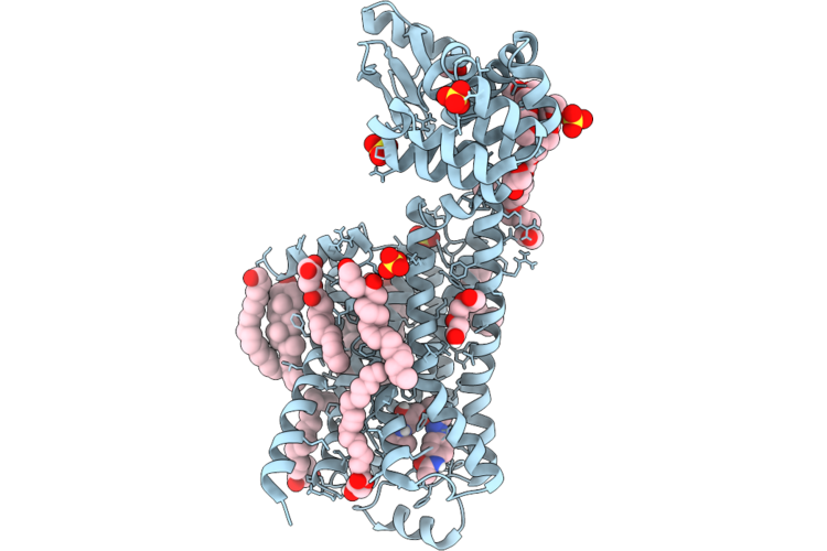
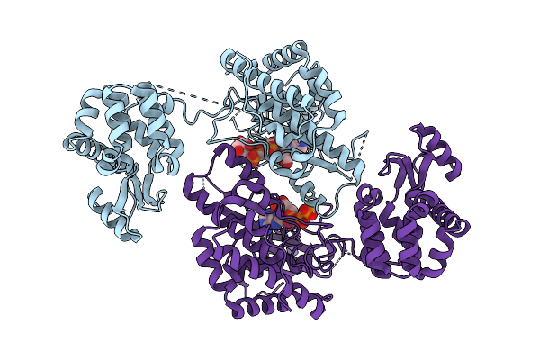
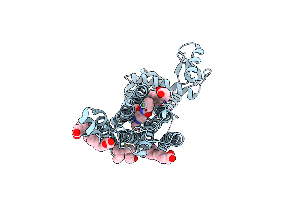
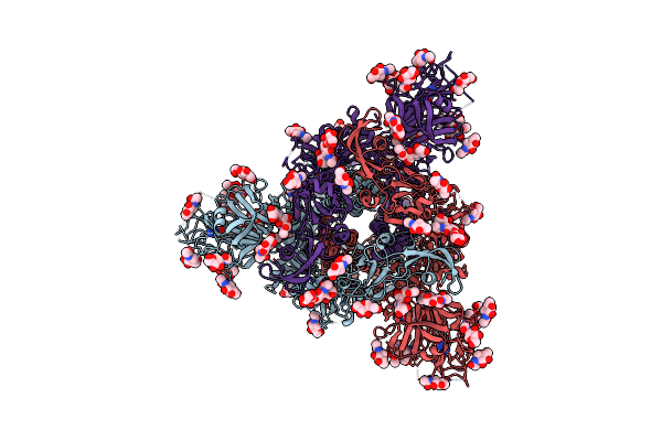
Cryo-Em Structure Of The Human Beta2-Adrenergic Receptor In Complex With A Novel Antagonist
Organism: Homo sapiens, Enterobacteria phage t4, Lama glama
Method: ELECTRON MICROSCOPY Resolution:3.04 Å Release Date: 2026-06-10 Classification: MEMBRANE PROTEIN Ligands: BER |
 |
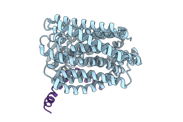
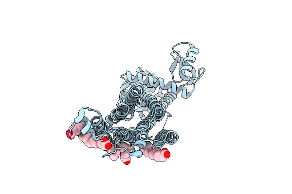
Dark Structure Of Beta-2 Adrenergic Receptor With Photoazolol In Dark State Recorded At Swissfel
Organism: Homo sapiens, Enterobacteria phage t4
Method: X-RAY DIFFRACTION Resolution:2.45 Å Release Date: 2026-04-22 Classification: MEMBRANE PROTEIN Ligands: SO4, CLR, 12P, OLC, 1PE, EDO, GOL, A1JHU, PLM |
 |
Dark Structure Of Beta-2 Adrenergic Receptor With Photoazolol In Dark State Recorded At Lcls
Organism: Homo sapiens, Enterobacteria phage t4
Method: X-RAY DIFFRACTION Resolution:2.50 Å Release Date: 2026-04-22 Classification: MEMBRANE PROTEIN Ligands: SO4, CLR, PLM, 12P, A1JHU, OLC, EDO, GOL |
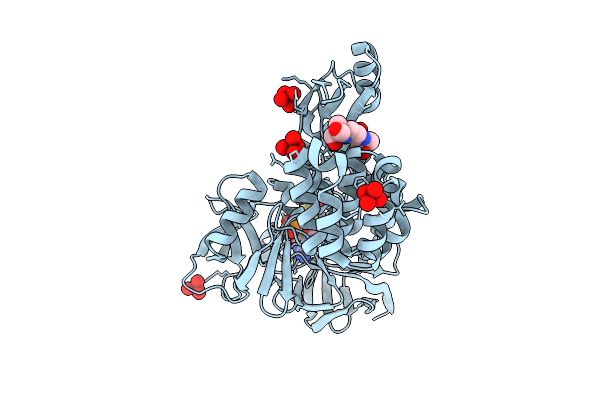 |
Organism: Homo sapiens, Enterobacteria phage t4
Method: X-RAY DIFFRACTION Resolution:1.63 Å Release Date: 2026-03-18 Classification: METAL BINDING PROTEIN Ligands: MTE, B3P, MOO, EFK, CL |
 |
Organism: Escherichia coli k-12, Enterobacteria phage m
Method: ELECTRON MICROSCOPY Resolution:3.60 Å Release Date: 2026-02-11 Classification: TRANSPORT PROTEIN |
 |
Cryo-Em Structure Of The Escherichia Phage Hk446 Rip1 In Complex With The Enterobacteria Phage T6 Small Terminase
Organism: Enterobacteria phage t6, Escherichia phage hk446
Method: ELECTRON MICROSCOPY Release Date: 2026-02-04 Classification: VIRAL PROTEIN |
 |
Organism: Feline infectious peritonitis virus (strain 79-1146), Enterobacteria phage t6
Method: ELECTRON MICROSCOPY Resolution:2.78 Å Release Date: 2026-01-28 Classification: PROTEIN BINDING Ligands: NAG |
 |
Organism: Human respiratory syncytial virus, Enterobacteria phage t6
Method: X-RAY DIFFRACTION Resolution:2.92 Å Release Date: 2025-12-17 Classification: VIRAL PROTEIN |
 |
Organism: Respiratory syncytial virus, Enterobacteria phage t6
Method: X-RAY DIFFRACTION Resolution:2.77 Å Release Date: 2025-12-17 Classification: VIRAL PROTEIN |
 |
Crystal Structure Of Phosphatidyl Inositol 4-Kinase Ii Beta In Complex With Hh5129
Organism: Homo sapiens, Enterobacteria phage t4
Method: X-RAY DIFFRACTION Resolution:2.25 Å Release Date: 2025-12-03 Classification: TRANSFERASE Ligands: A1IVA |
 |
Organism: Enterobacteria phage t4
Method: ELECTRON MICROSCOPY Release Date: 2025-11-26 Classification: ISOMERASE |
 |
Organism: Escherichia coli, Enterobacteria phage m
Method: ELECTRON MICROSCOPY Release Date: 2025-09-10 Classification: VIRAL PROTEIN |
 |
Pre-Fusion Herv-K Envelope Protein Trimer Ectodomain In Complex With Kenv-6 Fab
Organism: Homo sapiens, Enterobacteria phage t4, Mus musculus
Method: ELECTRON MICROSCOPY Release Date: 2025-07-30 Classification: VIRAL PROTEIN Ligands: NAG |
 |
Organism: Homo sapiens, Enterobacteria phage t4
Method: ELECTRON MICROSCOPY Release Date: 2025-07-30 Classification: VIRAL PROTEIN Ligands: NAG |
 |
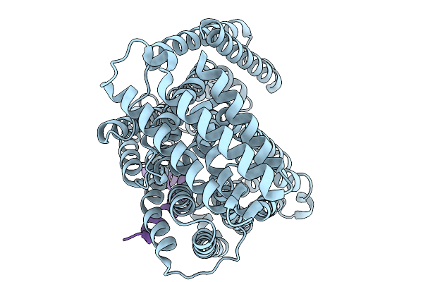
Dark Structure Of The Human Metabotropic Glutamate Receptor 5 Transmembrane Domain Bound To Photoswitchable Ligand Alloswitch-1
Organism: Homo sapiens, Enterobacteria phage t4
Method: X-RAY DIFFRACTION Resolution:2.33 Å Release Date: 2025-06-25 Classification: SIGNALING PROTEIN Ligands: OLA, 4YI |
 |
Apo-State Structure Of The Human Metabotropic Glutamate Receptor 5 Transmembrane Domain Freeze-Trapped After Light Activation Of Photoswitchable Ligand Alloswitch-1
Organism: Homo sapiens, Enterobacteria phage t4
Method: X-RAY DIFFRACTION Resolution:2.90 Å Release Date: 2025-06-25 Classification: SIGNALING PROTEIN Ligands: OLA |
 |
Organism: Severe acute respiratory syndrome coronavirus 2, Enterobacteria phage t4, Homo sapiens
Method: ELECTRON MICROSCOPY Release Date: 2025-06-18 Classification: VIRAL PROTEIN |
 |
Organism: Coronavirus neoromicia/pml-phe1/rsa/2011, Enterobacteria phage t4
Method: ELECTRON MICROSCOPY Release Date: 2025-06-18 Classification: VIRAL PROTEIN Ligands: NAG, EIC |
 |
Cryo-Em Structure Of Eu-Hedgehogcov (Erinaceus/Vmc/Deu/2012) S-Trimer In A Locked-2 Conformation
Organism: Betacoronavirus erinaceus/vmc/deu/2012, Enterobacteria phage t4 (bacteriophage t4)
Method: ELECTRON MICROSCOPY Release Date: 2025-06-18 Classification: VIRAL PROTEIN Ligands: NAG, FOL, EIC |
 |
Cryo-Em Structure Of Cn-Hedgehogcov (Hku31/Erinaceus Amurensis/China/2014) S-Trimer In A Locked-2 Conformation
Organism: Erinaceus hedgehog coronavirus hku31, Enterobacteria phage t4
Method: ELECTRON MICROSCOPY Release Date: 2025-06-18 Classification: VIRAL PROTEIN Ligands: FOL, NAG |