
Search Count: 4,502
 |
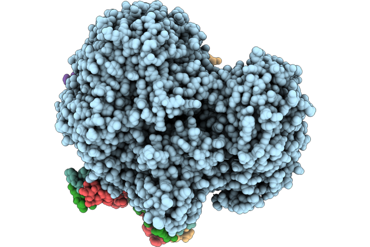
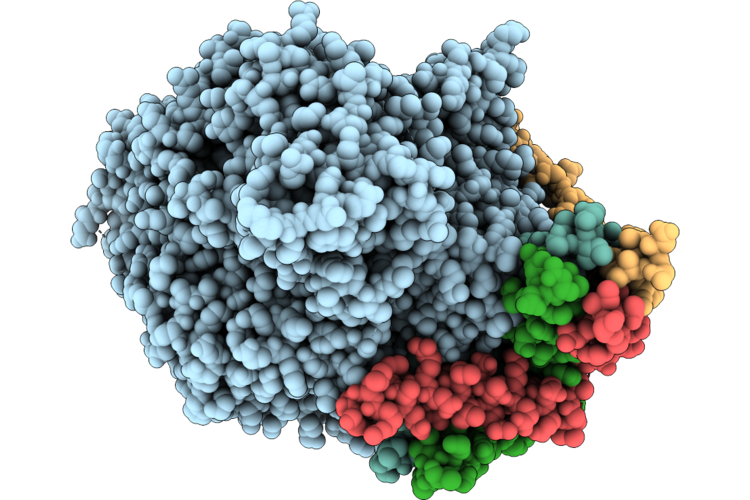
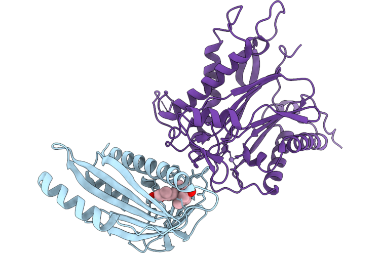
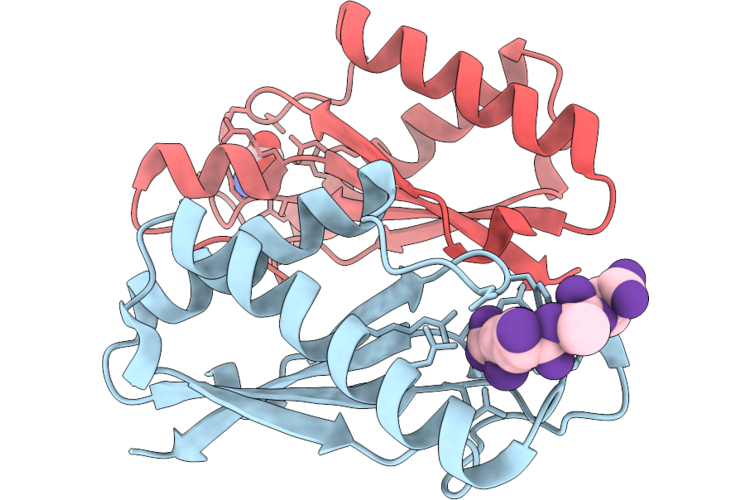
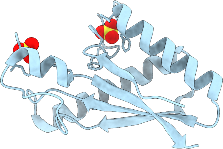
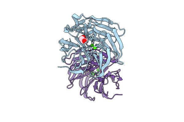
Cryo-Em Structure Of Vx77 Fab In Complex With Gii.4 Norovirus P Domain
Organism: Norovirus hu/gii.4/sydney/nsw0514/2012/au, Homo sapiens
Method: ELECTRON MICROSCOPY Resolution:3.08 Å Release Date: 2026-07-01 Classification: VIRAL PROTEIN/Immune System |
Organism: Norovirus hu/gii.4/sydney/nsw0514/2012/au, Homo sapiens
Method: ELECTRON MICROSCOPY
Release Date: 2026-07-01
 |
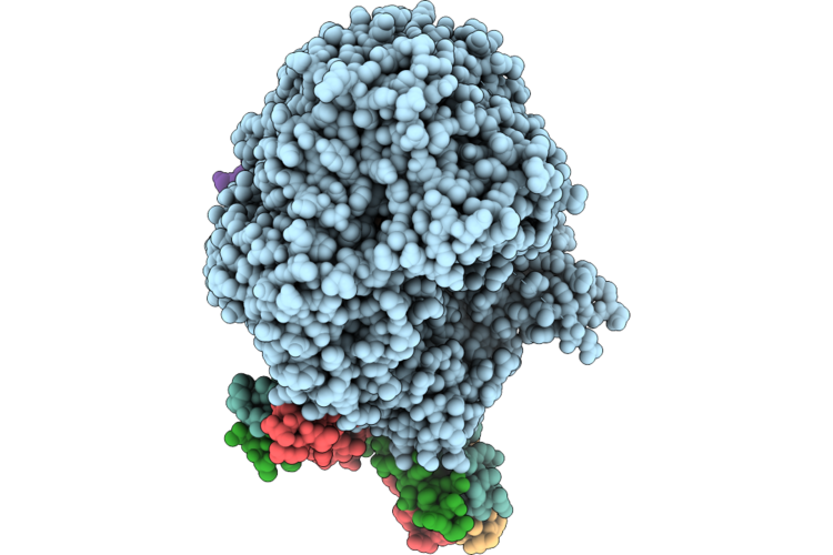
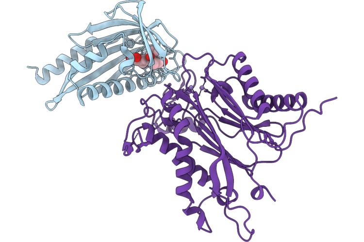
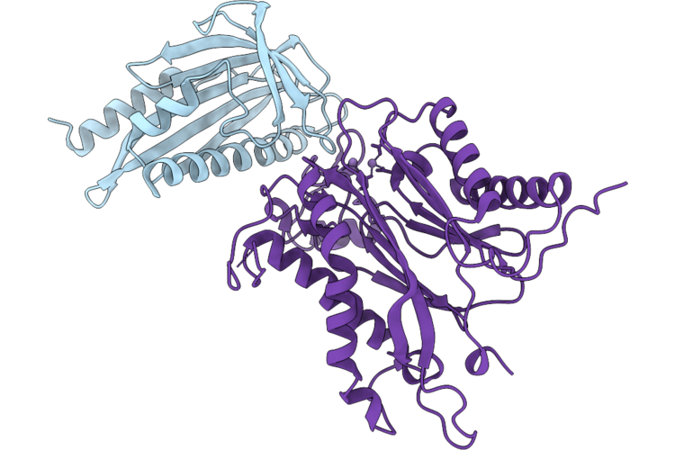
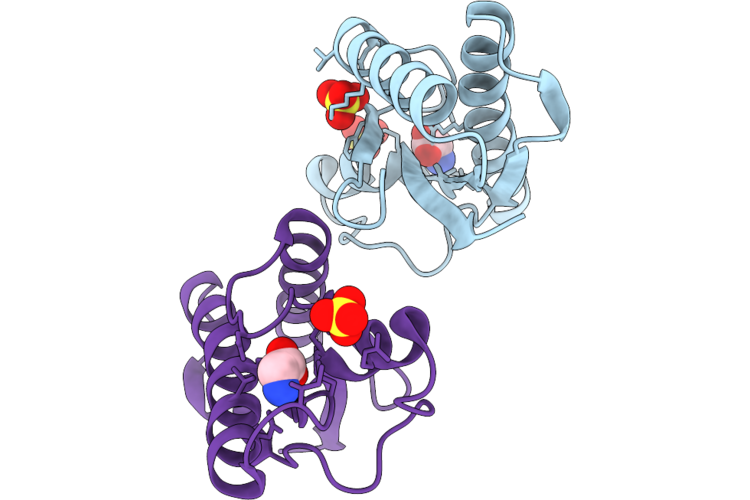
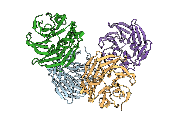
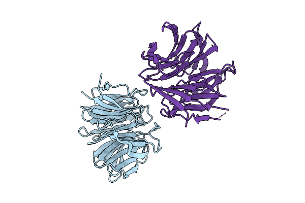
Cryo-Em Structure Of Vx93 Fab In Complex With Gii.4 Norovirus P Domain
Organism: Norovirus hu/gii.4/sydney/nsw0514/2012/au, Homo sapiens
Method: ELECTRON MICROSCOPY Release Date: 2026-07-01 Classification: VIRAL PROTEIN/Immune System |
Organism: Norovirus hu/gii.4/sydney/nsw0514/2012/au, Homo sapiens
Method: ELECTRON MICROSCOPY
Release Date: 2026-07-01
 |
Cryo-Em Structure Of Napa, The Periplasmic Nitrate Reductase From Campylobacter Jejuni
Organism: Campylobacter jejuni
Method: ELECTRON MICROSCOPY Release Date: 2026-06-17 Classification: OXIDOREDUCTASE Ligands: SF4, MO, MGD |
Organism: Campylobacter jejuni
Method: ELECTRON MICROSCOPY
Release Date: 2026-06-17
Ligands: SF4, MO, MGD
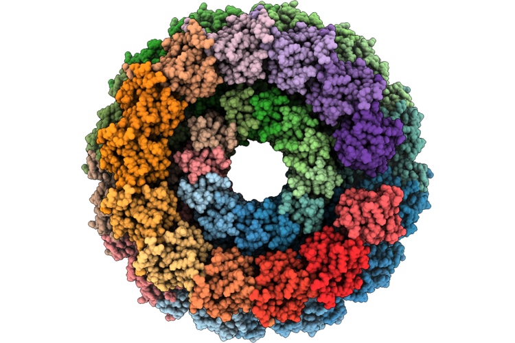 |
Kpswz In Complex With Bacteriophage Bas14 Portal
Organism: Escherichia phage theodorherzl, Klebsiella pneumoniae
Method: ELECTRON MICROSCOPY Release Date: 2026-05-20 Classification: HYDROLASE Ligands: MG |
Organism: Escherichia phage theodorherzl, Klebsiella pneumoniae
Method: ELECTRON MICROSCOPY
Release Date: 2026-05-20
Ligands: MG
 |
Cryo-Em Structure Of The Respiratory Syncytial Virus Polymerase (L:P) In Ntp-Bound Elongation State
Organism: Respiratory syncytial virus, Respiratory syncytial virus a2
Method: ELECTRON MICROSCOPY Resolution:3.08 Å Release Date: 2026-05-06 Classification: VIRAL PROTEIN,TRANSFERASE/RNA Ligands: MG, ZAN |
Organism: Respiratory syncytial virus, Respiratory syncytial virus a2
Method: ELECTRON MICROSCOPY
Release Date: 2026-05-06
Ligands: MG, ZAN
 |
Cryo-Em Structure Of The Respiratory Syncytial Virus Polymerase (L:P) In Pre-Reaction Elongation State
Organism: Respiratory syncytial virus, Respiratory syncytial virus a2
Method: ELECTRON MICROSCOPY Resolution:2.75 Å Release Date: 2026-05-06 Classification: VIRAL PROTEIN,TRANSFERASE/RNA Ligands: MG, ZAN |
Organism: Respiratory syncytial virus, Respiratory syncytial virus a2
Method: ELECTRON MICROSCOPY
Release Date: 2026-05-06
Ligands: MG, ZAN
 |
Cryo-Em Structure Of The Respiratory Syncytial Virus Polymerase (L:P) In Pre-Translocation Elongation State
Organism: Respiratory syncytial virus, Respiratory syncytial virus a2
Method: ELECTRON MICROSCOPY Resolution:3.07 Å Release Date: 2026-05-06 Classification: VIRAL PROTEIN,TRANSFERASE/RNA |
Organism: Respiratory syncytial virus, Respiratory syncytial virus a2
Method: ELECTRON MICROSCOPY
Release Date: 2026-05-06
 |
Cryo-Em Structure Of The Respiratory Syncytial Virus Polymerase (L:P) In Post-Translocation Elongation State
Organism: Respiratory syncytial virus a2
Method: ELECTRON MICROSCOPY Resolution:3.54 Å Release Date: 2026-05-06 Classification: VIRAL PROTEIN,TRANSFERASE/RNA |
Organism: Respiratory syncytial virus a2
Method: ELECTRON MICROSCOPY
Release Date: 2026-05-06
 |
Crystal Structure Of The Mppyl1-Aba-Hab1 Ternary Complex
Organism: Marchantia polymorpha, Arabidopsis thaliana
Method: X-RAY DIFFRACTION Resolution:1.98 Å Release Date: 2026-05-06 Classification: PLANT PROTEIN Ligands: A8S, MN, CL |
Organism: Marchantia polymorpha, Arabidopsis thaliana
Method: X-RAY DIFFRACTION
Release Date: 2026-05-06
Ligands: A8S, MN, CL
 |
Crystal Structure Of The Mppyl1-Hab1 Complex
Organism: Marchantia polymorpha, Arabidopsis thaliana
Method: X-RAY DIFFRACTION Resolution:2.39 Å Release Date: 2026-05-06 Classification: PLANT PROTEIN Ligands: MN, CL |
Organism: Marchantia polymorpha, Arabidopsis thaliana
Method: X-RAY DIFFRACTION
Release Date: 2026-05-06
Ligands: MN, CL
 |
Crystal Structure Of The Mppyl1(L91I-L111V-L191V-C194L-S198K)-Aba-Hab1 Ternary Complex
Organism: Marchantia polymorpha, Arabidopsis thaliana
Method: X-RAY DIFFRACTION Resolution:2.30 Å Release Date: 2026-05-06 Classification: PLANT PROTEIN Ligands: A8S, MN, CL |
Organism: Marchantia polymorpha, Arabidopsis thaliana
Method: X-RAY DIFFRACTION
Release Date: 2026-05-06
Ligands: A8S, MN, CL
 |
Crystal Structure Of The Periplasmic Domain Of Cadf From Campylobacter Jejuni In Complex With A Peptidoglycan Peptide
Organism: Campylobacter jejuni, Escherichia coli
Method: X-RAY DIFFRACTION Resolution:1.80 Å Release Date: 2026-05-06 Classification: PEPTIDE BINDING PROTEIN Ligands: API |
Organism: Campylobacter jejuni, Escherichia coli
Method: X-RAY DIFFRACTION
Release Date: 2026-05-06
Ligands: API
 |
Crystal Structure Of The Periplasmic Domain Of Cadf From Campylobacter Jejuni In Complex With Glycine
Organism: Campylobacter jejuni
Method: X-RAY DIFFRACTION Resolution:1.75 Å Release Date: 2026-05-06 Classification: PEPTIDE BINDING PROTEIN Ligands: SO4, GLY |
Organism: Campylobacter jejuni
Method: X-RAY DIFFRACTION
Release Date: 2026-05-06
Ligands: SO4, GLY
 |
Crystal Structure Of The Periplasmic Domain Of Campylobacter Jejuni Cadf R268E
Organism: Campylobacter jejuni
Method: X-RAY DIFFRACTION Resolution:1.50 Å Release Date: 2026-05-06 Classification: PEPTIDE BINDING PROTEIN Ligands: SO4 |
Organism: Campylobacter jejuni
Method: X-RAY DIFFRACTION
Release Date: 2026-05-06
Ligands: SO4
 |
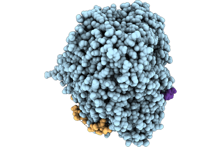
Crystal Structure Of Apoform Human Usp18
Organism: Homo sapiens, Myxococcus xanthus (strain dk1622)
Method: X-RAY DIFFRACTION Resolution:1.92 Å Release Date: 2026-04-29 Classification: HYDROLASE Ligands: ZN |
Organism: Homo sapiens, Myxococcus xanthus (strain dk1622)
Method: X-RAY DIFFRACTION
Release Date: 2026-04-29
Ligands: ZN
 |
Crystal Structure Of The Cj1041C Protein From Campylobacter Jejuni In Complex With Ca2+ In Space Group P212121
Organism: Campylobacter jejuni
Method: X-RAY DIFFRACTION Resolution:1.50 Å Release Date: 2026-04-01 Classification: METAL BINDING PROTEIN Ligands: CA, GOL |
Organism: Campylobacter jejuni
Method: X-RAY DIFFRACTION
Release Date: 2026-04-01
Ligands: CA, GOL
 |
Crystal Structure Of The Cj1041C Protein From Campylobacter Jejuni In Complex With Ca2+ In Space Group P1
Organism: Campylobacter jejuni
Method: X-RAY DIFFRACTION Resolution:2.00 Å Release Date: 2026-04-01 Classification: METAL BINDING PROTEIN Ligands: CA, GOL |
Organism: Campylobacter jejuni
Method: X-RAY DIFFRACTION
Release Date: 2026-04-01
Ligands: CA, GOL
 |
Crystal Structure Of The Cj1041C Protein From Campylobacter Jejuni In The Apo Form In Space Group P2
Organism: Campylobacter jejuni
Method: X-RAY DIFFRACTION Resolution:2.00 Å Release Date: 2026-04-01 Classification: METAL BINDING PROTEIN |
Organism: Campylobacter jejuni
Method: X-RAY DIFFRACTION
Release Date: 2026-04-01
 |
Crystal Structure Of The Cj1041C Protein From Campylobacter Jejuni In The Apo Form In Space Group C2
Organism: Campylobacter jejuni
Method: X-RAY DIFFRACTION Resolution:1.95 Å Release Date: 2026-04-01 Classification: METAL BINDING PROTEIN |
Organism: Campylobacter jejuni
Method: X-RAY DIFFRACTION
Release Date: 2026-04-01
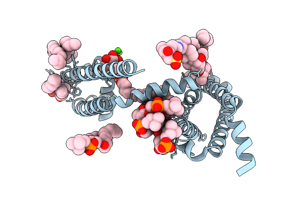 |
Crystal Structure Of Voltage-Gated Sodium Channel Navab N49K/S178T Mutant
Organism: Aliarcobacter butzleri
Method: X-RAY DIFFRACTION Resolution:2.50 Å Release Date: 2026-02-18 Classification: MEMBRANE PROTEIN Ligands: CA, 1N7, LMT, PX4 |
Organism: Aliarcobacter butzleri
Method: X-RAY DIFFRACTION
Release Date: 2026-02-18
Ligands: CA, 1N7, LMT, PX4