9V9A image
Deposition Date 2025-05-30
Release Date 2026-05-06
Last Version Date 2026-05-06
Entry Detail
PDB ID:
9V9A
Title:
Crystal structure of the periplasmic domain of CadF from Campylobacter jejuni in complex with glycine
Biological Source:
Source Organism(s):
Expression System(s):
Method Details:
Experimental Method:
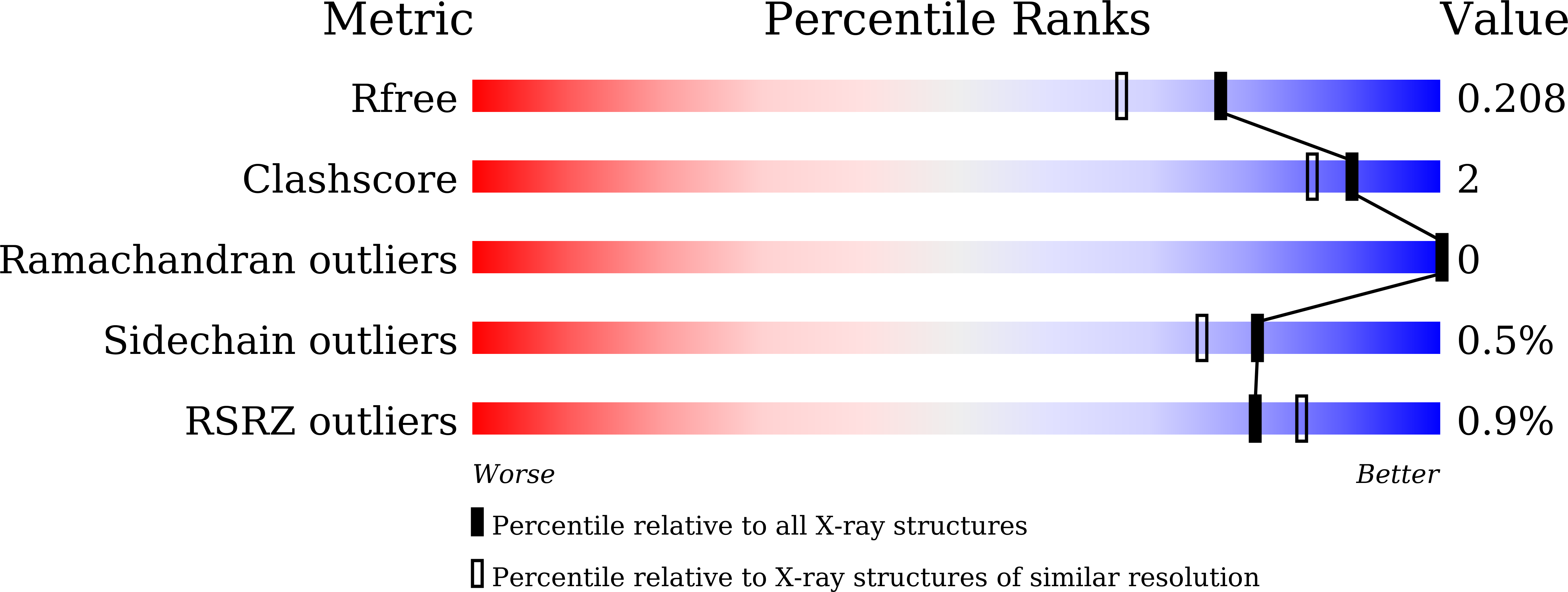
Resolution:
1.75 Å
R-Value Free:
0.20
R-Value Work:
0.17
R-Value Observed:
0.17
Space Group:
P 1
Macromolecular Entities
Structures with similar UniProt ID
Protein Blast
Polymer Type:polypeptide(L)
Molecule:Outer membrane beta-barrel do
Chain IDs:A, B
Chain Length:118
Number of Molecules:2
Biological Source:Campylobacter jejuni
Primary Citation
Crystal structures of Campylobacter jejuni CadF reveal a potential role for the arginine residue R268 in peptidoglycan recognition and pocket formation.
Sci Rep 15 36733 36733 (2025)
PMID: 41120626 DOI: 10.1038/s41598-025-20604-7

Abstact

Campylobacter jejuni is a pathogenic gram-negative bacterium that primarily causes foodborne enteritis in humans. The C. jejuni protein CadF is located in the outer membrane and is essential for the adhesion of C. jejuni to human cells. CadF contains an outer membrane-embedded domain and a periplasmic domain (CadF(PD)). The transmembrane domain is well known to bind fibronectin, contributing to host cell recognition. However, the exact function of the CadF(PD) has never been assessed experimentally. In this study, we present the crystal structures of the CadF(PD) and demonstrate that CadF may function as a peptidoglycan-binding protein. The CadF(PD) adopts a two-layer alpha/beta structure and harbors a narrow pocket. The CadF(PD) interacts with a peptidoglycan-derived peptide by accommodating m-diaminopimelic acid (m-DAP), a non-proteinogenic amino acid of the gram-negative peptidoglycan peptide, into the pocket. The positively charged R268 residue of CadF forms the base of the pocket and specifically interacts with the carboxyl group of m-DAP via salt bridges. This observation combined with the ligand-less structure of the CadF(PD) mutant R268E highlights the potential role of the R268 residue in maintaining the intact pocket in the CadF(PD) structure and mediating peptidoglycan recognition.

Legend

Protein

Chemical

Disease

Primary Citation of related structures
Feedback Form
Name
Email
Institute
Feedback