
Search Count: 6,268
 |
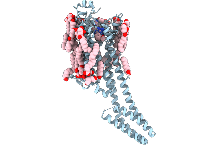
Crystal Structure Of Human Orexin Type 2 Receptor In Complex With Vornorexant
Organism: Homo sapiens
Method: X-RAY DIFFRACTION Resolution:3.29 Å Release Date: 2026-06-03 Classification: MEMBRANE PROTEIN Ligands: A1MCY, SO4 |
 |
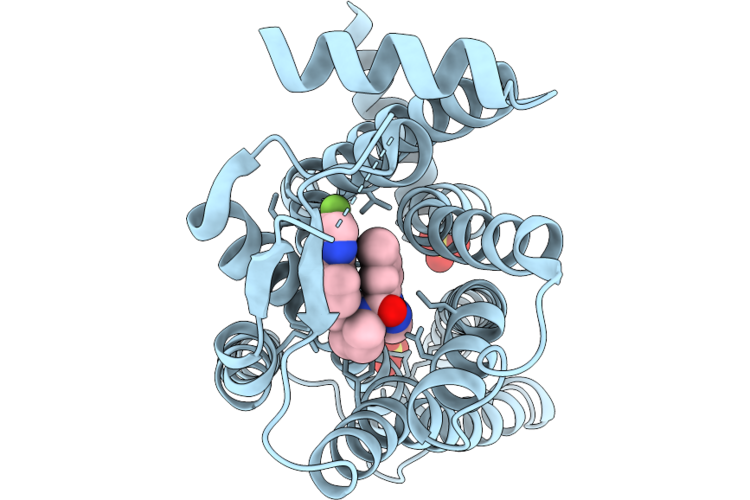
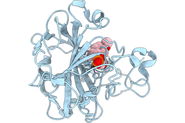
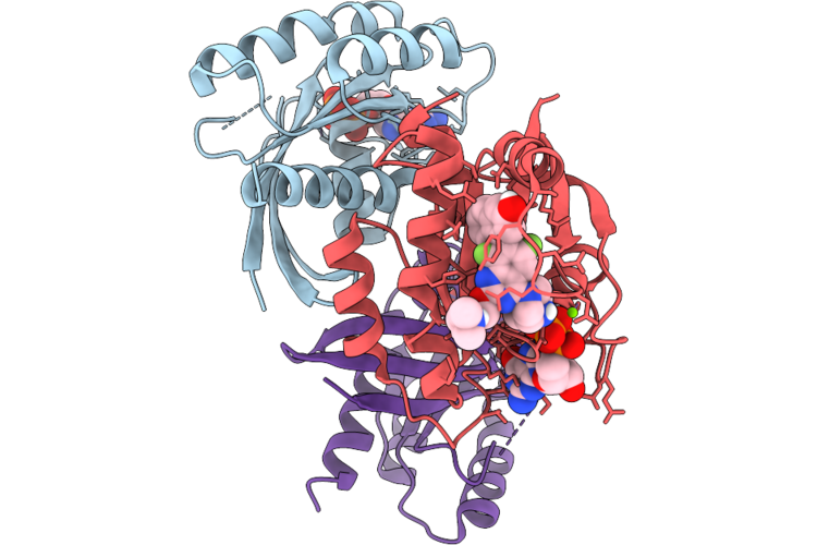
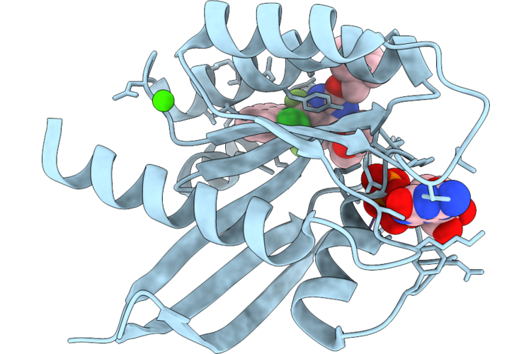
Structure Of Ptp1B Complexed With Difluoromethylphosphonate Inhibitor Compound 2
Organism: Homo sapiens
Method: X-RAY DIFFRACTION Resolution:1.73 Å Release Date: 2026-05-27 Classification: HYDROLASE/INHIBITOR Ligands: A1C3A |
 |
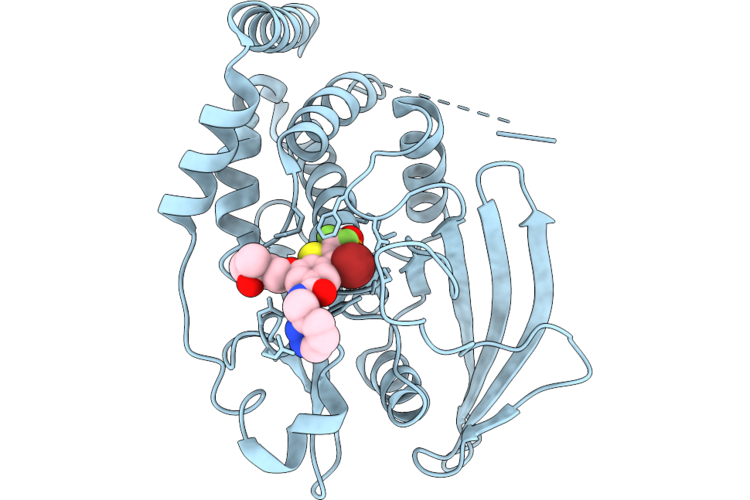
Structure Of Ptp1B Complexed With Difluoromethylphosphonate Inhibitor Compound 10
Organism: Homo sapiens
Method: X-RAY DIFFRACTION Resolution:2.51 Å Release Date: 2026-05-27 Classification: HYDROLASE/INHIBITOR Ligands: A1C3B |
 |
Structure Of Ptp1B Complexed With Difluoromethylphosphonate Inhibitor Compound 15
Organism: Homo sapiens
Method: X-RAY DIFFRACTION Resolution:2.15 Å Release Date: 2026-05-27 Classification: HYDROLASE/INHIBITOR Ligands: A1C3C, DMS |
 |
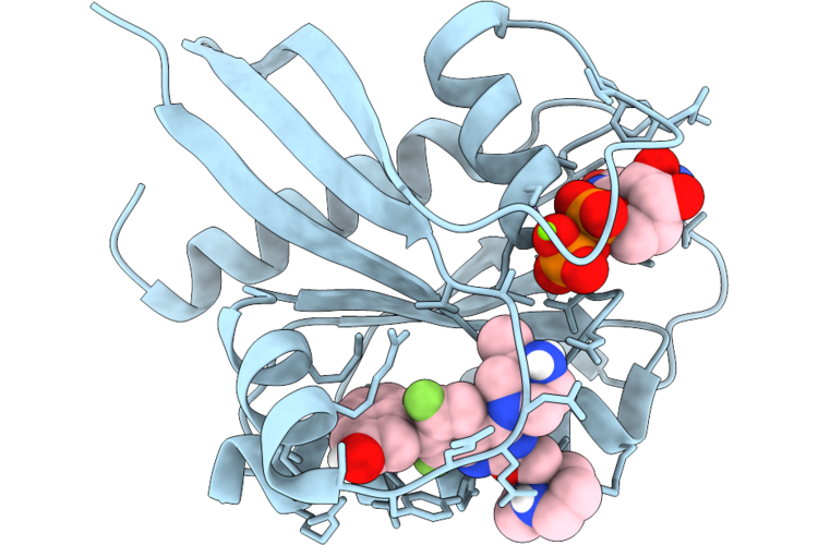
Structure Of Ptp1B Complexed With Difluoromethylphosphonate Inhibitor Compound 30
Organism: Homo sapiens
Method: X-RAY DIFFRACTION Resolution:1.94 Å Release Date: 2026-05-27 Classification: HYDROLASE/INHIBITOR Ligands: A1C3D |
 |
Crystal Structure Of A2A Adenosine Receptor A2Ar-Bril In Complex With Compound50
Organism: Homo sapiens, Escherichia coli
Method: X-RAY DIFFRACTION Resolution:2.59 Å Release Date: 2026-05-20 Classification: MEMBRANE PROTEIN |
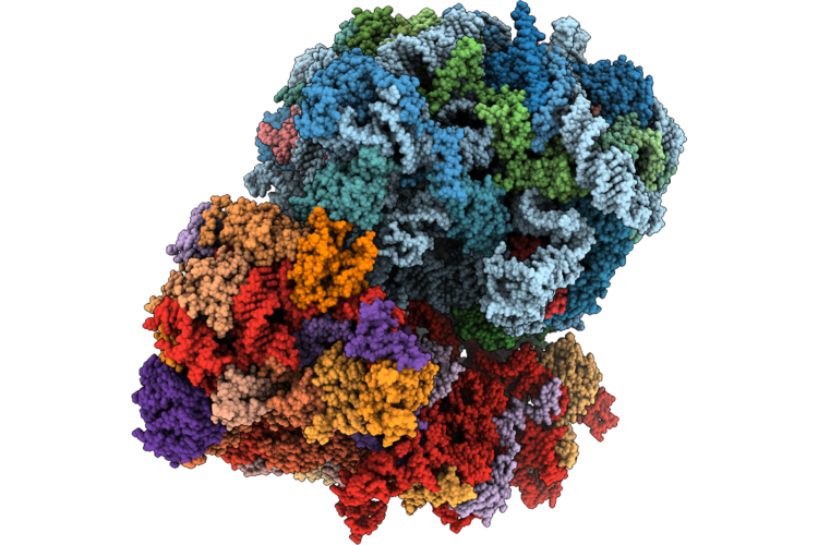 |
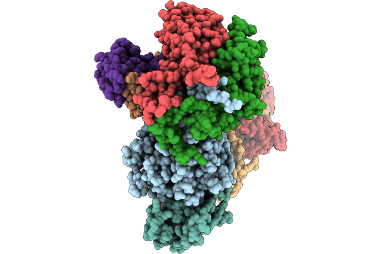
Organism: Saccharomyces cerevisiae by4741
Method: ELECTRON MICROSCOPY Release Date: 2026-05-20 Classification: RIBOSOME Ligands: MG, K, ZN |
 |
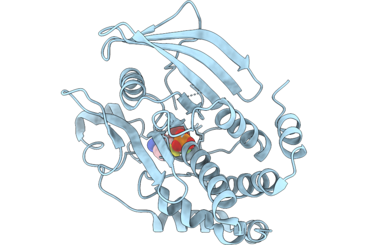
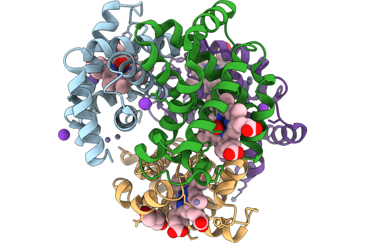
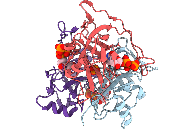
Crystal Structure Of A Phosphocoumarin Derivative In Complex With Human Carbonic Anhydrase Ii
Organism: Homo sapiens
Method: X-RAY DIFFRACTION Resolution:1.65 Å Release Date: 2026-05-13 Classification: LYASE Ligands: ZN, A1JX9, GOL |
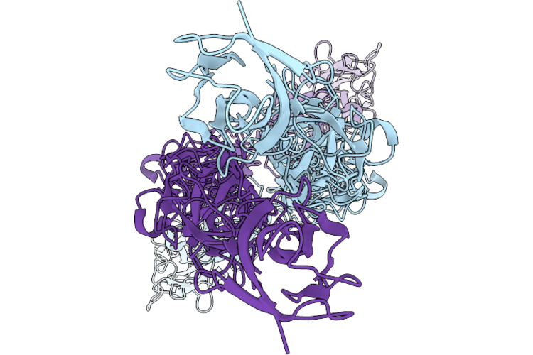 |
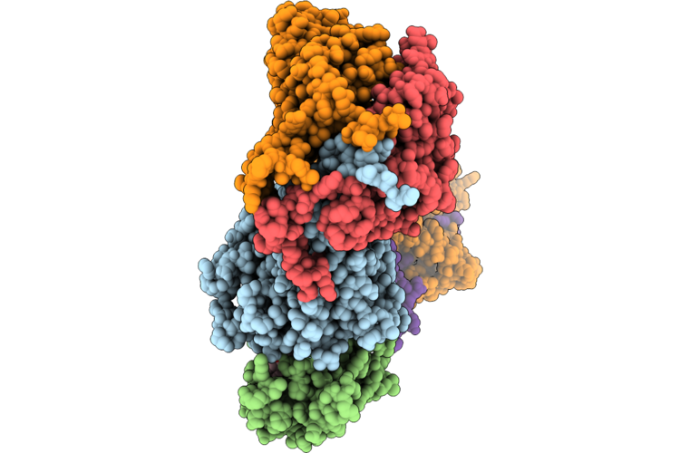
Organism: Mus musculus
Method: ELECTRON MICROSCOPY Resolution:3.57 Å Release Date: 2026-05-06 Classification: CELL ADHESION |
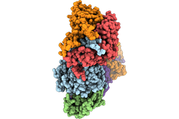 |
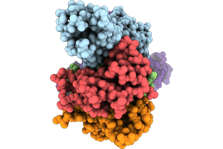
Cryo-Em Structure Of The Cul1-Rbx1-Skp1-Fbxo22 Scf Ubiquition Ligase In Complex With Nsd2 Via Unc10088
Organism: Homo sapiens
Method: ELECTRON MICROSCOPY Release Date: 2026-05-06 Classification: LIGASE Ligands: A1J20 |
 |
Cryo-Em Structure Of The Cul1-Rbx1-Skp1-Fbxo22 Scf Ubiquition Ligase In Complex With Nsd2, Unc10088 And Bach1
Organism: Homo sapiens
Method: ELECTRON MICROSCOPY Release Date: 2026-05-06 Classification: LIGASE Ligands: A1J20 |
 |
Cryo-Em Structure Of The Cul1-Rbx1-Skp1-Fbxo22 Scf Ubiquition Ligase In Complex With Nsd2 Via Unc10415667
Organism: Homo sapiens
Method: ELECTRON MICROSCOPY Release Date: 2026-05-06 Classification: LIGASE Ligands: A1J21 |
 |
Crystal Structure Of Sheep (Ovis Aries) Oxyhemoglobin At 2.1 Angstrom Resolution
Organism: Ovis aries
Method: X-RAY DIFFRACTION Resolution:2.08 Å Release Date: 2026-05-06 Classification: OXYGEN TRANSPORT Ligands: HEM, OXY, K, ZN, PEG |
 |
Organism: Homo sapiens
Method: ELECTRON MICROSCOPY Release Date: 2026-04-29 Classification: CYTOKINE |
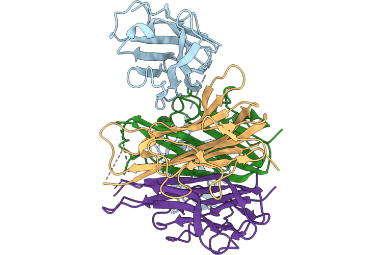 |
Cryo-Em Structure Of Tnf-Alpha In Complex With Two Anti-Tnf-Alpha Nanobodies, Tnf30, Derived From The Tnf-Alpha Inhibitor Ozoralizumab (Ozr)
Organism: Homo sapiens
Method: ELECTRON MICROSCOPY Release Date: 2026-04-29 Classification: CYTOKINE |
 |
Organism: Methanosarcina mazei
Method: X-RAY DIFFRACTION Resolution:1.45 Å Release Date: 2026-04-29 Classification: HYDROLASE Ligands: PO4 |
 |
Organism: Methanosarcina mazei
Method: X-RAY DIFFRACTION Resolution:1.53 Å Release Date: 2026-04-29 Classification: HYDROLASE Ligands: DUT, PO4 |
 |
Organism: Homo sapiens
Method: X-RAY DIFFRACTION Resolution:1.60 Å Release Date: 2026-04-22 Classification: SIGNALING PROTEIN Ligands: MG, GDP, A1JU5 |
 |
Organism: Homo sapiens
Method: X-RAY DIFFRACTION Resolution:1.65 Å Release Date: 2026-04-22 Classification: SIGNALING PROTEIN Ligands: GDP, MG, A1JU6 |
 |
Organism: Homo sapiens
Method: X-RAY DIFFRACTION Resolution:1.54 Å Release Date: 2026-04-22 Classification: SIGNALING PROTEIN Ligands: MG, GDP, CA, A1JU7 |