
Search Count: 5,458
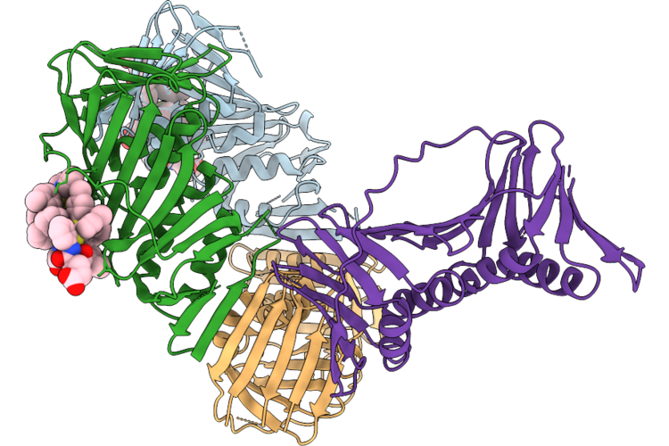 |
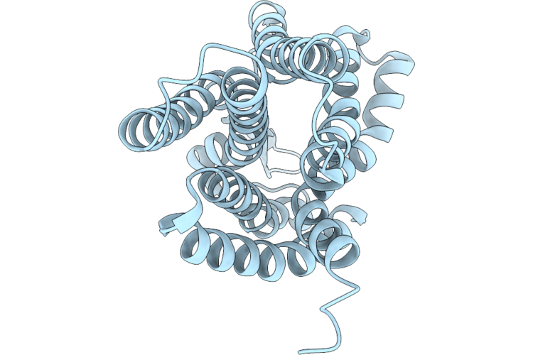
Co-Crystal Structure Of Capcna Bound To The Aoh1996 Derivative, Aoh2.29-Le
Organism: Homo sapiens
Method: X-RAY DIFFRACTION Resolution:2.87 Å Release Date: 2026-07-15 Classification: DNA BINDING PROTEIN Ligands: XEU, CL |
Organism: Homo sapiens
Method: X-RAY DIFFRACTION
Release Date: 2026-07-15
Ligands: XEU, CL
 |
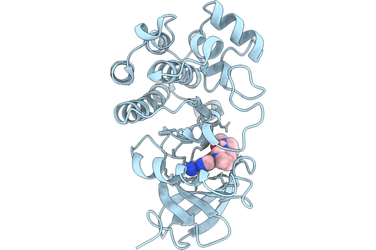
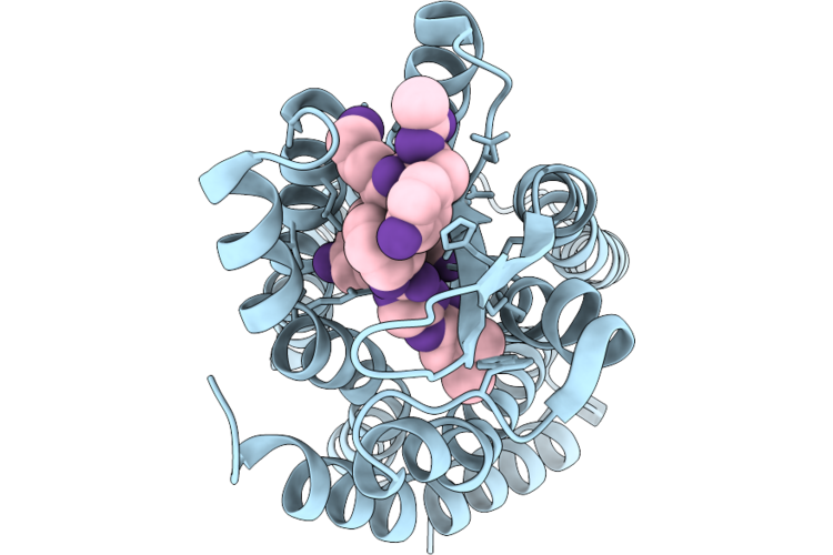
Co-Crystal Structure Of Btk Kinase Domain With Non-Covalent Inhibitor
Organism: Homo sapiens
Method: X-RAY DIFFRACTION Resolution:1.60 Å Release Date: 2026-07-15 Classification: TRANSFERASE Ligands: IMD, A1CSI |
Organism: Homo sapiens
Method: X-RAY DIFFRACTION
Release Date: 2026-07-15
Ligands: IMD, A1CSI
 |
Co-Crystal Structure Of Btk Kinase Domain With Non-Covalent Inhibitor
Organism: Homo sapiens
Method: X-RAY DIFFRACTION Resolution:1.40 Å Release Date: 2026-07-15 Classification: TRANSFERASE Ligands: IMD, A1CSJ |
Organism: Homo sapiens
Method: X-RAY DIFFRACTION
Release Date: 2026-07-15
Ligands: IMD, A1CSJ
 |
Co-Crystal Structure Of Btk Kinase Domain With Non-Covalent Inhibitor
Organism: Homo sapiens
Method: X-RAY DIFFRACTION Resolution:1.70 Å Release Date: 2026-07-15 Classification: TRANSFERASE Ligands: IMD, PG4, A1CSK, EDO |
Organism: Homo sapiens
Method: X-RAY DIFFRACTION
Release Date: 2026-07-15
Ligands: IMD, PG4, A1CSK, EDO
 |
Co-Crystal Structure Of Btk Kinase Domain With Non-Covalent Inhibitor
Organism: Homo sapiens
Method: X-RAY DIFFRACTION Resolution:1.90 Å Release Date: 2026-07-15 Classification: TRANSFERASE Ligands: A1CSL |
Organism: Homo sapiens
Method: X-RAY DIFFRACTION
Release Date: 2026-07-15
Ligands: A1CSL
 |
Co-Crystal Structure Of Btk Kinase Domain With Non-Covalent Inhibitor
Organism: Homo sapiens
Method: X-RAY DIFFRACTION Resolution:1.66 Å Release Date: 2026-07-15 Classification: TRANSFERASE Ligands: IMD, 1PE, DMS, A1CSM |
Organism: Homo sapiens
Method: X-RAY DIFFRACTION
Release Date: 2026-07-15
Ligands: IMD, 1PE, DMS, A1CSM
 |
Co-Crystal Structure Of Btk Kinase Domain With Non-Covalent Inhibitor
Organism: Homo sapiens
Method: X-RAY DIFFRACTION Resolution:2.51 Å Release Date: 2026-07-15 Classification: TRANSFERASE Ligands: A1CSG |
Organism: Homo sapiens
Method: X-RAY DIFFRACTION
Release Date: 2026-07-15
Ligands: A1CSG
 |
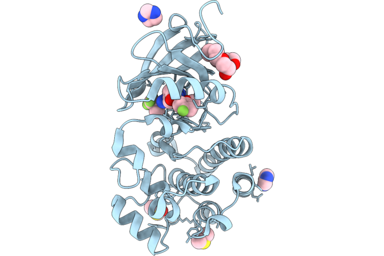
Crystal Structure Of S-Adenosyl-L-Homocysteine Hydrolase From P. Aeruginosa: F324A Mutant Complexed With Adenosine And Rubidium Ions Replacing Native Potassium
Organism: Pseudomonas aeruginosa pao1
Method: X-RAY DIFFRACTION Resolution:2.06 Å Release Date: 2026-07-08 Classification: HYDROLASE Ligands: NAD, ADN, PO4, RB |
Organism: Pseudomonas aeruginosa pao1
Method: X-RAY DIFFRACTION
Release Date: 2026-07-08
Ligands: NAD, ADN, PO4, RB
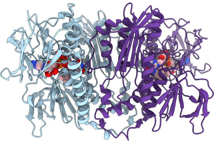 |
Structure Of Trypanothione Reductase From Leishmania Donovani
Organism: Leishmania donovani
Method: X-RAY DIFFRACTION Resolution:2.38 Å Release Date: 2026-07-08 Classification: OXIDOREDUCTASE Ligands: FAD |
Organism: Leishmania donovani
Method: X-RAY DIFFRACTION
Release Date: 2026-07-08
Ligands: FAD
 |
Structure Of Mc5Ar2 In Complex With Mc5A-Desarg (Monomer)
Organism: Mus musculus
Method: ELECTRON MICROSCOPY Resolution:3.42 Å Release Date: 2026-07-01 Classification: SIGNALING PROTEIN |
Organism: Mus musculus
Method: ELECTRON MICROSCOPY
Release Date: 2026-07-01
 |
Structure Of C5A Anaphylatoxin Chemotactic Receptor 2, C5Ar2 In The Apo State
Organism: Homo sapiens
Method: ELECTRON MICROSCOPY Release Date: 2026-07-01 Classification: SIGNALING PROTEIN |
Organism: Homo sapiens
Method: ELECTRON MICROSCOPY
Release Date: 2026-07-01
 |
Structure Of C5A Anaphylatoxin Chemotactic Receptor 2, C5Ar2 Bound To Ep54
Organism: Homo sapiens
Method: ELECTRON MICROSCOPY Release Date: 2026-07-01 Classification: SIGNALING PROTEIN |
Organism: Homo sapiens
Method: ELECTRON MICROSCOPY
Release Date: 2026-07-01
 |
Structure Of C5A Anaphylatoxin Chemotactic Receptor 2, C5Ar2 Bound To C5A
Organism: Homo sapiens
Method: ELECTRON MICROSCOPY Release Date: 2026-07-01 Classification: SIGNALING PROTEIN |
Organism: Homo sapiens
Method: ELECTRON MICROSCOPY
Release Date: 2026-07-01
 |
Structure Of C5A Anaphylatoxin Chemotactic Receptor 2, C5Ar2 Bound To C5A-Pep
Organism: Homo sapiens
Method: ELECTRON MICROSCOPY Release Date: 2026-07-01 Classification: SIGNALING PROTEIN |
Organism: Homo sapiens
Method: ELECTRON MICROSCOPY
Release Date: 2026-07-01
 |
Structure Of C5A Anaphylatoxin Chemotactic Receptor 2, C5Ar2 Bound To R8Y
Organism: Homo sapiens
Method: ELECTRON MICROSCOPY Release Date: 2026-07-01 Classification: SIGNALING PROTEIN |
Organism: Homo sapiens
Method: ELECTRON MICROSCOPY
Release Date: 2026-07-01
 |
Structure Of Mc5Ar2 In Complex With Mc5A-Desarg
Organism: Mus musculus
Method: ELECTRON MICROSCOPY Release Date: 2026-07-01 Classification: SIGNALING PROTEIN |
Organism: Mus musculus
Method: ELECTRON MICROSCOPY
Release Date: 2026-07-01
 |
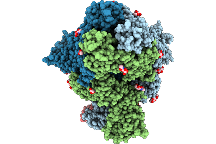
Hucov-Hku1 C S 2P In Complex With H501-018 Fab (State 1, Global Cryoem)
Organism: Human coronavirus hku1, Homo sapiens
Method: ELECTRON MICROSCOPY Release Date: 2026-07-01 Classification: VIRAL PROTEIN/IMMUNE SYSTEM Ligands: NAG |
Organism: Human coronavirus hku1, Homo sapiens
Method: ELECTRON MICROSCOPY
Release Date: 2026-07-01
Ligands: NAG
 |
Hucov-Hku1 C S 2P In Complex With H501-018 Fab (State 2, Global Cryoem)
Organism: Human coronavirus hku1 (isolate n5), Homo sapiens
Method: ELECTRON MICROSCOPY Release Date: 2026-07-01 Classification: VIRAL PROTEIN/IMMUNE SYSTEM Ligands: NAG |
Organism: Human coronavirus hku1 (isolate n5), Homo sapiens
Method: ELECTRON MICROSCOPY
Release Date: 2026-07-01
Ligands: NAG
 |
Hcov-Hku1 C S 2P In Complex With H501-018 Fab (Local Cryoem)
Organism: Human coronavirus hku1, Homo sapiens
Method: ELECTRON MICROSCOPY Release Date: 2026-07-01 Classification: VIRAL PROTEIN/IMMUNE SYSTEM Ligands: NAG |
Organism: Human coronavirus hku1, Homo sapiens
Method: ELECTRON MICROSCOPY
Release Date: 2026-07-01
Ligands: NAG
 |
Hcov-Hku1 C S 2P In Complex With H501-022 Fab (Global Cryoem)
Organism: Human coronavirus hku1 (isolate n5), Homo sapiens
Method: ELECTRON MICROSCOPY Release Date: 2026-07-01 Classification: VIRAL PROTEIN/IMMUNE SYSTEM Ligands: NAG |
Organism: Human coronavirus hku1 (isolate n5), Homo sapiens
Method: ELECTRON MICROSCOPY
Release Date: 2026-07-01
Ligands: NAG