
Search Count: 15,589
All
Selected
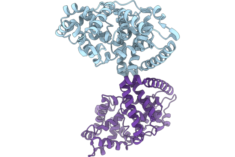 |
Organism: Mus musculus
Method: X-RAY DIFFRACTION Resolution:2.20 Å Release Date: 2026-05-06 Classification: HYDROLASE ACTIVATOR |
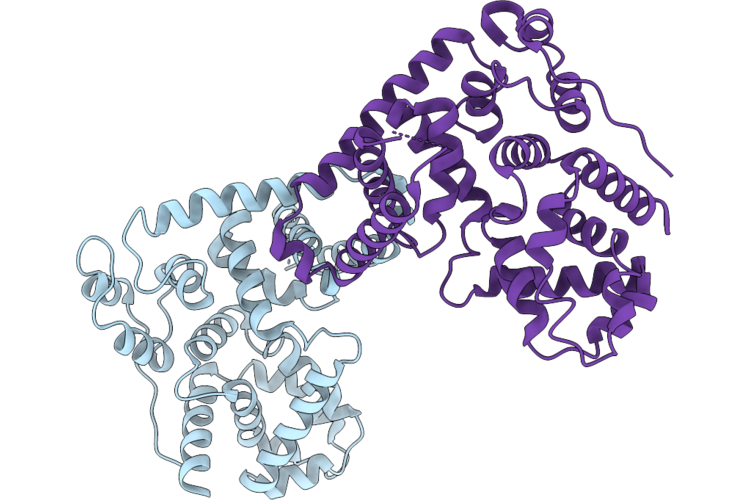 |
Organism: Homo sapiens
Method: X-RAY DIFFRACTION Resolution:3.34 Å Release Date: 2026-05-06 Classification: PROTEIN BINDING |
 |
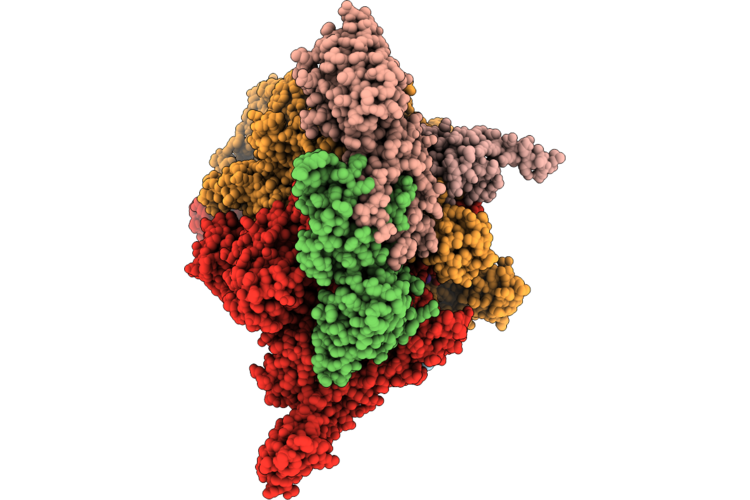
Closed Eco-Epec: Cryo-Em Structure Of Eco Rnap His-Elemental Paused Elongation Complex With A Closed Active Site (Closed Tl, Si3 And Rh-Fl)
Organism: Escherichia coli
Method: ELECTRON MICROSCOPY Resolution:2.90 Å Release Date: 2026-04-29 Classification: Transcription/DNA/RNA Ligands: 1N7, MG, ZN |
 |
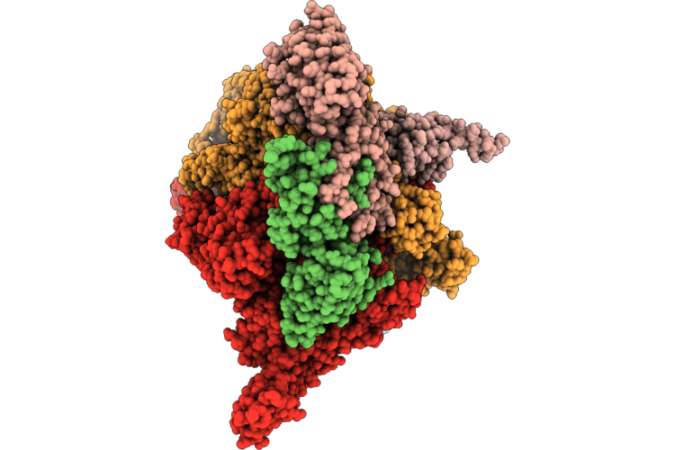
Open1 Eco-Epec: Cryo-Em Structure Of Eco Rnap His-Elemental Paused Elongation Complex With An Open Active Site (Open Tl, Si3 And Rh-Fl)
Organism: Escherichia coli
Method: ELECTRON MICROSCOPY Resolution:2.90 Å Release Date: 2026-04-29 Classification: TRANSCRIPTION/DNA/RNA Ligands: 1N7, MG, ZN |
 |
Open2 Eco-Epec: Cryo-Em Structure Of Eco Rnap His-Elemental Paused Elongation Complex With An Open Active Site (Open Tl, Si3 And Rh-Fl)
Organism: Escherichia coli
Method: ELECTRON MICROSCOPY Resolution:2.80 Å Release Date: 2026-04-29 Classification: Transcription/DNA/RNA Ligands: 1N7, MG, ZN |
 |
Open3 Eco-Epec: Cryo-Em Structure Of Eco Rnap His-Elemental Paused Elongation Complex With An Open Active Site (Open Tl, Si3 And Rh-Fl)
Organism: Escherichia coli
Method: ELECTRON MICROSCOPY Resolution:2.80 Å Release Date: 2026-04-29 Classification: TRANSCRIPTION Ligands: 1N7, MG, ZN |
 |
Organism: Burkholderia pseudomallei
Method: X-RAY DIFFRACTION Resolution:1.93 Å Release Date: 2026-04-29 Classification: METAL BINDING PROTEIN Ligands: FE |
 |
Organism: Burkholderia pseudomallei
Method: X-RAY DIFFRACTION Resolution:1.96 Å Release Date: 2026-04-29 Classification: METAL BINDING PROTEIN Ligands: FE |
 |
Organism: Human immunodeficiency virus 1, Macaca mulatta, Homo sapiens
Method: ELECTRON MICROSCOPY Resolution:2.70 Å Release Date: 2026-04-29 Classification: VIRAL PROTEIN Ligands: NAG |
 |
Organism: Human immunodeficiency virus 1, Macaca mulatta, Homo sapiens
Method: ELECTRON MICROSCOPY Resolution:2.70 Å Release Date: 2026-04-29 Classification: VIRAL PROTEIN Ligands: NAG |
 |
Organism: Mus musculus
Method: ELECTRON MICROSCOPY Release Date: 2026-04-29 Classification: RIBOSOME Ligands: MG, ZN, B8N, 4AC |
 |
Crystal Structure Of The Staphylococcal Efflux Pump Qaca In The Outward Open State
Organism: Staphylococcus aureus
Method: X-RAY DIFFRACTION Resolution:2.83 Å Release Date: 2026-04-22 Classification: MEMBRANE PROTEIN Ligands: PGT, LMU, PTY, 3PH, GOL, NA |
 |
Crystal Structure Of The Staphylococcal Efflux Pump Qaca In The Inward Open State In Complex With Nanobody 89
Organism: Staphylococcus aureus, Lama glama
Method: X-RAY DIFFRACTION Resolution:3.32 Å Release Date: 2026-04-22 Classification: MEMBRANE PROTEIN Ligands: PTY, PGT, LMU, OXM |
 |
Crystal Structure Of The Staphylococcal Efflux Pump Qaca In The Outward Open State Bound To Ethidium
Organism: Staphylococcus aureus
Method: X-RAY DIFFRACTION Resolution:2.79 Å Release Date: 2026-04-22 Classification: MEMBRANE PROTEIN Ligands: ET, GOL, TLA, CL, LMU, OXM |
 |
Re-Refinement Of Damage Free Ferric State Of Dye Type Peroxidase Aa From Streptomyces Lividans.
Organism: Streptomyces lividans 1326
Method: X-RAY DIFFRACTION Resolution:1.88 Å Release Date: 2026-04-22 Classification: OXIDOREDUCTASE Ligands: HEM |
 |
Reprocessing And Re-Refinement Of Damage Free Ferric State Of Dye Type Peroxidase Aa From Streptomyces Lividans
Organism: Streptomyces lividans 1326
Method: X-RAY DIFFRACTION Resolution:1.86 Å Release Date: 2026-04-22 Classification: OXIDOREDUCTASE Ligands: HEM |
 |
Organism: Homo sapiens, Mus musculus
Method: ELECTRON MICROSCOPY Resolution:2.90 Å Release Date: 2026-04-22 Classification: SIGNALING PROTEIN Ligands: GOQ |
 |
Organism: Homo sapiens
Method: ELECTRON MICROSCOPY Resolution:3.00 Å Release Date: 2026-04-22 Classification: SIGNALING PROTEIN Ligands: GOQ |
 |
Dark Structure Of Beta-2 Adrenergic Receptor With Photoazolol In Dark State Recorded At Swissfel
Organism: Homo sapiens, Enterobacteria phage t4
Method: X-RAY DIFFRACTION Resolution:2.45 Å Release Date: 2026-04-22 Classification: MEMBRANE PROTEIN Ligands: SO4, CLR, 12P, OLC, 1PE, EDO, GOL, A1JHU, PLM |
 |
Mixed Model Refinement Of Beta-2 Adrenergic Receptor With Photoazolol In Dark State And Light State, 10 Seconds After Light Activation, Recorded At Swissfel
Organism: Homo sapiens, Tequatrovirus t4
Method: X-RAY DIFFRACTION Resolution:2.45 Å Release Date: 2026-04-22 Classification: MEMBRANE PROTEIN Ligands: SO4, ACM, CLR, PLM, 12P, OLC, 1PE, EDO, GOL, A1JHU |