
Search Count: 8,628
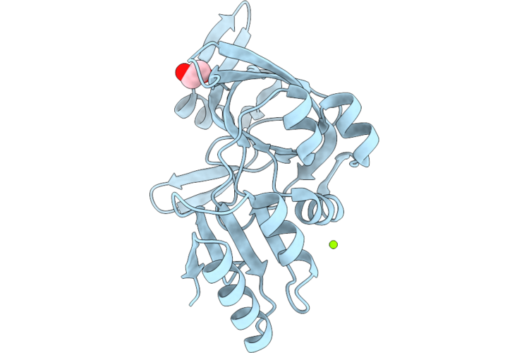 |
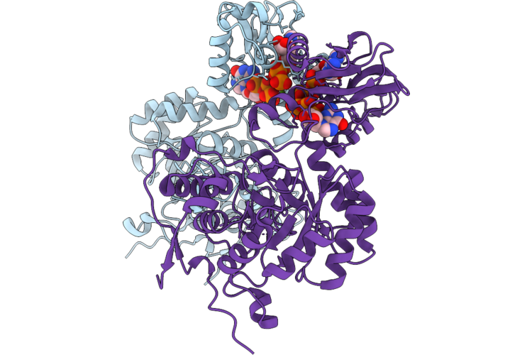
Nucleotide Binding Domain (Residues 475-720) Of Abc3 Transporter Permease From Clostridioides Difficile Strain 630
Organism: Clostridioides difficile 630
Method: X-RAY DIFFRACTION Resolution:2.00 Å Release Date: 2026-06-24 Classification: TRANSPORT PROTEIN Ligands: MG, CL, EDO |
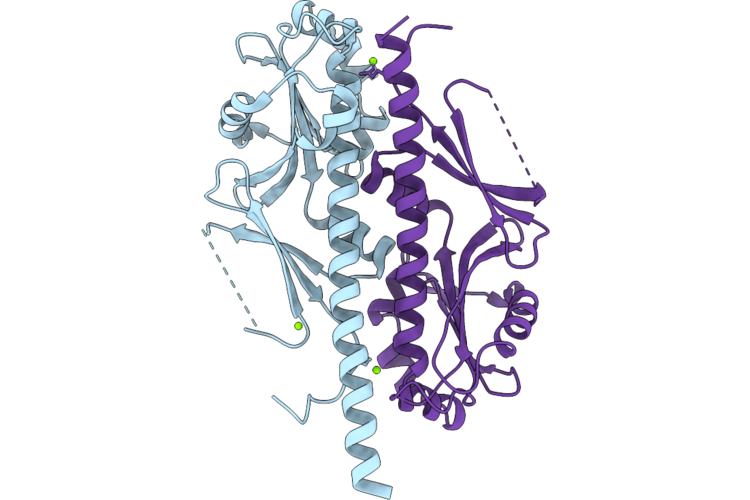 |
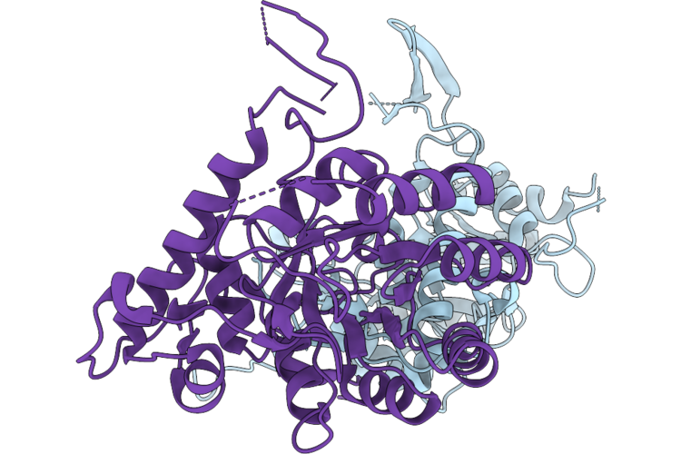
Crystal Structure Of The Ggdef Domain (Residues 31-260) Of Diguanylate Cyclase From Vibrio Cholerae Serotype O1
Organism: Vibrio cholerae o1 biovar el tor str. n16961
Method: X-RAY DIFFRACTION Resolution:2.00 Å Release Date: 2026-06-24 Classification: OXIDOREDUCTASE Ligands: MG, CL |
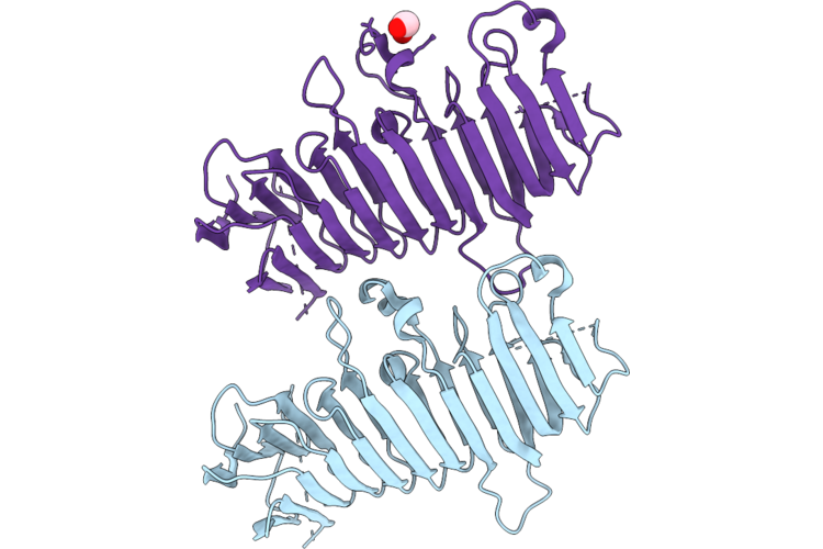 |
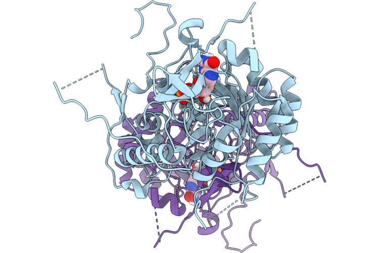
Crystal Structure Of Duf4097 Domain-Containing Protein From Clostridium Difficile Strain 630
Organism: Clostridioides difficile 630
Method: X-RAY DIFFRACTION Resolution:2.00 Å Release Date: 2026-06-24 Classification: UNKNOWN FUNCTION Ligands: EDO |
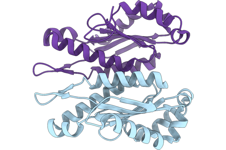 |
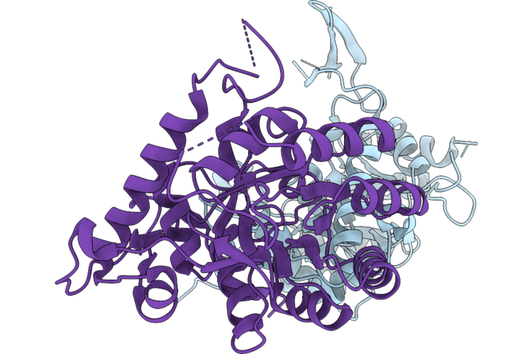
Crystal Structure Of The Ggdef Domain Of The Diguanylate Cyclase From Vibrio Vulnificus Cmcp6
Organism: Vibrio vulnificus cmcp6
Method: X-RAY DIFFRACTION Resolution:2.60 Å Release Date: 2026-06-24 Classification: TRANSFERASE |
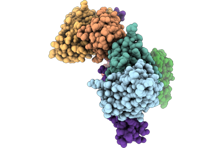 |
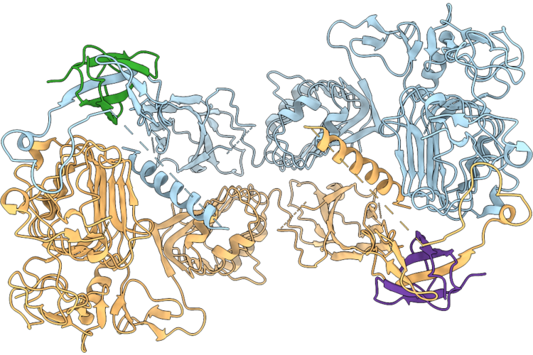
Cryo-Electron Microscopy Structure Of Pfripr Bound To Monoclonal Antibodies Rp.047, Rp.057 And Rp.035
Organism: Mus musculus, Plasmodium falciparum 3d7
Method: ELECTRON MICROSCOPY Resolution:3.35 Å Release Date: 2026-06-24 Classification: IMMUNE SYSTEM |
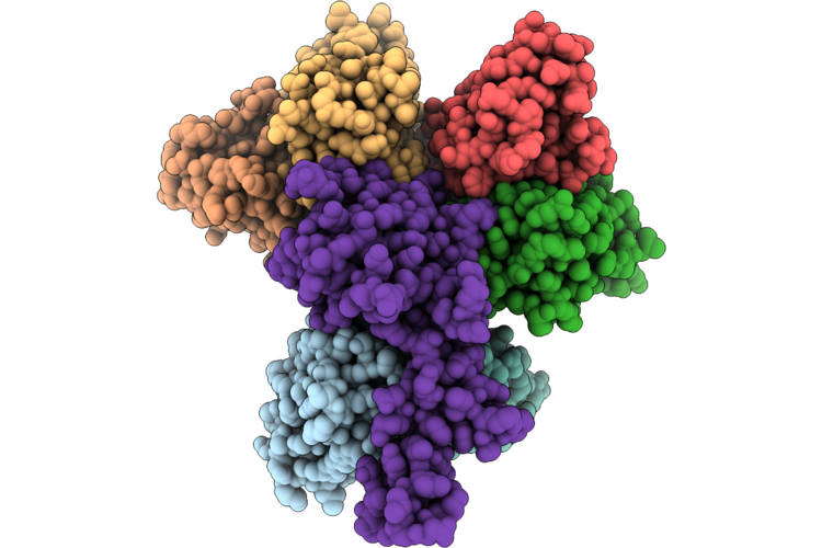 |
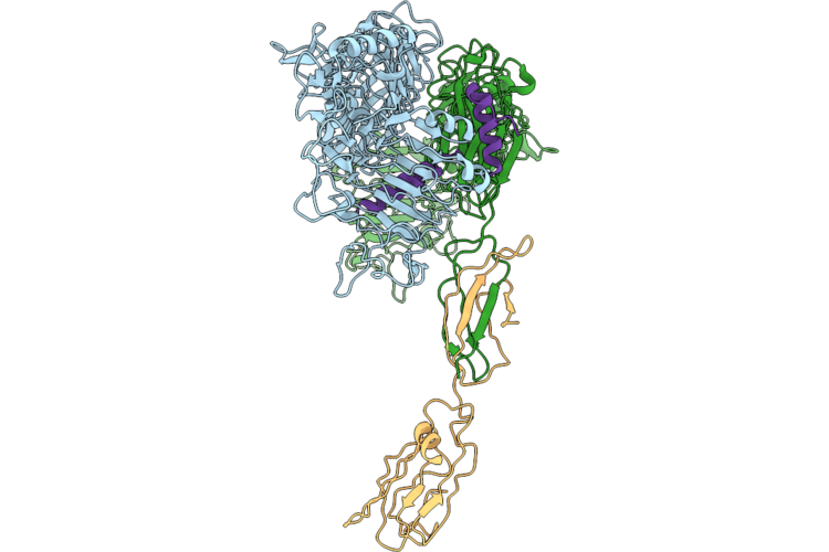
Cryo-Electron Microscopy Structure Of Pfripr Bound To Monoclonal Antibodies Rp.093, Rp.073 And Rp.063
Organism: Mus musculus, Plasmodium falciparum 3d7
Method: ELECTRON MICROSCOPY Resolution:2.97 Å Release Date: 2026-06-24 Classification: IMMUNE SYSTEM |
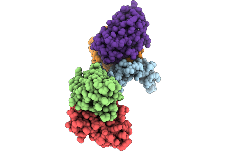 |
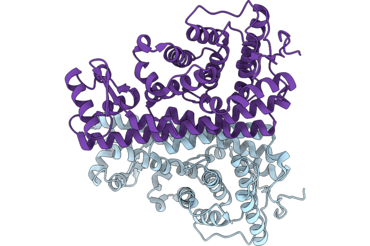
Cryo-Electron Microscopy Structure Of Pfripr Bound To Monoclonal Antibodies Rp.092 And Rp.052
Organism: Plasmodium falciparum 3d7, Mus musculus
Method: ELECTRON MICROSCOPY Resolution:4.01 Å Release Date: 2026-06-24 Classification: IMMUNE SYSTEM |
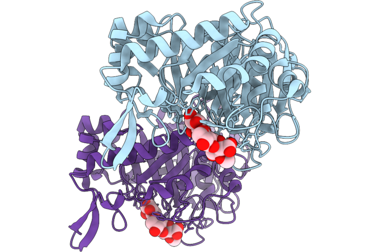 |
Glucuronoxylan-Specific Gh30_8 Family Xylanase Ctxyn30A From Clostridium Thermocellum Complex With Glucuronic Acid Epoxide Inhibitor
Organism: Acetivibrio thermocellus dsm 2360
Method: X-RAY DIFFRACTION Resolution:1.50 Å Release Date: 2026-06-24 Classification: HYDROLASE Ligands: HQ8 |
 |
Gh10 Family Xylanase Xyna From Bacillus Sp. Kw1 Complex With Xylobiosyl-Configured Cyclophellitol Probe Bearing An Alpha-1,3 - Araf Decoration
Organism: Bacillus sp. (in: firmicutes)
Method: X-RAY DIFFRACTION Resolution:2.02 Å Release Date: 2026-06-24 Classification: HYDROLASE Ligands: A1J01 |
 |
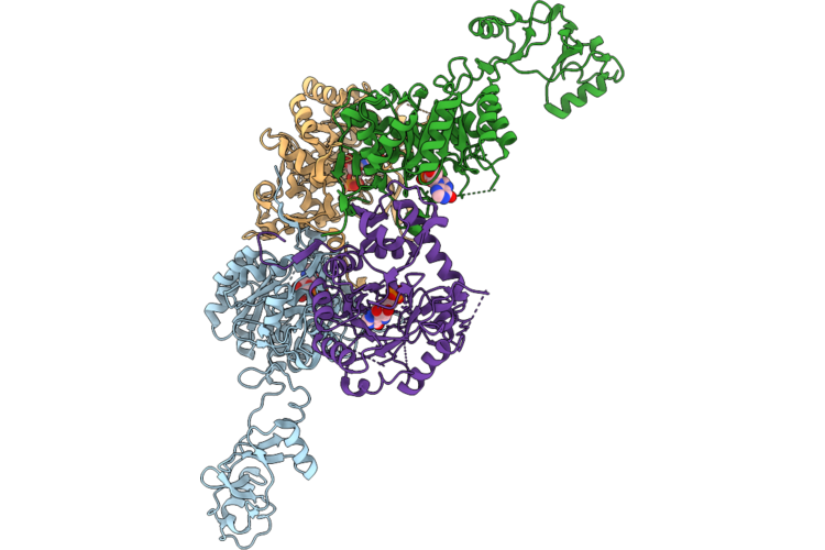
Organism: Homo sapiens
Method: ELECTRON MICROSCOPY Release Date: 2026-06-24 Classification: MEMBRANE PROTEIN |
 |
Organism: Homo sapiens, Synthetic construct
Method: ELECTRON MICROSCOPY Release Date: 2026-06-24 Classification: MEMBRANE PROTEIN |
 |
Organism: Escherichia coli 'bl21-gold(de3)plyss ag'
Method: X-RAY DIFFRACTION Resolution:2.28 Å Release Date: 2026-06-24 Classification: TOXIN |
 |
Organism: Burkholderia thailandensis
Method: X-RAY DIFFRACTION Resolution:3.35 Å Release Date: 2026-06-17 Classification: OXIDOREDUCTASE Ligands: IMP |
 |
Organism: Burkholderia thailandensis
Method: X-RAY DIFFRACTION Resolution:1.78 Å Release Date: 2026-06-17 Classification: OXIDOREDUCTASE Ligands: GTP, MG, ATP |
 |
Organism: Burkholderia thailandensis
Method: X-RAY DIFFRACTION Resolution:2.75 Å Release Date: 2026-06-17 Classification: OXIDOREDUCTASE Ligands: NAD, ATP, GTP, MG, IMP, K |
 |
Organism: Burkholderia thailandensis
Method: X-RAY DIFFRACTION Resolution:1.86 Å Release Date: 2026-06-17 Classification: OXIDOREDUCTASE Ligands: ATP, GTP, MG, IMP, K |
 |
Organism: Burkholderia thailandensis
Method: X-RAY DIFFRACTION Resolution:2.15 Å Release Date: 2026-06-17 Classification: OXIDOREDUCTASE |
 |
Organism: Burkholderia thailandensis
Method: X-RAY DIFFRACTION Resolution:1.44 Å Release Date: 2026-06-17 Classification: OXIDOREDUCTASE Ligands: 5GP, CL |
 |
Organism: Burkholderia thailandensis
Method: X-RAY DIFFRACTION Resolution:1.90 Å Release Date: 2026-06-17 Classification: OXIDOREDUCTASE |
 |
Crystal Structure Of Flagellar Assembly Protein Fliw From Campylobacter Jejuni Subsp. Jejuni 81-176-Drh212
Organism: Campylobacter jejuni subsp. jejuni 81-176-drh212
Method: X-RAY DIFFRACTION Resolution:2.95 Å Release Date: 2026-06-17 Classification: TRANSLATION Ligands: CL |