
Search Count: 55,099
 |
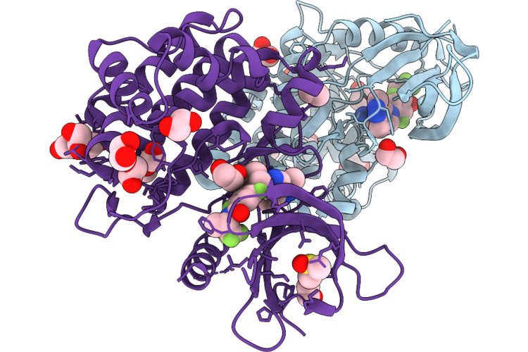
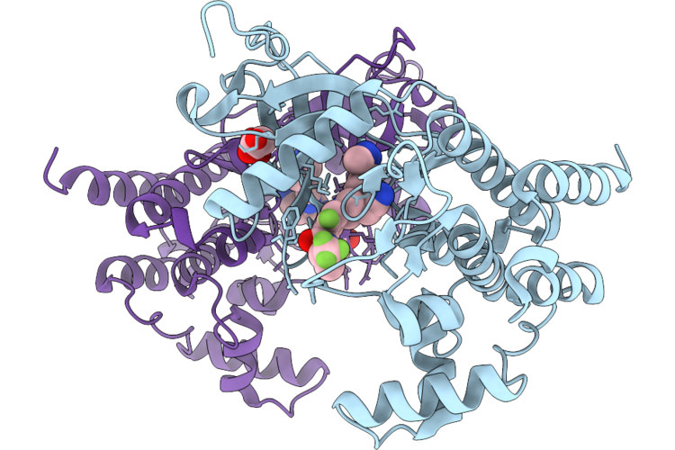
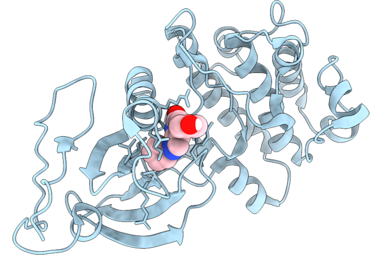
X-Ray Crystal Structure Of A High-Affinity Monoclonal Antibody Sequesters Xylazine
Organism: Homo sapiens
Method: X-RAY DIFFRACTION Resolution:1.99 Å Release Date: 2026-06-10 Classification: IMMUNE SYSTEM Ligands: A1C4R, CL |
 |
Organism: Homo sapiens
Method: X-RAY DIFFRACTION Resolution:1.54 Å Release Date: 2026-06-10 Classification: TRANSFERASE Ligands: EDO, A1C68, DMS |
 |
Organism: Homo sapiens
Method: X-RAY DIFFRACTION Resolution:1.59 Å Release Date: 2026-06-10 Classification: TRANSFERASE Ligands: MLA, A1C68 |
 |
Organism: Homo sapiens
Method: X-RAY DIFFRACTION Resolution:1.96 Å Release Date: 2026-06-10 Classification: TRANSFERASE Ligands: A1DAS, MN, CL |
 |
Crystal Structure Of Usp7 Traf Domain In Complex With Magel2 Peptide (968-980)
Organism: Homo sapiens
Method: X-RAY DIFFRACTION Resolution:1.63 Å Release Date: 2026-06-10 Classification: PEPTIDE BINDING PROTEIN Ligands: MG, GOL |
 |
Organism: Homo sapiens
Method: X-RAY DIFFRACTION Resolution:2.22 Å Release Date: 2026-06-10 Classification: TRANSFERASE/TRANSFERASE INHIBITOR Ligands: A1DG4 |
 |
Organism: Homo sapiens
Method: X-RAY DIFFRACTION Resolution:1.95 Å Release Date: 2026-06-10 Classification: TRANSFERASE/TRANSFERASE INHIBITOR Ligands: A1DG6 |
 |
Organism: Homo sapiens
Method: X-RAY DIFFRACTION Resolution:2.20 Å Release Date: 2026-06-10 Classification: TRANSFERASE/TRANSFERASE INHIBITOR Ligands: A1DG5 |
 |
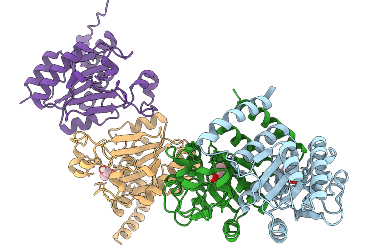
Crystal Structure Of Monoalkyl Phthalate Hydrolase From Rhodococcus Sp. Eg-5
Organism: Rhodococcus sp. eg-5
Method: X-RAY DIFFRACTION Resolution:3.00 Å Release Date: 2026-06-10 Classification: HYDROLASE |
 |
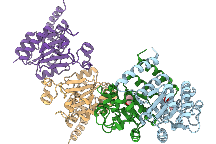
Crystal Structure Of Cysteine-Dependent Hydrolase (Csdh) From Rhodococcus Opacus In Complex With Monobutylphthalate (Mbp)
Organism: Rhodococcus opacus
Method: X-RAY DIFFRACTION Resolution:2.90 Å Release Date: 2026-06-10 Classification: HYDROLASE Ligands: PHT |
 |
Crystal Structure Of Cysteine-Dependent Hydrolase (Csdh) From Rhodococcus Opacus In Complex With Propylene Glycol
Organism: Rhodococcus opacus
Method: X-RAY DIFFRACTION Resolution:2.80 Å Release Date: 2026-06-10 Classification: HYDROLASE Ligands: PGO |
 |
Crystal Structure Of Cysteine-Dependent Hydrolase (Csdh) From Rhodococcus Opacus In Complex With Dibutylphthalate
Organism: Rhodococcus opacus
Method: X-RAY DIFFRACTION Resolution:2.90 Å Release Date: 2026-06-10 Classification: HYDROLASE Ligands: PHT, EOH |
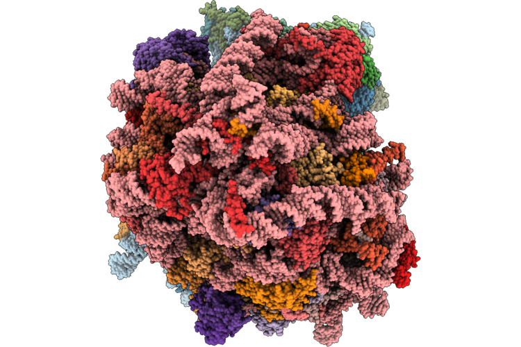 |
Organism: Homo sapiens
Method: ELECTRON MICROSCOPY Release Date: 2026-06-10 Classification: TRANSLATION Ligands: MG, ZN |
 |
Respiratory Syncytial Virus Fusion Protein N-Terminal Heptad Repeat Domain In Complex With Double Stapled Peptide 4/4G
Organism: Human respiratory syncytial virus a, Human respiratory syncytial virus
Method: X-RAY DIFFRACTION Resolution:1.33 Å Release Date: 2026-06-10 Classification: VIRAL PROTEIN |
 |
Organism: Homo sapiens
Method: X-RAY DIFFRACTION Resolution:2.11 Å Release Date: 2026-06-10 Classification: LYASE Ligands: TLA |
 |
Organism: Arabidopsis thaliana, Synthetic construct
Method: X-RAY DIFFRACTION Resolution:2.30 Å Release Date: 2026-06-10 Classification: DNA BINDING PROTEIN |
 |
Organism: Arabidopsis thaliana, Synthetic construct
Method: X-RAY DIFFRACTION Resolution:2.60 Å Release Date: 2026-06-10 Classification: DNA BINDING PROTEIN Ligands: EDO |
 |
Organism: Arabidopsis thaliana, Synthetic construct
Method: X-RAY DIFFRACTION Resolution:2.48 Å Release Date: 2026-06-10 Classification: DNA BINDING PROTEIN Ligands: EDO, SO4 |
 |
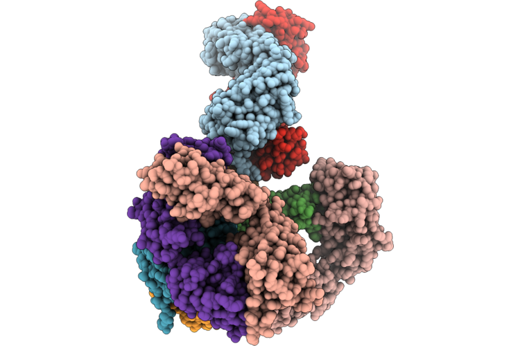
Structure Of Geobacillus Stearothermophilus Rnase P Ribozyme In Complex With Precursor Trna With Non-Complementary 5' Leader (Sub-Conformation 1 Of Trna Anticodon Arm Tilted)
Organism: Geobacillus stearothermophilus
Method: ELECTRON MICROSCOPY Release Date: 2026-06-10 Classification: RNA Ligands: CA |
 |
Structure Of Geobacillus Stearothermophilus Rnase P Ribozyme In Complex With Precursor Trna With Non-Complementary 5' Leader (Sub-Conformation 2 Of Trna Anticodon Arm Tilted)
Organism: Geobacillus stearothermophilus
Method: ELECTRON MICROSCOPY Release Date: 2026-06-10 Classification: RNA Ligands: CA |