

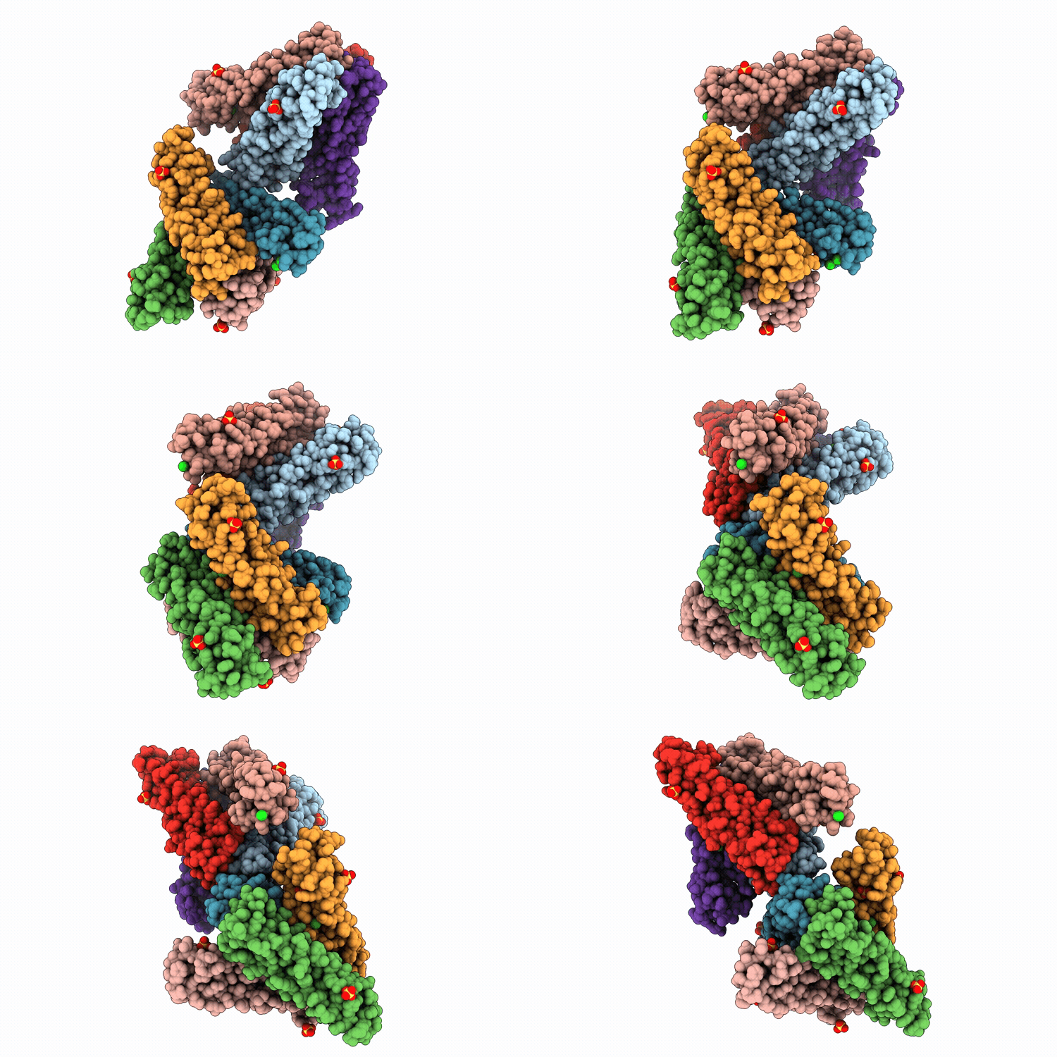
9EC1 image
Deposition Date 2024-11-13
Release Date 2026-04-22
Last Version Date 2026-05-13
Entry Detail
PDB ID:
9EC1
Title:
The Structure of Human Dystrophin Spectrin Repeat 24
Biological Source:
Source Organism(s):
Homo sapiens (Taxon ID: 9606)
Expression System(s):
Method Details:
Experimental Method:
Resolution:
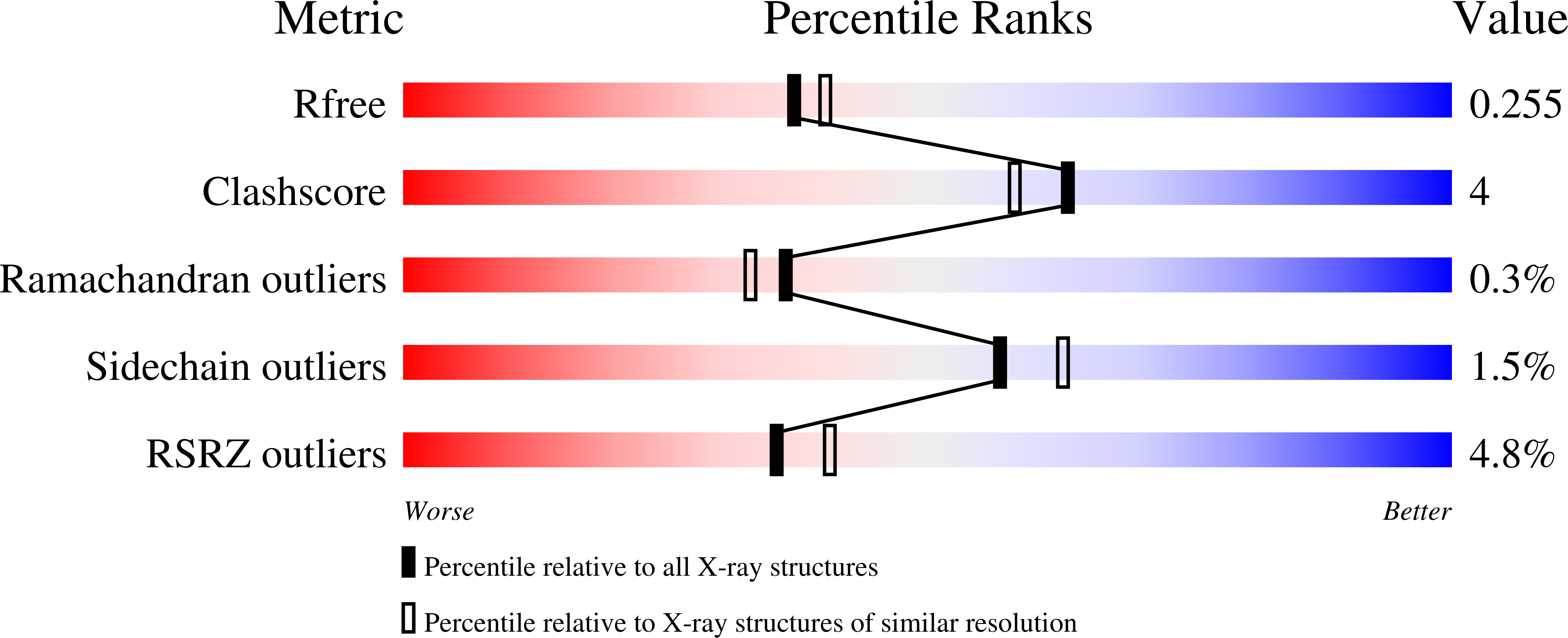
2.14 Å
R-Value Free:
0.25
R-Value Work:
0.21
R-Value Observed:
0.21
Space Group:
P 65 2 2
Macromolecular Entities
Structures with similar UniProt ID
Protein Blast
Polymer Type:polypeptide(L)
Molecule:Dystrophin
Gene (Uniprot):DMD
Chain IDs:A, B, C, D, E, F, G, H
Chain Length:117
Number of Molecules:8
Biological Source:Homo sapiens
Primary Citation
The atomic structure of human dystrophin spectrin-like repeat 24.
Acta Crystallogr.,Sect.F 82 184 193 (2026)
PMID: 42024151 DOI: 10.1107/S2053230X26003262

Abstact

The structure of spectrin-like repeat 24 of human dystrophin was determined at 2.5 A effective resolution. The structure exhibits a three-helix bundle fold, common to all spectrin-repeat family members, and shares a high degree of homology with existing structures of spectrin-like repeat 1 from dystrophin and utrophin. The structure provides molecular details of the atomic interactions that stabilize the repeat, including hydrophobic interactions and inter-helix and intra-helix salt bridges. AlphaFold models of the repeat are in excellent agreement with the structure, showing an all-atom r.m.s.d. of 1.13 A. Accurate modeling of SR24 supports AlphaFold modeling of all 24 of the dystrophin spectrin-like repeats and the use of these models in predicting the molecular determinants of dystrophin stability, a key aspect of its biological function as a structural protein that cross-links actin filaments to the dystrophin-glycoprotein complex to mediate a mechanical connection between the cytoskeleton and the extracellular matrix.

Legend

Protein

Chemical

Disease

Primary Citation of related structures
Feedback Form
Name
Email
Institute
Feedback