
Search Count: 21,757
 |
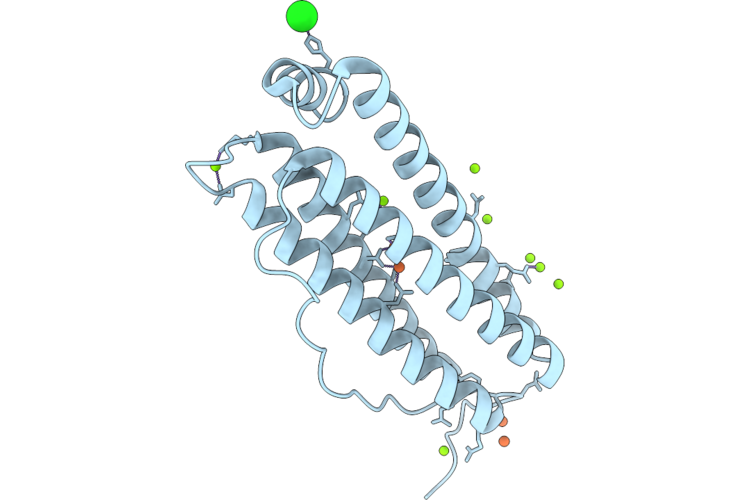
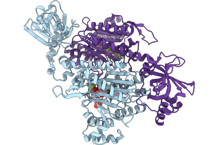
Organism: Homo sapiens
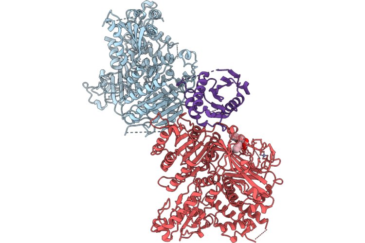
Method: X-RAY DIFFRACTION Resolution:1.60 Å Release Date: 2026-06-10 Classification: METAL BINDING PROTEIN Ligands: FE, MG, CL |
 |
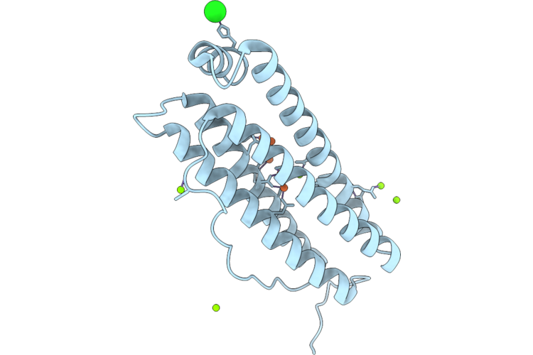
Organism: Homo sapiens
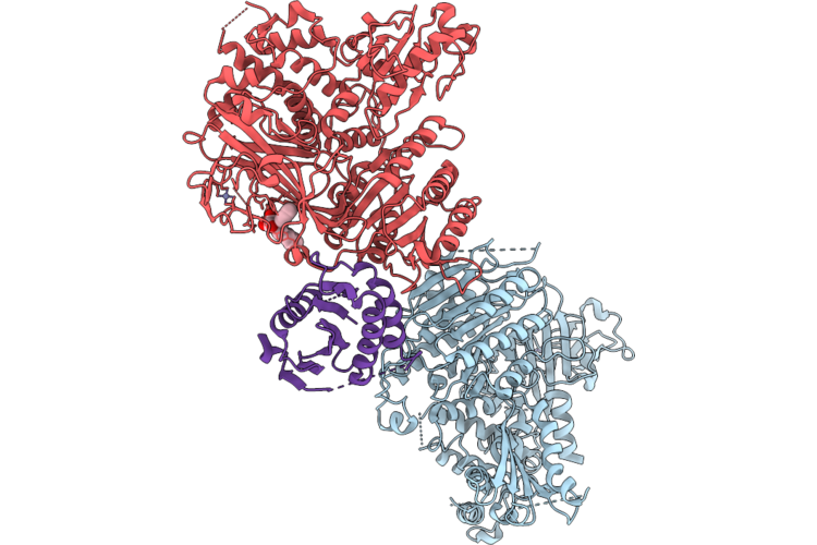
Method: X-RAY DIFFRACTION Resolution:2.41 Å Release Date: 2026-06-10 Classification: METAL BINDING PROTEIN Ligands: FE, CL, MG |
 |
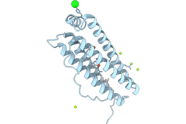
Organism: Homo sapiens
Method: X-RAY DIFFRACTION Resolution:2.11 Å Release Date: 2026-06-10 Classification: METAL BINDING PROTEIN Ligands: FE, MG, CL |
 |
Organism: Homo sapiens
Method: X-RAY DIFFRACTION Resolution:1.83 Å Release Date: 2026-06-10 Classification: METAL BINDING PROTEIN Ligands: FE, MG, CL |
 |
Organism: Homo sapiens
Method: X-RAY DIFFRACTION Resolution:1.94 Å Release Date: 2026-06-10 Classification: METAL BINDING PROTEIN Ligands: FE, CL, MG |
 |
Organism: Homo sapiens
Method: X-RAY DIFFRACTION Resolution:1.94 Å Release Date: 2026-06-10 Classification: METAL BINDING PROTEIN Ligands: FE, CL, MG |
 |
Organism: Homo sapiens
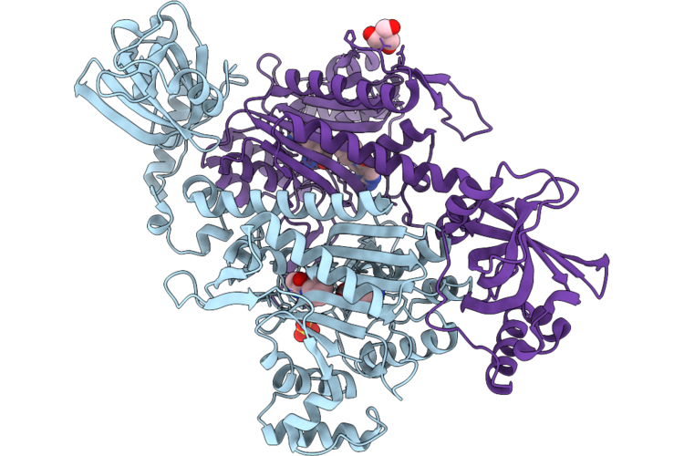
Method: X-RAY DIFFRACTION Resolution:2.72 Å Release Date: 2026-06-10 Classification: TRANSPORT PROTEIN Ligands: ZN, A1EOS |
 |
Organism: Homo sapiens
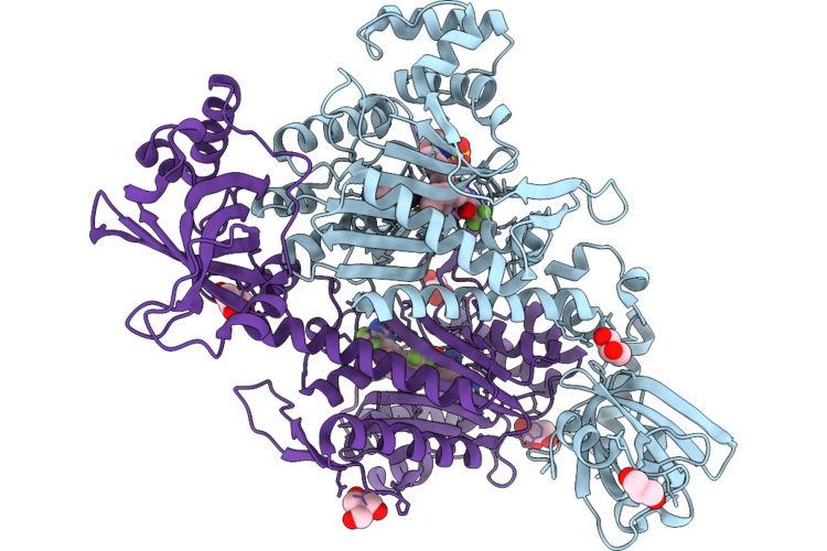
Method: X-RAY DIFFRACTION Resolution:2.80 Å Release Date: 2026-06-10 Classification: TRANSPORT PROTEIN Ligands: ZN, A1CE9 |
 |
Organism: Homo sapiens
Method: X-RAY DIFFRACTION Resolution:3.22 Å Release Date: 2026-06-10 Classification: TRANSPORT PROTEIN Ligands: ZN, A1EP8 |
 |
Organism: Homo sapiens
Method: X-RAY DIFFRACTION Resolution:2.64 Å Release Date: 2026-06-10 Classification: PROTEIN TRANSPORT Ligands: ZN, A1EP9 |
 |
Organism: Homo sapiens
Method: X-RAY DIFFRACTION Resolution:2.31 Å Release Date: 2026-06-10 Classification: PROTEIN TRANSPORT Ligands: ZN, A1EQA |
 |
Organism: Homo sapiens
Method: X-RAY DIFFRACTION Resolution:2.65 Å Release Date: 2026-06-10 Classification: PROTON TRANSPORT Ligands: ZN, A1CGA |
 |
Organism: Homo sapiens
Method: X-RAY DIFFRACTION Resolution:2.95 Å Release Date: 2026-06-10 Classification: PROTEIN TRANSPORT Ligands: ZN, A1EQC |
 |
Organism: Homo sapiens
Method: X-RAY DIFFRACTION Resolution:2.49 Å Release Date: 2026-06-10 Classification: PROTEIN TRANSPORT Ligands: ZN, A1CF9 |
 |
Organism: Homo sapiens
Method: X-RAY DIFFRACTION Resolution:2.53 Å Release Date: 2026-06-10 Classification: PROTEIN TRANSPORT Ligands: ZN, A1EQD |
 |
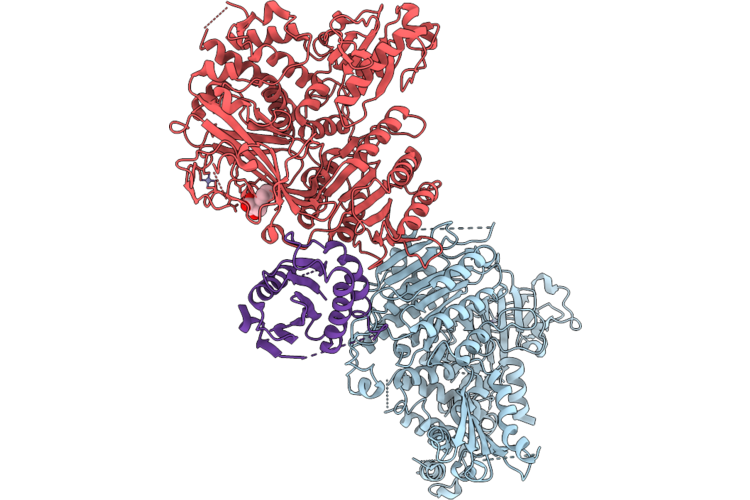
Organism: Cryptosporidium parvum iowa ii
Method: X-RAY DIFFRACTION Resolution:1.84 Å Release Date: 2026-06-10 Classification: LIGASE Ligands: LYS, A1JCS, SO4, TRS |
 |
Crystal Structure Of Lysyl-Trna Synthetase From Cryptosporidium Parvum Complexed With L-Lysine And Inhibitor Ddd01887015
Organism: Cryptosporidium parvum iowa ii
Method: X-RAY DIFFRACTION Resolution:1.90 Å Release Date: 2026-06-10 Classification: LIGASE Ligands: A1JCT, EDO, TRS, GOL, LYS, SO4 |
 |
Crystal Structure Of Lysyl-Trna Synthetase From Cryptosporidium Parvum Complexed With L-Lysine And Inhibitor Ddd01932549
Organism: Cryptosporidium parvum iowa ii
Method: X-RAY DIFFRACTION Resolution:2.30 Å Release Date: 2026-06-10 Classification: LIGASE Ligands: LYS, A1JC7, SO4 |
 |
Crystal Structure Of Lysyl-Trna Synthetase From Cryptosporidium Parvum / Plasmodium Falciparum Chimera Complexed With L-Lysine And Inhibitor Ddd02174286
Organism: Cryptosporidium parvum iowa ii
Method: X-RAY DIFFRACTION Resolution:1.90 Å Release Date: 2026-06-10 Classification: LIGASE Ligands: LYS, A1JC4, GOL, SO4, TRS |
 |
Crystal Structure Of Lysyl-Trna Synthetase From Cryptosporidium Parvum Complexed With L-Lysine And Inhibitor Ddd01827593
Organism: Cryptosporidium parvum iowa ii
Method: X-RAY DIFFRACTION Resolution:1.50 Å Release Date: 2026-06-10 Classification: LIGASE Ligands: LYS, A1JC9, SO4 |