| Project Accession: | IBIAP_1000000029 |
| Title: | 3D medical imaging data in head and neck cancer patients |
| Representative Image: | 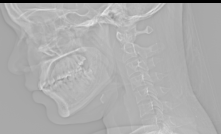 |
| Description: | The comprehensive dataset specifically includes three-dimensional (3D) medical imaging data acquired from head and neck cancer patients. The primary imaging modality utilized for this collection is Computed Tomography (CT) scanning. CT scans provide detailed cross-sectional images essential for accurate diagnosis, tumor delineation, treatment planning (such as radiation therapy), and monitoring of therapeutic response in oncology. The dataset aims to serve as a high-quality resource for researchers and developers working on projects such as automated tumor segmentation, radiomics analysis, deep learning model training for prognosis prediction, and personalized treatment strategies within the domain of head and neck oncology. The detailed 3D structure captured by the CT scans allows for a more complete and spatial understanding of the disease, moving beyond traditional 2D assessments. |
| Publications: | N/A |
| Associated Codes (URL only): | N/A |
| Funding agency: | N/A |
| Grant Number: | N/A |
| Ethics Statement: | N/A |
| Any Other Information : | N/A |
| Additional File: | N/A |
| Acknowledgments: | The authors acknowledge the Department of Oncoradiology, AIIMS New Delhi, for technical and clinical support. |
| Sr.No | First name | Last name | Organization | Designation | |
|---|---|---|---|---|---|
| 1 | Amit | Gupta | amit.aiims2014@gmail.com | NCI Jhajjar, All India Institute of Medical Sciences (AIIMS), New Delhi, India | Co-Principal Investigator |
| 2 | Krithika | Rangarajan | krithikarangarajan@aiims.edu | All India Institute of Medical Sciences (AIIMS), New Delhi, India | Principal Investigator |
| 3 | Hema | Malhotra | hemamalhotraaiims.2020@gmail.com | All India Institute of Medical Sciences (AIIMS), New Delhi, India | Program Manager |
| 4 | Ashish | Rastogi | ashishrastogi.3150@gmail.com | All India Institute of Medical Sciences (AIIMS), New Delhi, India | Data Analyst |
| 5 | Rohit | Shah | rohitaiims.2024@gmail.com | All India Institute of Medical Sciences (AIIMS), New Delhi, India | D.E.O |
| Study Accession: | CTS_1000000034 |
| Title: | 3D medical imaging data in head and neck cancer patients |
| Imaging Type: | Computed Tomography (CT) |
| Imaging Sub-type: | Diagnostic Radiology |
| Summary: | The comprehensive dataset specifically includes three-dimensional (3D) medical imaging data acquired from head and neck cancer patients. The primary imaging modality utilized for this collection is Computed Tomography (CT) scanning. CT scans provide detailed cross-sectional images essential for accurate diagnosis, tumor delineation, treatment planning (such as radiation therapy), and monitoring of therapeutic response in oncology. The dataset aims to serve as a high-quality resource for researchers and developers working on projects such as automated tumor segmentation, radiomics analysis, deep learning model training for prognosis prediction, and personalized treatment strategies within the domain of head and neck oncology. The detailed 3D structure captured by the CT scans allows for a more complete and spatial understanding of the disease, moving beyond traditional 2D assessments. |
| Keywords: | Laryngeal cancer; Head and Neck cancer; CT scan |
| Additional / Any Other Information: | N/A |
| Release Date: | April 29, 2026 |
| Access Licence Type: | Open Access |
Table 1. The sample types registered under this study are as follows:
Table 2. The samples registered under this study are as follows:
| Sample Type ID | Organism | Taxon ID | Biological Entity | Laterality | Source Tissue | Source Cell/Cell-line | Cell Organelle |
|---|---|---|---|---|---|---|---|
| CTSMT_10000000064 | Homo sapiens | 9606 | Multiple body regions | Both | N/A | N/A | N/A |
Table 2. The samples registered under this study are as follows:
| Sample Type ID | Sample ID | Subject Type | Clinical Indication (Diagnosis) | ICD-11 Code (patient health condition) | Tumor Site | Image category/label | De-identified Patient ID | Procedure | Clinical Information |
|---|---|---|---|---|---|---|---|---|---|
| CTSMT_10000000064 | CTSM_10000298470 | Head and neck cancer | Suspicious of malignancy | 2B66 | Oral cavity (right RMT) | N/A | Patient_835 | CECT of Face-Neck-Chest | right MRM, Now right BM ulcer. |
| CTSMT_10000000064 | CTSM_10000297880 | Head and neck cancer | Known case of cancer - Followup | 2C23 | Larynx- supraglottis | N/A | Patient_245 | CECT of Face-Neck-Chest | Ca supraglottic larynx post CTRT |
| CTSMT_10000000064 | CTSM_10000297895 | Head and neck cancer | Known case of cancer - Followup | 2B62 | Oral cavity (left side of tongue) | N/A | Patient_260 | CECT of Face-Neck-Chest | K/C/O CA left side of tongue and BOT (T4 N0M0). Post RT/Ct for follow up |
| CTSMT_10000000064 | CTSM_10000298485 | Head and neck cancer | Known case of cancer - Followup | 2B66 | Oral cavity (right buccal mucosa) with and recurrent disease | N/A | Patient_850 | CECT of Face & Neck. | K/C/O CA right RMT, s/p palliative RT. |
| CTSMT_10000000064 | CTSM_10000297898 | Head and neck cancer | Known case of cancer - Followup | 2B66 | Oral cavity- right Buccal mucosa | N/A | Patient_263 | CECT of Face-Neck-Chest | Ca right buccal mucosa on chemotherapy. |
| CTSMT_10000000064 | CTSM_10000298488 | Head and neck cancer | Known case of cancer - Followup | 2B66 | Oral cavity- left upper alveolus | N/A | Patient_853 | CECT of Face-Neck-Chest | Ca left upper alveolus post surgery and post RT, for response evaluation. |
| CTSMT_10000000064 | CTSM_10000297910 | Head and neck cancer | Known case of cancer - Followup | 2B62 | Oral cavity- right lateral border of tongue | N/A | Patient_275 | CECT of Face & Neck. | C/o CA right lateral border of the tongue post surgery and RT |
| CTSMT_10000000064 | CTSM_10000298500 | Head and neck cancer | Known case of cancer - Followup | N/A | Unkown primary site | N/A | Patient_865 | CECT of Neck-Chest and abdomen | Follow up case of Neuroendocrine tumor; subtotal scapulectomy |
| CTSMT_10000000064 | CTSM_10000298503 | Head and neck cancer | Known case of cancer - Followup | 2B61 | Oropharynx - Base of the tongue | N/A | Patient_868 | CECT of Face & Neck. Chest | CA base of tongue, post CTRT in 2022, on follow up |
| CTSMT_10000000064 | CTSM_10000298528 | Head and neck cancer | Known case of cancer - Followup | 2B65 | Skin (sebaceous gland) | N/A | Patient_893 | CECT of Face-Neck-Chest | O/C/O left sebaceous cell carcinoma (left side of face). |
| CTSMT_10000000064 | CTSM_10000298530 | Head and neck cancer | Known case of cancer - Followup | 2C31 | Scalp | N/A | Patient_895 | CECT of Head, face-neck-chest | Marjolin ulcer of scalp. |
| CTSMT_10000000064 | CTSM_10000297946 | Head and neck cancer | Known case of cancer - Followup | 2B66 | Oral cavity- left Buccal mucosa | N/A | Patient_311 | CECT of Face-Neck-Chest | CA left BM-Post op -post RT |
| CTSMT_10000000064 | CTSM_10000297948 | Head and neck cancer | Known case of cancer - Followup | N/A | Unkown primary site | N/A | Patient_313 | CECT of Face-Neck-Chest | Recurrent head and neck SCC |
| CTSMT_10000000064 | CTSM_10000298534 | Head and neck cancer | Suspicious of malignancy | 2D10 | Thyroid gland | N/A | Patient_899 | CECT of neck,Chest, Abdomen & Pelvis. | throid swelling with multiple cervical LAP |
| CTSMT_10000000064 | CTSM_10000298538 | Head and neck cancer | Known case of cancer - Followup | 2B67 | Parotid gland | N/A | Patient_903 | CECT of Face & Neck. | Ca Parotid |
| CTSMT_10000000064 | CTSM_10000297954 | Head and neck cancer | Known case of cancer - Followup | 2C30 | cervical lymphnodes | N/A | Patient_319 | CECT of Face-Neck-Chest | case of hodgkin's on follow up. |
| CTSMT_10000000064 | CTSM_10000297930 | Head and neck cancer | Known case of cancer - Followup | N/A | Unkown primary site | N/A | Patient_295 | CECT of Face-Neck-Chest | Right maxillectomy with oro-nasal fistula |
| CTSMT_10000000064 | CTSM_10000297979 | Head and neck cancer | Known case of cancer - Followup | 2B66 | Oral cavity (upper alveolous) | N/A | Patient_344 | CECT of Face-Neck-Chest | C/O Ca upper alveolus post WLE B/L SOH ND Post CT.RT (Oct, 24) |
| CTSMT_10000000064 | CTSM_10000297982 | Head and neck cancer | Suspicious of malignancy | N/A | Unkown primary site | N/A | Patient_347 | CECT of Neck, Chest, Abdomen & Pelvis. | right conjunctival NHL post IFRT to right orbiton follow up |
| CTSMT_10000000064 | CTSM_10000297985 | Head and neck cancer | Suspicious of malignancy | 2B66 | Oral cavity (left gbs, left RMT) | N/A | Patient_350 | CECT of Face-Neck-Chest | CA left buccal mucosa |
Table 3. The experiment types registered under this study are as follows:
| Experiment Type ID | Instrument Name | Instrument Type | Manufacturer | Model |
|---|---|---|---|---|
| CTET_10000000030 | CT-Scan Machine | Multisclice | Siemens Healthineers | Somatom Definition Edge |
| Experimental Design Summary (CTET_10000000030) |
|---|
| The CT scans included in this dataset were performed for patients diagnosed with head and neck cancer at our institute. The CT images include various body regions including face, neck, chest, abdomen and pelvis. These scans were performed at various stages of management of the cancer, including pre-treatment scans, post-radiotherapy response assessment scans, and post-operative follow-up scans. All the scans were double reported by two radiologists - first, a resident radiologist (3-6 years of experience), and second reading by a specialist onco-radiologist (more than 10 years of experience). |
| Acquired Images Annotation Description (CTET_10000000030) |
|---|
| Each CT scan in the dataset is accompanied by the summary of main findings in the corresponding radiology reports. All the scans were double reported by two radiologists - first, a resident radiologist (3-6 years of experience), and second reading by a specialist onco-radiologist (more than 10 years of experience). The findings from these reports provided in this dataset, are robust scan-level labels for each of the CT exams included in the dataset. Image and pixel-level annotations are not included in this dataset. |
Table 4. The experiments registered under this study are as follows:
| Sample ID | Experiment Type ID | Experiment ID | Image type (Original / Derived / Unknown) | Any Other Information | Image plane | Slice Progression Direction | Slice thickness (in mm) | Image Count | Contrast / Bolus ingredient | Image file format | Image modality type (contrast-enhanced or non-contrast) | Implants or devices visible in scan (yes/no) | Motion artifacts noted (yes/no) | Type of contrast agent | Use of contrast agent (yes/no) |
|---|---|---|---|---|---|---|---|---|---|---|---|---|---|---|---|
| CTSM_10000297662 | CTET_10000000030 | CTE_10000271118 | Derived | N/A | Axial | superio-inferior | 1 | 759 | non ionic iodinated contrast | dcm | CECT | N/A | no | non ionic iodinated contrast | yes |
| CTSM_10000297663 | CTET_10000000030 | CTE_10000271119 | Derived | N/A | Axial | superio-inferior | 1 | 770 | non ionic iodinated contrast | dcm | CECT | N/A | no | non ionic iodinated contrast | yes |
| CTSM_10000297664 | CTET_10000000030 | CTE_10000271120 | Derived | N/A | Axial | superio-inferior | 1 | 616 | non ionic iodinated contrast | dcm | CECT | N/A | no | non ionic iodinated contrast | yes |
| CTSM_10000297665 | CTET_10000000030 | CTE_10000271121 | Derived | N/A | Axial | superio-inferior | 1 | 873 | non ionic iodinated contrast | dcm | CECT | N/A | no | non ionic iodinated contrast | yes |
| CTSM_10000297666 | CTET_10000000030 | CTE_10000271122 | Derived | N/A | Axial | superio-inferior | 1 | 390 | non ionic iodinated contrast | dcm | CECT | N/A | no | non ionic iodinated contrast | yes |
| CTSM_10000297667 | CTET_10000000030 | CTE_10000271123 | Derived | N/A | Axial | superio-inferior | 1 | 783 | non ionic iodinated contrast | dcm | CECT | N/A | no | non ionic iodinated contrast | yes |
| CTSM_10000297668 | CTET_10000000030 | CTE_10000271124 | Derived | N/A | Axial | superio-inferior | 1 | 1384 | non ionic iodinated contrast | dcm | CECT | N/A | no | non ionic iodinated contrast | yes |
| CTSM_10000297669 | CTET_10000000030 | CTE_10000271125 | Derived | N/A | Axial | superio-inferior | 1 | 802 | non ionic iodinated contrast | dcm | CECT | N/A | no | non ionic iodinated contrast | yes |
| CTSM_10000297670 | CTET_10000000030 | CTE_10000271126 | Derived | N/A | Axial | superio-inferior | 1 | 790 | non ionic iodinated contrast | dcm | CECT | N/A | no | non ionic iodinated contrast | yes |
| CTSM_10000297671 | CTET_10000000030 | CTE_10000271127 | Derived | N/A | Axial | superio-inferior | 1 | 873 | non ionic iodinated contrast | dcm | CECT | N/A | no | non ionic iodinated contrast | yes |
| CTSM_10000297674 | CTET_10000000030 | CTE_10000271130 | Derived | N/A | Axial | superio-inferior | 1 | 820 | non ionic iodinated contrast | dcm | CECT | N/A | no | non ionic iodinated contrast | yes |
| CTSM_10000297675 | CTET_10000000030 | CTE_10000271131 | Derived | N/A | Axial | superio-inferior | 1 | 795 | non ionic iodinated contrast | dcm | CECT | N/A | no | non ionic iodinated contrast | yes |
| CTSM_10000297676 | CTET_10000000030 | CTE_10000271132 | Derived | N/A | Axial | superio-inferior | 1 | 735 | non ionic iodinated contrast | dcm | CECT | N/A | no | non ionic iodinated contrast | yes |
| CTSM_10000297677 | CTET_10000000030 | CTE_10000271133 | Derived | N/A | Axial | superio-inferior | 1 | 899 | non ionic iodinated contrast | dcm | CECT | N/A | no | non ionic iodinated contrast | yes |
| CTSM_10000297678 | CTET_10000000030 | CTE_10000271134 | Derived | N/A | Axial | superio-inferior | 1 | 895 | non ionic iodinated contrast | dcm | CECT | N/A | no | non ionic iodinated contrast | yes |
| CTSM_10000297679 | CTET_10000000030 | CTE_10000271135 | Derived | N/A | Axial | superio-inferior | 1 | 54 | non ionic iodinated contrast | dcm | CECT | N/A | no | non ionic iodinated contrast | yes |
| CTSM_10000297680 | CTET_10000000030 | CTE_10000271136 | Derived | N/A | Axial | superio-inferior | 1 | 773 | non ionic iodinated contrast | dcm | CECT | N/A | no | non ionic iodinated contrast | yes |
| CTSM_10000297681 | CTET_10000000030 | CTE_10000271137 | Derived | N/A | Axial | superio-inferior | 1 | 722 | non ionic iodinated contrast | dcm | CECT | N/A | no | non ionic iodinated contrast | yes |
| CTSM_10000297682 | CTET_10000000030 | CTE_10000271138 | Derived | N/A | Axial | superio-inferior | 1 | 897 | non ionic iodinated contrast | dcm | CECT | N/A | no | non ionic iodinated contrast | yes |
| CTSM_10000298026 | CTET_10000000030 | CTE_10000270553 | Derived | N/A | Axial | superio-inferior | 1 | 1062 | non ionic iodinated contrast | dcm | CECT | N/A | no | non ionic iodinated contrast | yes |
| Experiment ID | Image File Name (with path) | Image Preview | Image Size |
|---|---|---|---|
| CTE_10000270588 | HNC_data/Patient_426.zip |  Download Image |
179M |
| CTE_10000270589 | HNC_data/Patient_427.zip | Download Image |
214M |
| CTE_10000270590 | HNC_data/Patient_428.zip | Download Image |
206M |
| CTE_10000270591 | HNC_data/Patient_429.zip | Download Image |
179M |
| CTE_10000270592 | HNC_data/Patient_430.zip | Download Image |
194M |
| CTE_10000270593 | HNC_data/Patient_431.zip | Download Image |
188M |
| CTE_10000270594 | HNC_data/Patient_432.zip | Download Image |
198M |
| CTE_10000270595 | HNC_data/Patient_433.zip | Download Image |
204M |
| CTE_10000270597 | HNC_data/Patient_435.zip | Download Image |
176M |
| CTE_10000270599 | HNC_data/Patient_437.zip | Download Image |
97M |