

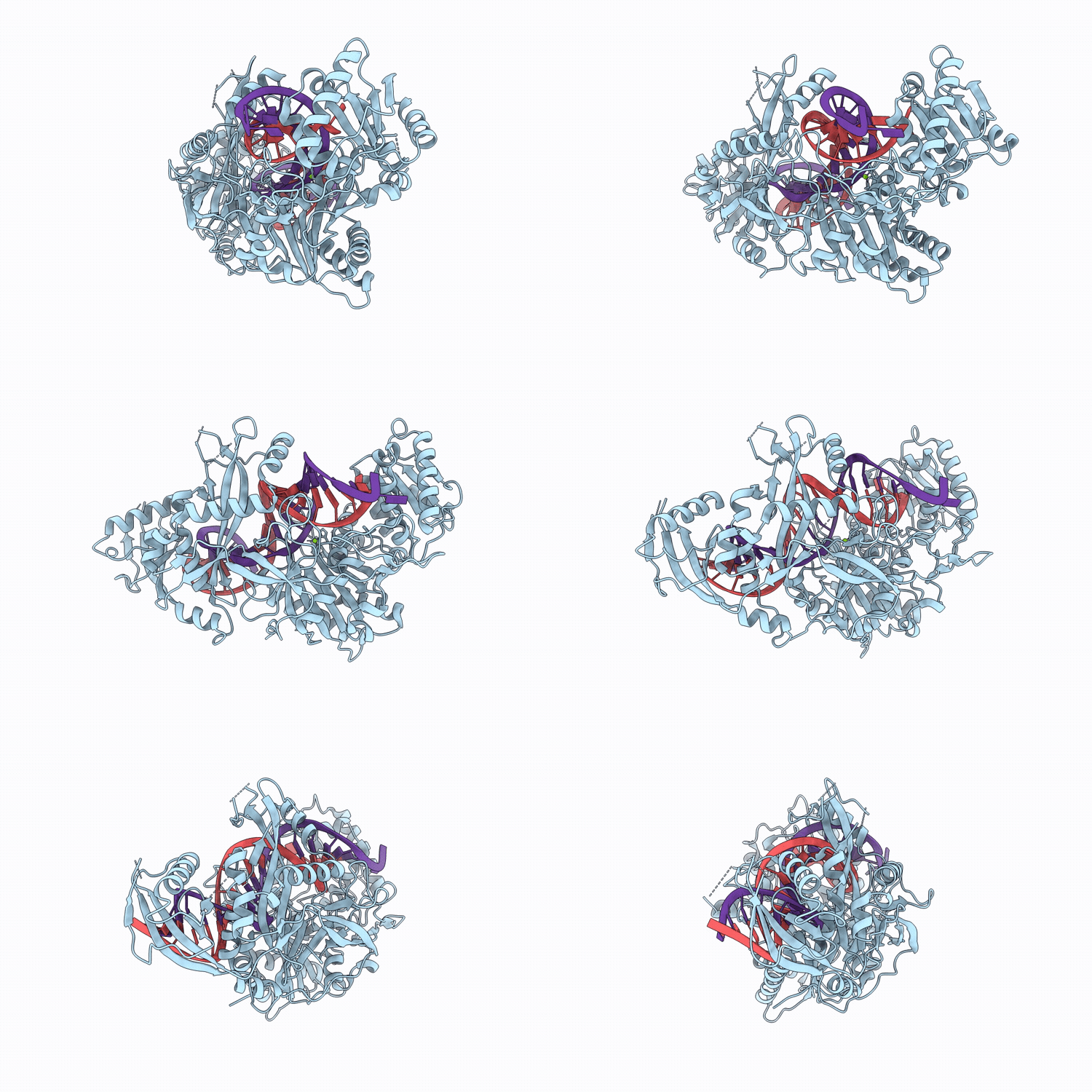
11GI image
Deposition Date 2026-02-23
Release Date 2026-05-13
Last Version Date 2026-08-26
Entry Detail
PDB ID:
11GI
Title:
Human Argonaute2 WT - guide(3 prime-amino) RNA in complex with a fully complementary target
Biological Source:
Source Organism(s):
Expression System(s):
Method Details:
Experimental Method:
Resolution:
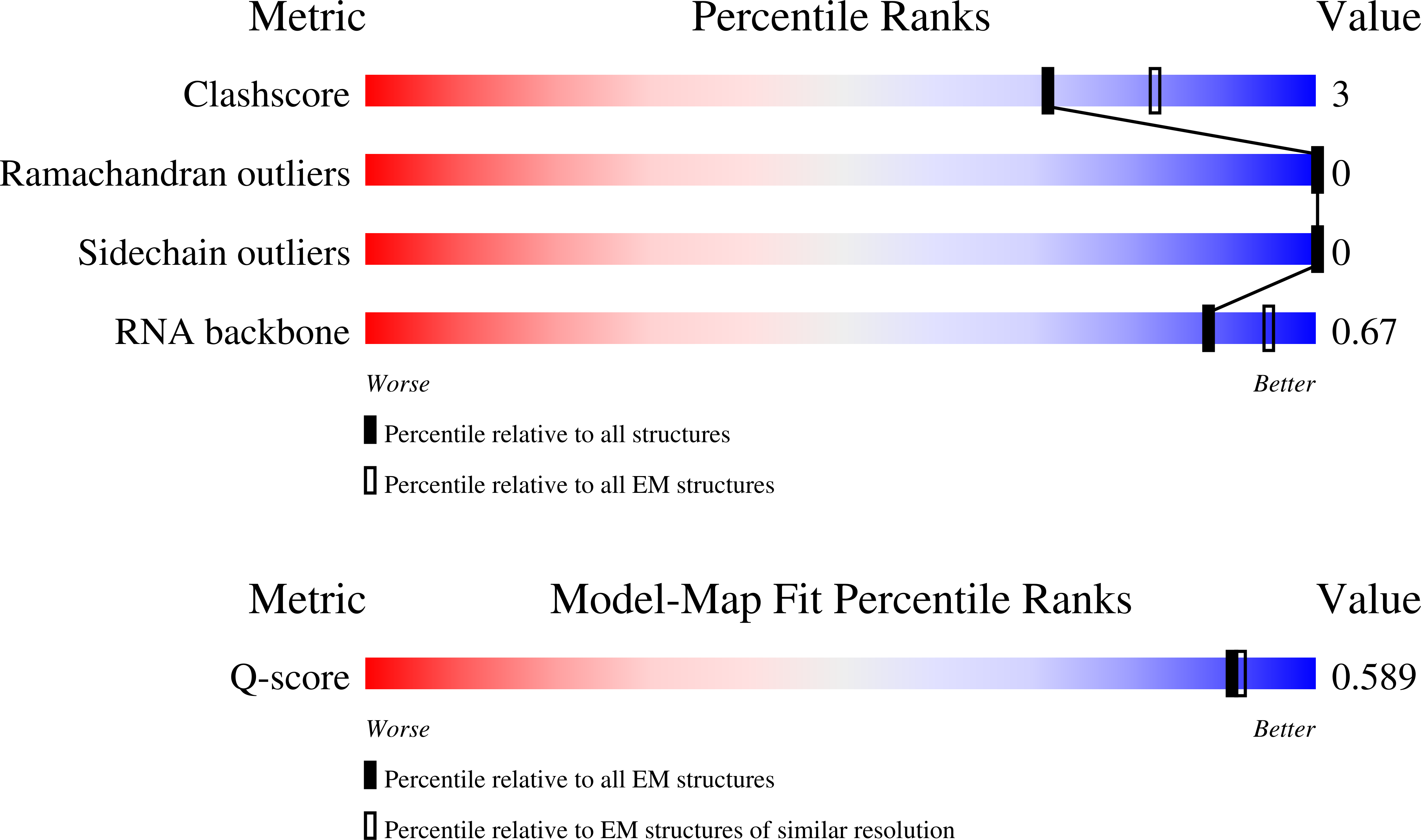
3.00 Å
Aggregation State:
PARTICLE
Reconstruction Method:
SINGLE PARTICLE
Macromolecular Entities
Polymer Type:polypeptide(L)
Molecule:Argonaute2 WT
Gene (Uniprot):AGO2
Mutagens:S387D, S824A, S828D, S831D, S834A
Chain IDs:A
Chain Length:859
Number of Molecules:1
Biological Source:Homo sapiens
Polymer Type:polyribonucleotide
Molecule:guide(3 prime-amino) RNA
Chain IDs:B
Chain Length:20
Number of Molecules:1
Biological Source:synthetic construct
Polymer Type:polyribonucleotide
Molecule:target RNA
Chain IDs:C
Chain Length:23
Number of Molecules:1
Biological Source:synthetic construct
Ligand Molecules
Primary Citation
Catalytic activation of human Argonaute 2 requires RNA duplex deformation.
Nat.Struct.Mol.Biol. 33 1214 1223 (2026)
PMID: 42342972 DOI: 10.1038/s41594-026-01840-5

Abstact

Small interfering RNAs (siRNAs) are an expanding class of RNA therapeutics, with seven drugs approved by the US Food and Drug Administration and many more in development. Rational design, however, has been limited by incomplete understanding of how human Argonaute 2 (hAgo2) catalyzes target cleavage. Here we report high-resolution cryo-electron microscopy structures of hAgo2 bound to a target RNA in catalytic and noncatalytic conformations. The structures reveal that guide-target pairing alone is insufficient for slicing and catalysis requires deformation of the duplex through a coordinated network of RNA-protein interactions. Expansion of the central major groove positions the scissile phosphate, while compression toward the supplementary region docks the duplex into the hAgo2 cleft. A kink after guide nucleotide g6 disrupts seed-only pairing conformation and promotes the extended pairing required for catalysis. This rearrangement enables repositioning of K709 within the active site, while a pyrimidine at target position t10 optimally aligns R710 to accelerate cleavage. These findings provide a structural framework linking siRNA duplex geometry to catalytic efficiency and inform rational design of siRNAs with improved potency and specificity.

Legend

Protein

Chemical

Disease

Primary Citation of related structures
Feedback Form
Name
Email
Institute
Feedback