

Search Count: 2,016
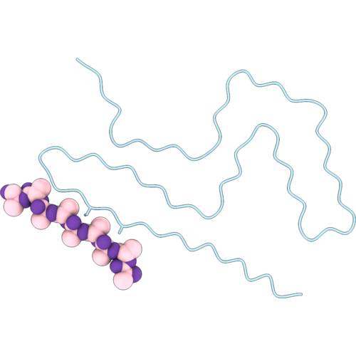 |
Cryo-Em Structure Of Alpha-Synuclein Filaments (Singlet) From Parkinson'S Disease With A53T Mutation In Snca
Organism: Homo sapiens
Method: ELECTRON MICROSCOPY Resolution:2.10 Å Release Date: 2026-10-07 Classification: PROTEIN FIBRIL |
Organism: Homo sapiens
Method: ELECTRON MICROSCOPY
Release Date: 2026-10-07
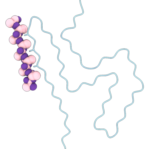 |
Cryo-Em Structure Of Alpha-Synuclein Filaments (Doublet) From Parkinson'S Disease With A53T Mutation In Snca
Organism: Homo sapiens
Method: ELECTRON MICROSCOPY Resolution:2.10 Å Release Date: 2026-10-07 Classification: PROTEIN FIBRIL |
Organism: Homo sapiens
Method: ELECTRON MICROSCOPY
Release Date: 2026-10-07
 |
Cryo-Em Structure Of Alpha-Synuclein Filaments (Singlet) From M83(+/+) Mouse Brains With A53T Mutation
Organism: Homo sapiens
Method: ELECTRON MICROSCOPY Resolution:2.90 Å Release Date: 2026-10-07 Classification: PROTEIN FIBRIL |
Organism: Homo sapiens
Method: ELECTRON MICROSCOPY
Release Date: 2026-10-07
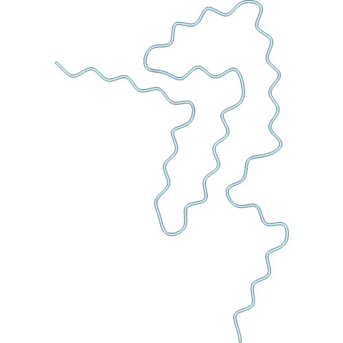 |
Cryo-Em Structure Of Alpha-Synuclein Filaments (Doublet) From M83(+/+) Mouse Brains With A53T Mutation
Organism: Homo sapiens
Method: ELECTRON MICROSCOPY Resolution:3.10 Å Release Date: 2026-10-07 Classification: PROTEIN FIBRIL |
Organism: Homo sapiens
Method: ELECTRON MICROSCOPY
Release Date: 2026-10-07
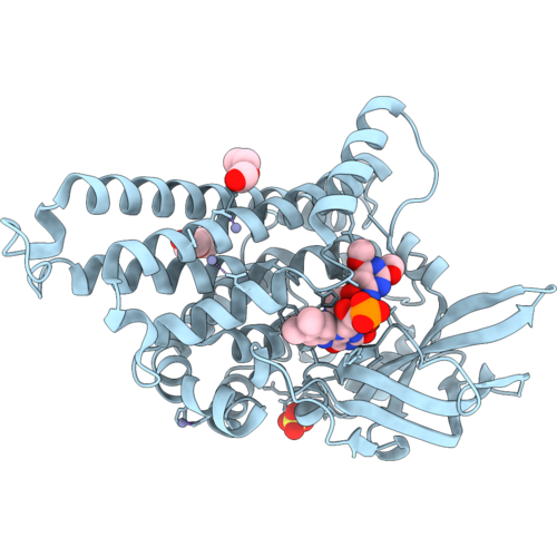 |
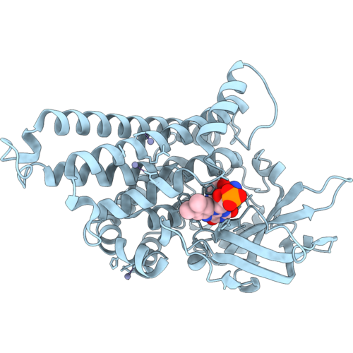
Crystal Structure Of Sulx In Complex With Fmn And Sulfadimethoxine
Organism: Microbacterium sp. cj77
Method: X-RAY DIFFRACTION Resolution:2.37 Å Release Date: 2026-09-30 Classification: OXIDOREDUCTASE Ligands: FMN, A1E1Y, ZN, PEG, SO4 |
Organism: Microbacterium sp. cj77
Method: X-RAY DIFFRACTION
Release Date: 2026-09-30
Ligands: FMN, A1E1Y, ZN, PEG, SO4
 |
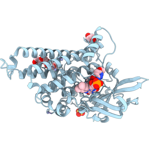
Crystal Structure Of Sulx In Complex With Fmn And Sulfanilamide
Organism: Microbacterium sp. cj77
Method: X-RAY DIFFRACTION Resolution:2.83 Å Release Date: 2026-09-30 Classification: OXIDOREDUCTASE Ligands: FMN, SAN, ZN |
Organism: Microbacterium sp. cj77
Method: X-RAY DIFFRACTION
Release Date: 2026-09-30
Ligands: FMN, SAN, ZN
 |
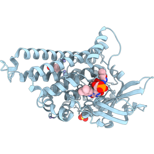
Crystal Structure Of Sulx In Complex With Fmn And Sulfamethoxypyridazine
Organism: Microbacterium sp. cj77
Method: X-RAY DIFFRACTION Resolution:2.09 Å Release Date: 2026-09-30 Classification: OXIDOREDUCTASE Ligands: FMN, PEG, SO4, PGE, ZN, A1E1X |
Organism: Microbacterium sp. cj77
Method: X-RAY DIFFRACTION
Release Date: 2026-09-30
Ligands: FMN, PEG, SO4, PGE, ZN, A1E1X
 |
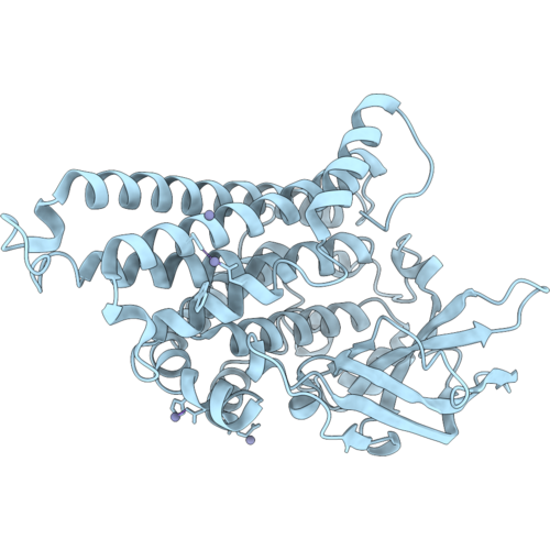
Crystal Structure Of Sulx In Complex With Fmn And Sulfamethoxazole
Organism: Microbacterium sp. cj77
Method: X-RAY DIFFRACTION Resolution:2.46 Å Release Date: 2026-09-30 Classification: OXIDOREDUCTASE Ligands: PEG, FMN, SO4, ZN, 08D |
Organism: Microbacterium sp. cj77
Method: X-RAY DIFFRACTION
Release Date: 2026-09-30
Ligands: PEG, FMN, SO4, ZN, 08D
 |
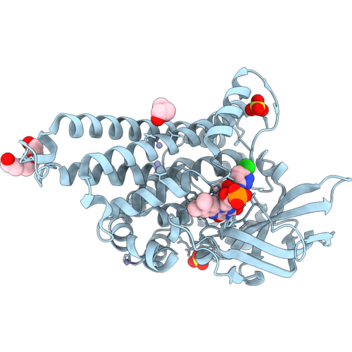
Crystal Structure Of Sulx
Organism: Microbacterium sp. cj77
Method: X-RAY DIFFRACTION Resolution:3.03 Å Release Date: 2026-09-30 Classification: OXIDOREDUCTASE Ligands: ZN |
Organism: Microbacterium sp. cj77
Method: X-RAY DIFFRACTION
Release Date: 2026-09-30
Ligands: ZN
 |
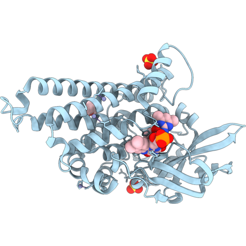
Crystal Structure Of Sulx In Complex With Fmn And Sulfachloropyridazine
Organism: Microbacterium sp. cj77
Method: X-RAY DIFFRACTION Resolution:2.71 Å Release Date: 2026-09-30 Classification: OXIDOREDUCTASE Ligands: FMN, A1E3B, ZN, SO4, PGE, PEG |
Organism: Microbacterium sp. cj77
Method: X-RAY DIFFRACTION
Release Date: 2026-09-30
Ligands: FMN, A1E3B, ZN, SO4, PGE, PEG
 |
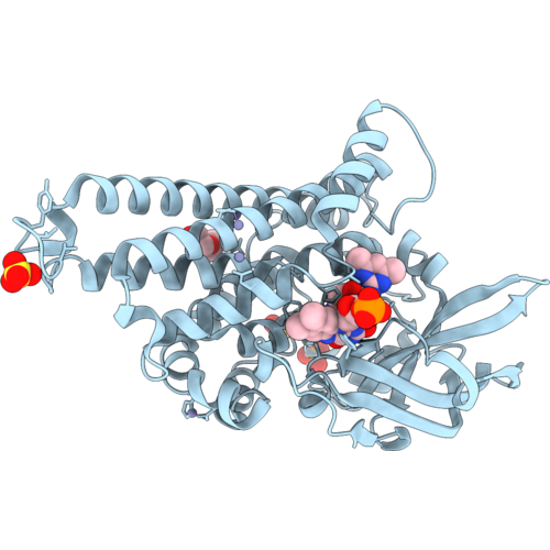
Crystal Structure Of Sulx Mutant-A399S In Complex With Fmn And Sulfamethazine
Organism: Microbacterium sp. cj77
Method: X-RAY DIFFRACTION Resolution:2.47 Å Release Date: 2026-09-30 Classification: OXIDOREDUCTASE Ligands: FMN, A1E1F, SO4, PEG, ZN |
Organism: Microbacterium sp. cj77
Method: X-RAY DIFFRACTION
Release Date: 2026-09-30
Ligands: FMN, A1E1F, SO4, PEG, ZN
 |
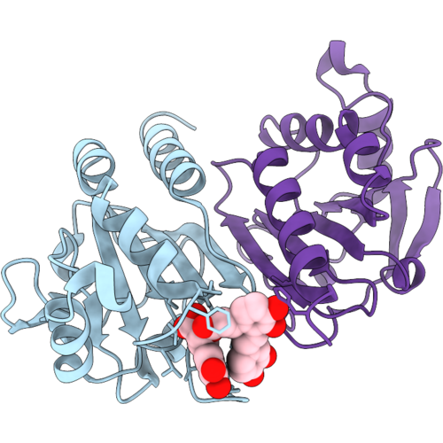
Crystal Structure Of Sulx Mutant-S375A In Complex With Fmn And Sulfamethazine
Organism: Microbacterium sp. cj77
Method: X-RAY DIFFRACTION Resolution:2.62 Å Release Date: 2026-09-30 Classification: OXIDOREDUCTASE Ligands: FMN, A1E1F, PEG, SO4, ZN |
Organism: Microbacterium sp. cj77
Method: X-RAY DIFFRACTION
Release Date: 2026-09-30
Ligands: FMN, A1E1F, PEG, SO4, ZN
 |
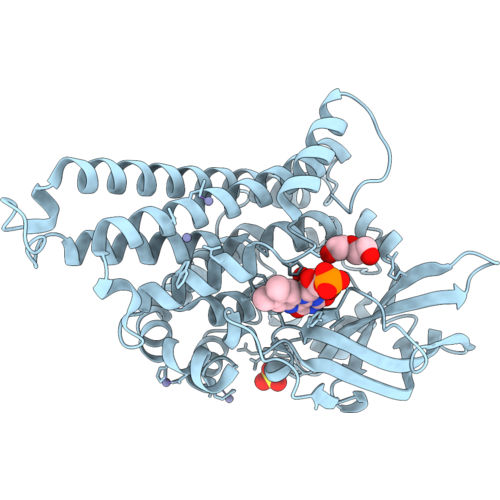
Crystal Structure Of Peroxiredoxin I In Complex With Saa
Organism: Homo sapiens
Method: X-RAY DIFFRACTION Resolution:1.83 Å Release Date: 2026-09-30 Classification: OXIDOREDUCTASE Ligands: 4N4 |
Organism: Homo sapiens
Method: X-RAY DIFFRACTION
Release Date: 2026-09-30
Ligands: 4N4
 |
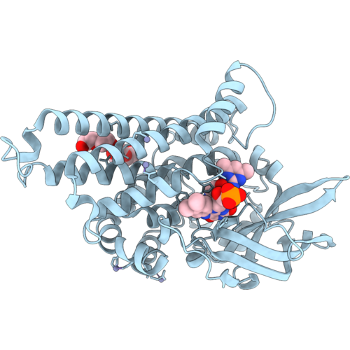
Crystal Structure Of Sulx In Complex With Fmn
Organism: Microbacterium sp. cj77
Method: X-RAY DIFFRACTION Resolution:2.44 Å Release Date: 2026-09-30 Classification: OXIDOREDUCTASE Ligands: FMN, ZN, SO4, PEG |
Organism: Microbacterium sp. cj77
Method: X-RAY DIFFRACTION
Release Date: 2026-09-30
Ligands: FMN, ZN, SO4, PEG
 |
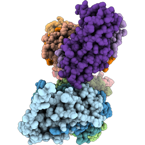
Crystal Structure Of Sulx In Complex With Fmn And Sulfamethazine
Organism: Microbacterium sp. cj77
Method: X-RAY DIFFRACTION Resolution:2.52 Å Release Date: 2026-09-30 Classification: OXIDOREDUCTASE Ligands: FMN, A1E1F, ZN, PEG |
Organism: Microbacterium sp. cj77
Method: X-RAY DIFFRACTION
Release Date: 2026-09-30
Ligands: FMN, A1E1F, ZN, PEG
 |
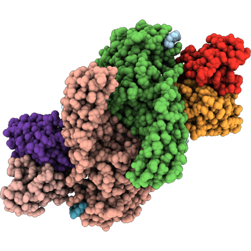
Cryo-Em Of Chaperon Usher Pathway Pilus From Stenotrophomonas Maltophilia
Organism: Stenotrophomonas maltophilia
Method: ELECTRON MICROSCOPY Resolution:4.00 Å Release Date: 2026-09-09 Classification: PROTEIN FIBRIL |
Organism: Stenotrophomonas maltophilia
Method: ELECTRON MICROSCOPY
Release Date: 2026-09-09
 |
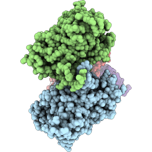
Cryo-Em Structure Of The Mglu5-Beta-Arrestin 1 Complex
Organism: Homo sapiens, Mus musculus
Method: ELECTRON MICROSCOPY Release Date: 2026-09-09 Classification: MEMBRANE PROTEIN/IMMUNE SYSTEM Ligands: GLU, NAG |
Organism: Homo sapiens, Mus musculus
Method: ELECTRON MICROSCOPY
Release Date: 2026-09-09
Ligands: GLU, NAG
 |
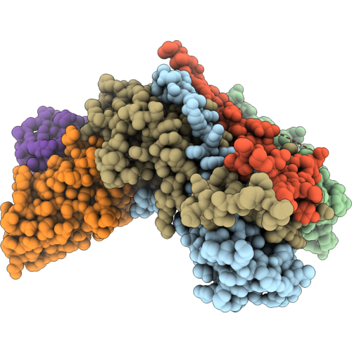
Cryo-Em Structure Of The Mglu7-Beta-Arrestin 1 Complex
Organism: Homo sapiens, Mus musculus
Method: ELECTRON MICROSCOPY Release Date: 2026-09-09 Classification: MEMBRANE PROTEIN/IMMUNE SYSTEM Ligands: GLU, CLR |
Organism: Homo sapiens, Mus musculus
Method: ELECTRON MICROSCOPY
Release Date: 2026-09-09
Ligands: GLU, CLR
 |
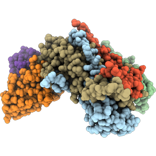
Complex Of Fmdv O/Hn/Cha/93 And Porcine-Derived Neutralizing Monoclonal Antibody Po18-29
Organism: Sus scrofa, Foot-and-mouth disease virus o
Method: ELECTRON MICROSCOPY Resolution:2.24 Å Release Date: 2026-09-09 Classification: VIRUS |
Organism: Sus scrofa, Foot-and-mouth disease virus o
Method: ELECTRON MICROSCOPY
Release Date: 2026-09-09
 |
Complex Of Fmdv O/18074 And Porcine-Derived Neutralizing Monoclonal Antibody Po18-29
Organism: Sus scrofa, Foot-and-mouth disease virus o
Method: ELECTRON MICROSCOPY Resolution:2.34 Å Release Date: 2026-09-09 Classification: VIRUS |
Organism: Sus scrofa, Foot-and-mouth disease virus o
Method: ELECTRON MICROSCOPY
Release Date: 2026-09-09