
Search Count: 363
All
Selected
 |
Organism: Penicillium simplicissimum
Method: X-RAY DIFFRACTION Resolution:2.55 Å Release Date: 2026-03-11 Classification: OXIDOREDUCTASE Ligands: CO, GOL |
 |
Organism: Penicillium simplicissimum
Method: X-RAY DIFFRACTION Resolution:2.72 Å Release Date: 2026-03-11 Classification: OXIDOREDUCTASE Ligands: CO, AKG, GOL |
 |
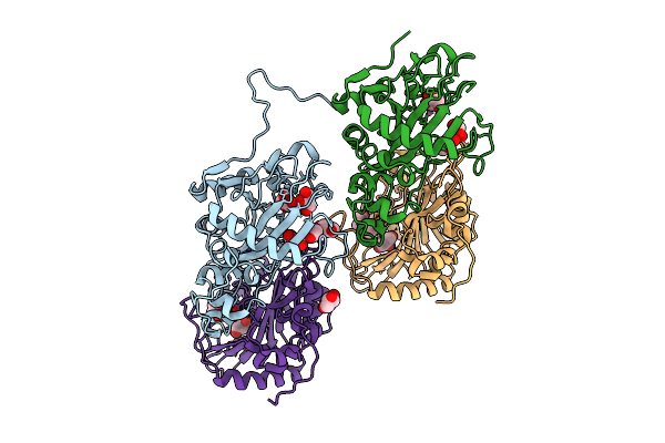
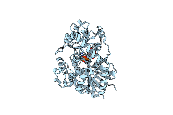
Crystal Structure Of The Okae-M64A Mutant With A-Ketoglutarate And Okaramine A
Organism: Penicillium simplicissimum
Method: X-RAY DIFFRACTION Resolution:2.44 Å Release Date: 2026-03-11 Classification: OXIDOREDUCTASE Ligands: CO, AKG, A1EMZ |
 |
Organism: Penicillium simplicissimum
Method: X-RAY DIFFRACTION Resolution:2.40 Å Release Date: 2026-03-11 Classification: OXIDOREDUCTASE Ligands: CO, GOL |
 |
Organism: Penicillium simplicissimum
Method: X-RAY DIFFRACTION Resolution:2.54 Å Release Date: 2026-03-11 Classification: OXIDOREDUCTASE Ligands: CO, AKG, GOL |
 |
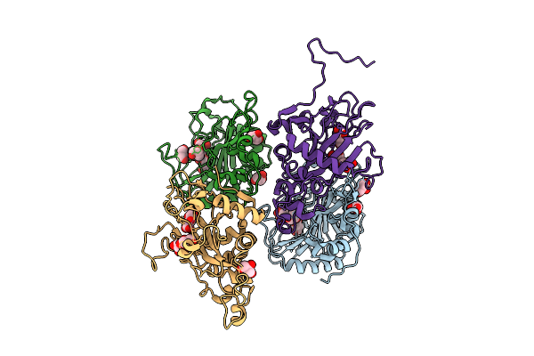
Crystal Structure Of The Okae-M71A Mutant With A-Ketoglutarate And Okaramine A
Organism: Penicillium simplicissimum
Method: X-RAY DIFFRACTION Resolution:2.55 Å Release Date: 2026-03-11 Classification: OXIDOREDUCTASE Ligands: CO, AKG, A1EMZ |
 |
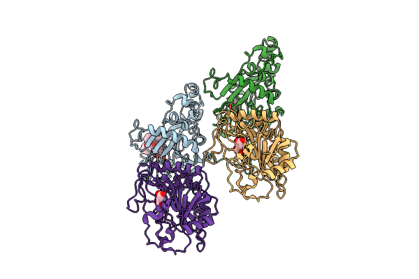
Crystal Structure Of The Okae-W79A Mutant With A-Ketoglutarate And Okaramine A
Organism: Penicillium simplicissimum
Method: X-RAY DIFFRACTION Resolution:2.50 Å Release Date: 2026-03-11 Classification: OXIDOREDUCTASE Ligands: CO, AKG, A1EMZ |
 |
Organism: Penicillium simplicissimum
Method: X-RAY DIFFRACTION Resolution:2.15 Å Release Date: 2026-03-04 Classification: ANTIBIOTIC Ligands: CO, AKG, GOL |
 |
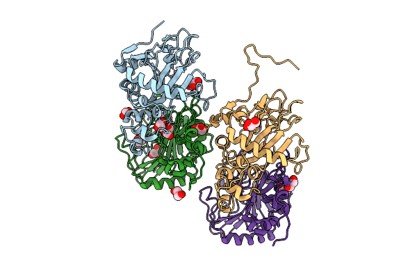
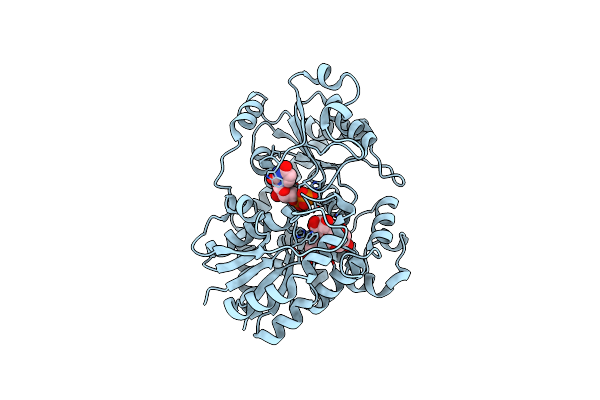
Crystal Structure Of Okae In Complex With A-Ketoglutarate And Okaramine A At 2.5 Angstroms Resolution.
Organism: Penicillium simplicissimum
Method: X-RAY DIFFRACTION Resolution:2.55 Å Release Date: 2026-03-04 Classification: OXIDOREDUCTASE Ligands: CO, A1EMZ, AKG |
 |
Organism: Penicillium simplicissimum
Method: X-RAY DIFFRACTION Resolution:2.20 Å Release Date: 2026-03-04 Classification: OXIDOREDUCTASE Ligands: CO |
 |
Organism: Arabidopsis thaliana
Method: X-RAY DIFFRACTION Resolution:1.91 Å Release Date: 2026-03-04 Classification: TRANSFERASE |
 |
Organism: Penicillium expansum
Method: X-RAY DIFFRACTION Resolution:1.99 Å Release Date: 2025-10-22 Classification: LYASE Ligands: POP, BTM, MG |
 |
Organism: Synthetic construct
Method: X-RAY DIFFRACTION Resolution:2.40 Å Release Date: 2025-10-15 Classification: DE NOVO PROTEIN Ligands: ALL |
 |
Organism: Synthetic construct
Method: X-RAY DIFFRACTION Resolution:1.80 Å Release Date: 2025-10-15 Classification: DE NOVO PROTEIN Ligands: CIT |
 |
Organism: Synthetic construct
Method: X-RAY DIFFRACTION Resolution:1.90 Å Release Date: 2025-10-15 Classification: DE NOVO PROTEIN |
 |
Organism: Synthetic construct
Method: X-RAY DIFFRACTION Resolution:1.60 Å Release Date: 2025-10-15 Classification: DE NOVO PROTEIN Ligands: ALL |
 |
Crystal Structure Of A Phage Catechol 1,2-Dioxygenase Identified From A Soil Metagenomic Survey
Organism: Metagenome
Method: X-RAY DIFFRACTION Resolution:1.65 Å Release Date: 2025-10-08 Classification: OXIDOREDUCTASE Ligands: FE |
 |
Organism: Cicer arietinum
Method: X-RAY DIFFRACTION Resolution:1.74 Å Release Date: 2025-06-25 Classification: PLANT PROTEIN Ligands: UDP |
 |
Organism: Cicer arietinum
Method: X-RAY DIFFRACTION Resolution:1.64 Å Release Date: 2025-06-25 Classification: PLANT PROTEIN Ligands: UDP, A1EGV |
 |
Organism: Cicer arietinum
Method: X-RAY DIFFRACTION Resolution:1.78 Å Release Date: 2025-06-25 Classification: PLANT PROTEIN Ligands: HW2, UDP |