

Search Count: 42
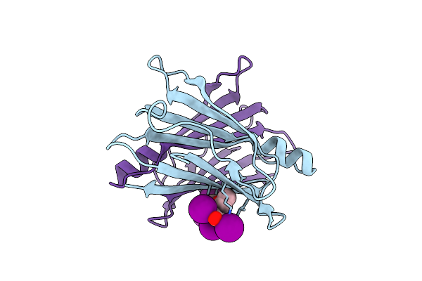 |
Crystal Structure Of V30M-Ttr In Complex With Compound 4
Organism: Homo sapiens
Method: X-RAY DIFFRACTION Resolution:1.80 Å Release Date: 2024-05-15 Classification: TRANSPORT PROTEIN Ligands: WGH |
Organism: Homo sapiens
Method: X-RAY DIFFRACTION
Release Date: 2024-05-15
Ligands: WGH
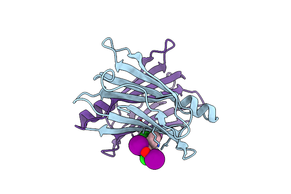 |
Crystal Structure Of V30M-Ttr In Complex With Compound 7
Organism: Homo sapiens
Method: X-RAY DIFFRACTION Resolution:1.70 Å Release Date: 2024-05-15 Classification: TRANSPORT PROTEIN Ligands: WGJ |
Organism: Homo sapiens
Method: X-RAY DIFFRACTION
Release Date: 2024-05-15
Ligands: WGJ
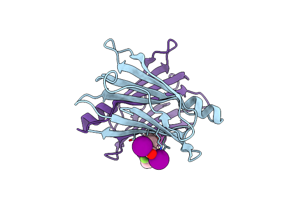 |
Crystal Structure Of V30M-Ttr In Complex With Compound 20
Organism: Homo sapiens
Method: X-RAY DIFFRACTION Resolution:1.51 Å Release Date: 2024-05-15 Classification: TRANSPORT PROTEIN Ligands: WH0 |
Organism: Homo sapiens
Method: X-RAY DIFFRACTION
Release Date: 2024-05-15
Ligands: WH0
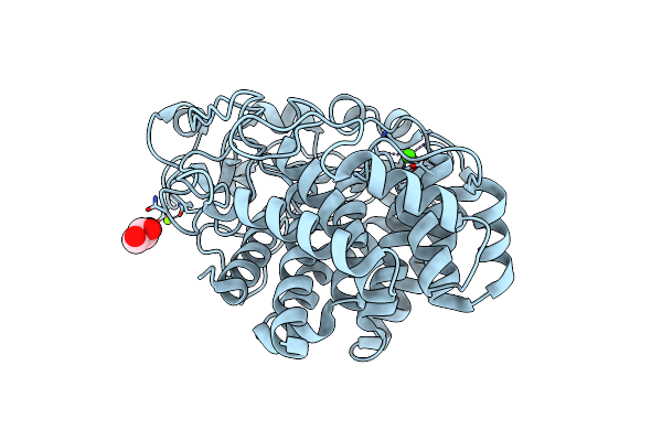 |
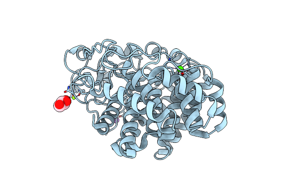
X-Ray Crystal Structure Of D43R Mutant Of Endo-1,4-Beta Glucanase From Eisenia Fetida
Organism: Eisenia fetida
Method: X-RAY DIFFRACTION Resolution:1.70 Å Release Date: 2023-12-06 Classification: HYDROLASE Ligands: GOL, MG, CA |
Organism: Eisenia fetida
Method: X-RAY DIFFRACTION
Release Date: 2023-12-06
Ligands: GOL, MG, CA
 |
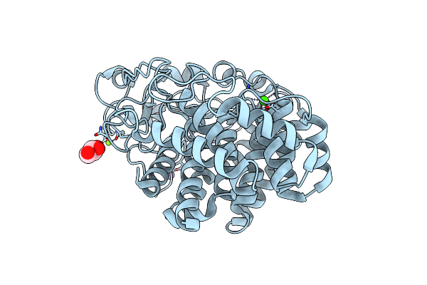
X-Ray Crystal Structure Of N372D Mutant Of Endo-1,4-Beta Glucanase From Eisenia Fetida
Organism: Eisenia fetida
Method: X-RAY DIFFRACTION Resolution:1.60 Å Release Date: 2023-12-06 Classification: HYDROLASE Ligands: MG, CA, NA, GOL |
Organism: Eisenia fetida
Method: X-RAY DIFFRACTION
Release Date: 2023-12-06
Ligands: MG, CA, NA, GOL
 |
X-Ray Crystal Structure Of Q387E Mutant Of Endo-1,4-Beta Glucanase From Eisenia Fetida
Organism: Eisenia fetida
Method: X-RAY DIFFRACTION Resolution:1.60 Å Release Date: 2023-12-06 Classification: HYDROLASE Ligands: MG, CA, NA, GOL |
Organism: Eisenia fetida
Method: X-RAY DIFFRACTION
Release Date: 2023-12-06
Ligands: MG, CA, NA, GOL
 |
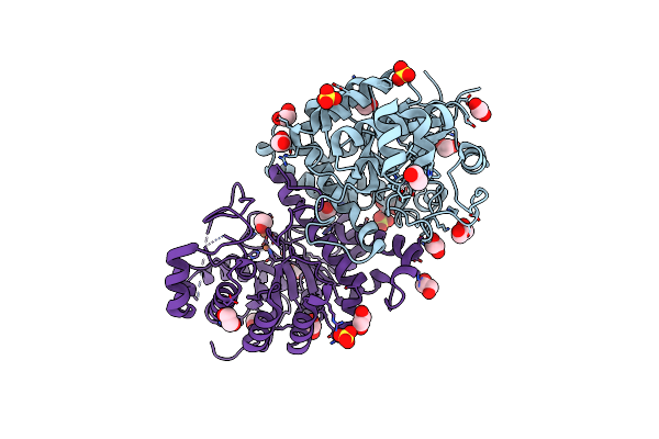
Structure Of The 2-Aminoisobutyric Acid Monooxygenase Hydroxylase
Organism: Rhodococcus wratislaviensis
Method: X-RAY DIFFRACTION Resolution:2.75 Å Release Date: 2021-01-06 Classification: OXIDOREDUCTASE Ligands: ZN, GOL, SO4, FE, CL |
Organism: Rhodococcus wratislaviensis
Method: X-RAY DIFFRACTION
Release Date: 2021-01-06
Ligands: ZN, GOL, SO4, FE, CL
 |
Structure Of The 2-Aminoisobutyric Acid Monooxygenase Hydroxylase
Organism: Rhodococcus wratislaviensis
Method: X-RAY DIFFRACTION Resolution:2.45 Å Release Date: 2021-01-06 Classification: OXIDOREDUCTASE Ligands: ZN, SO4, EDO, FE |
Organism: Rhodococcus wratislaviensis
Method: X-RAY DIFFRACTION
Release Date: 2021-01-06
Ligands: ZN, SO4, EDO, FE
 |
Crystal Structure Of The Catalytic Domain Of Chitiniphilus Shinanonensis Chitinase Chil (Cschil) Complexed With N,N'-Diacetylchitobiose
Organism: Chitiniphilus shinanonensis
Method: X-RAY DIFFRACTION Resolution:1.35 Å Release Date: 2020-09-16 Classification: HYDROLASE Ligands: ACT, MXE, MLI, PGE, EDO, NAG, PG4 |
Organism: Chitiniphilus shinanonensis
Method: X-RAY DIFFRACTION
Release Date: 2020-09-16
Ligands: ACT, MXE, MLI, PGE, EDO, NAG, PG4
 |
Crystal Structure Of D157N Mutant Of Chitiniphilus Shinanonensis Chitinase Chil (Cschil) Complexed With N,N'-Diacetylchitobiose
Organism: Chitiniphilus shinanonensis
Method: X-RAY DIFFRACTION Resolution:1.95 Å Release Date: 2020-09-16 Classification: HYDROLASE |
Organism: Chitiniphilus shinanonensis
Method: X-RAY DIFFRACTION
Release Date: 2020-09-16
 |
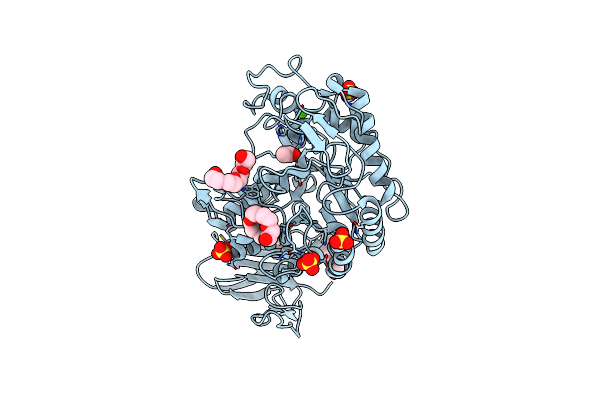
Crystal Structure Of W50A Mutant Of Chitiniphilus Shinanonensis Chitinase Chil (Cschil) Complexed With N,N'-Diacetylchitobiose
Organism: Chitiniphilus shinanonensis
Method: X-RAY DIFFRACTION Resolution:1.50 Å Release Date: 2020-09-16 Classification: HYDROLASE Ligands: SO4, PEG, GOL, EDO |
Organism: Chitiniphilus shinanonensis
Method: X-RAY DIFFRACTION
Release Date: 2020-09-16
Ligands: SO4, PEG, GOL, EDO
 |
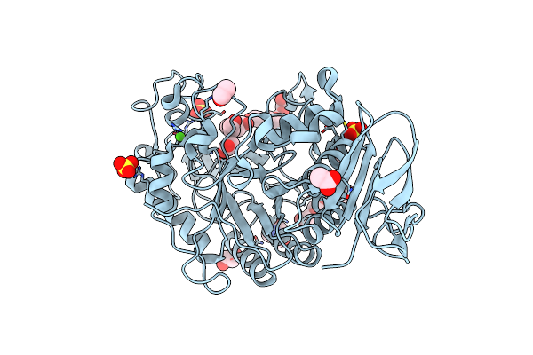
X-Ray Crystal Structure Of Wild Type Alpha-Amylase I From Eisenia Fetida
Organism: Eisenia fetida
Method: X-RAY DIFFRACTION Resolution:1.30 Å Release Date: 2020-09-16 Classification: HYDROLASE Ligands: CA, CL, ACT, SO4, 1PE, PG4 |
Organism: Eisenia fetida
Method: X-RAY DIFFRACTION
Release Date: 2020-09-16
Ligands: CA, CL, ACT, SO4, 1PE, PG4
 |
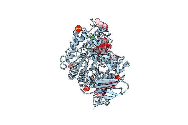
X-Ray Crystal Structure Of The E249Q Mutant Of Alpha-Amylase I From Eisenia Fetida
Organism: Eisenia fetida
Method: X-RAY DIFFRACTION Resolution:1.60 Å Release Date: 2020-09-16 Classification: HYDROLASE Ligands: CA, CL, SO4, ACT, TRS, PG4, EDO, PGE |
Organism: Eisenia fetida
Method: X-RAY DIFFRACTION
Release Date: 2020-09-16
Ligands: CA, CL, SO4, ACT, TRS, PG4, EDO, PGE
 |
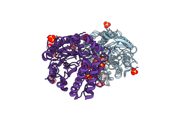
X-Ray Crystal Structure Of The E249Q Mutan Of Alpha-Amylase I And Maltohexaose Complex From Eisenia Fetida
Organism: Eisenia fetida
Method: X-RAY DIFFRACTION Resolution:1.70 Å Release Date: 2020-09-16 Classification: HYDROLASE Ligands: CA, CL, SO4, PG4, PGE |
Organism: Eisenia fetida
Method: X-RAY DIFFRACTION
Release Date: 2020-09-16
Ligands: CA, CL, SO4, PG4, PGE
 |
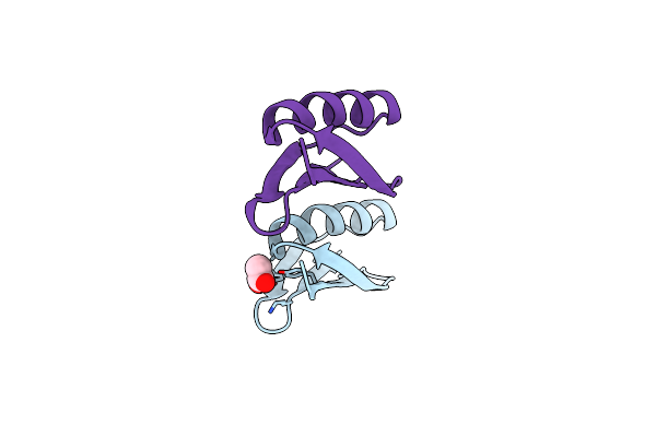
Crystal Structure Of The Catalytic Domain Of Chitinase Chil From Chitiniphilus Shinanonensis (Cschil)
Organism: Chitiniphilus shinanonensis
Method: X-RAY DIFFRACTION Resolution:1.25 Å Release Date: 2020-08-26 Classification: HYDROLASE Ligands: GOL, SO4, EDO, MXE, CL |
Organism: Chitiniphilus shinanonensis
Method: X-RAY DIFFRACTION
Release Date: 2020-08-26
Ligands: GOL, SO4, EDO, MXE, CL
 |
Crystal Structure Of A Streptococcal Protein G B1 Mutant
Organism: Streptococcus sp. 'group g'
Method: X-RAY DIFFRACTION Resolution:1.84 Å Release Date: 2019-10-23 Classification: IMMUNE SYSTEM Ligands: ACY |
Organism: Streptococcus sp. 'group g'
Method: X-RAY DIFFRACTION
Release Date: 2019-10-23
Ligands: ACY
 |
Crystal Structure Of C Terminal Region Of G-Protein Interacting Protein 1 (Gip1) From Dictyostelium Discoideum
Organism: Dictyostelium discoideum
Method: X-RAY DIFFRACTION Resolution:1.95 Å Release Date: 2018-10-17 Classification: PROTEIN BINDING Ligands: PEF, LHG, NA |
Organism: Dictyostelium discoideum
Method: X-RAY DIFFRACTION
Release Date: 2018-10-17
Ligands: PEF, LHG, NA
 |
Crystal Structure Of C Terminal Region Of G-Protein Interacting Protein 1 (Gip1) From Dictyostelium Discoideum Form Ii
Organism: Dictyostelium discoideum
Method: X-RAY DIFFRACTION Resolution:2.74 Å Release Date: 2018-10-17 Classification: PROTEIN BINDING Ligands: PEF, LHG, NA |
Organism: Dictyostelium discoideum
Method: X-RAY DIFFRACTION
Release Date: 2018-10-17
Ligands: PEF, LHG, NA
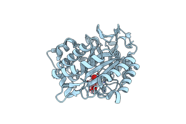 |
Crystal Structure Of Endo-1,4-Beta-Mannanase From Eisenia Fetida
Organism: Eisenia fetida
Method: X-RAY DIFFRACTION Resolution:1.70 Å Release Date: 2018-06-27 Classification: CARBOHYDRATE Ligands: TRS, IPA |
Organism: Eisenia fetida
Method: X-RAY DIFFRACTION
Release Date: 2018-06-27
Ligands: TRS, IPA
 |
Crystal Structure Of A Pl 26 Exo-Rhamnogalacturonan Lyase From Penicillium Chrysogenum Complexed With Unsaturated Galacturonosyl Rhamnose Substituted With Galactose
Organism: Penicillium chrysogenum
Method: X-RAY DIFFRACTION Resolution:2.75 Å Release Date: 2018-04-04 Classification: LYASE Ligands: CA |
Organism: Penicillium chrysogenum
Method: X-RAY DIFFRACTION
Release Date: 2018-04-04
Ligands: CA