

Search Count: 28
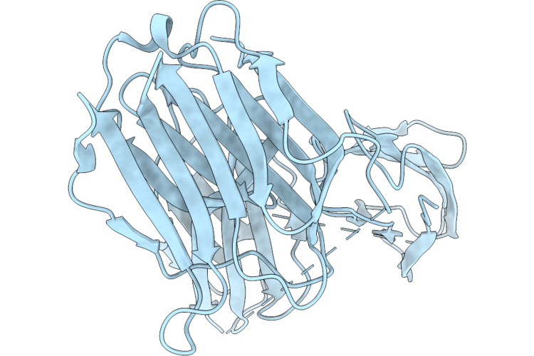 |
Crystal Structure Of Galectin-3 Bound To Fn3Con-9 And Fn3Con-41, A Cooperative Binder That Recognises The Galectin-3-Fn3Con-9 Interface
Organism: Synthetic construct, Homo sapiens
Method: X-RAY DIFFRACTION Resolution:1.96 Å Release Date: 2026-05-20 Classification: CARBOHYDRATE |
Organism: Synthetic construct, Homo sapiens
Method: X-RAY DIFFRACTION
Release Date: 2026-05-20
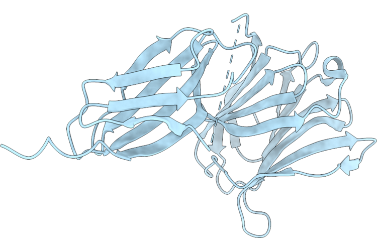 |
Crystal Structure Of The Galectin-3 Complex With Fn3Con-7, An Mrna Display-Derived Binding Protein
Organism: Homo sapiens, Synthetic construct
Method: X-RAY DIFFRACTION Resolution:1.92 Å Release Date: 2026-05-20 Classification: CARBOHYDRATE |
Organism: Homo sapiens, Synthetic construct
Method: X-RAY DIFFRACTION
Release Date: 2026-05-20
 |
Crystal Structure Of Gh30_8 Xylanase From Bacillus Pumilus
Organism: Bacillus pumilus atcc 7061
Method: X-RAY DIFFRACTION Resolution:2.16 Å Release Date: 2026-04-29 Classification: HYDROLASE Ligands: GOL, CIT |
Organism: Bacillus pumilus atcc 7061
Method: X-RAY DIFFRACTION
Release Date: 2026-04-29
Ligands: GOL, CIT
 |
Crystal Structure Of Galectin-3 In Complex With Fn3
Organism: Homo sapiens
Method: X-RAY DIFFRACTION Resolution:2.04 Å Release Date: 2026-02-18 Classification: PROTEIN BINDING Ligands: SO4 |
Organism: Homo sapiens
Method: X-RAY DIFFRACTION
Release Date: 2026-02-18
Ligands: SO4
 |
Crystal Structure Of S1007A In Complex With Fn3
Organism: Homo sapiens
Method: X-RAY DIFFRACTION Resolution:2.33 Å Release Date: 2025-12-17 Classification: PROTEIN BINDING |
Organism: Homo sapiens
Method: X-RAY DIFFRACTION
Release Date: 2025-12-17
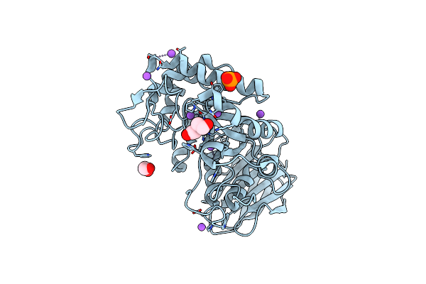 |
Crystallographic Structure Of Ruminococcus Champanellensis Xylanase (Rcxyn30A)
Organism: Ruminococcus champanellensis 18p13 = jcm 17042
Method: X-RAY DIFFRACTION Resolution:1.37 Å Release Date: 2024-12-25 Classification: HYDROLASE Ligands: EDO, PEG, NA, F, PO4 |
Organism: Ruminococcus champanellensis 18p13 = jcm 17042
Method: X-RAY DIFFRACTION
Release Date: 2024-12-25
Ligands: EDO, PEG, NA, F, PO4
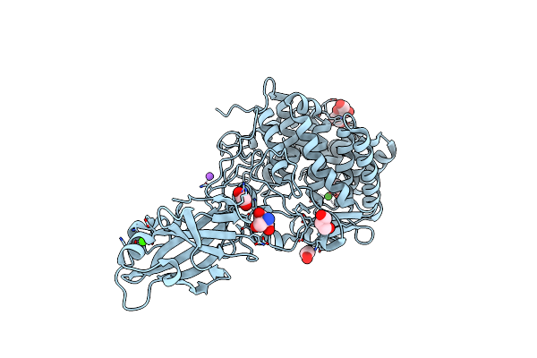 |
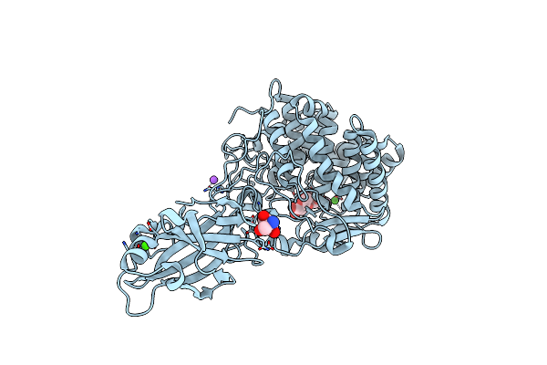
The Apo Crystal Structure Of Blcel9A From Glycoside Hydrolase Family 9
Organism: Bacillus licheniformis dsm 13 = atcc 14580
Method: X-RAY DIFFRACTION Resolution:1.80 Å Release Date: 2024-02-14 Classification: HYDROLASE Ligands: TRS, GOL, CL, EDO, NA, CA |
Organism: Bacillus licheniformis dsm 13 = atcc 14580
Method: X-RAY DIFFRACTION
Release Date: 2024-02-14
Ligands: TRS, GOL, CL, EDO, NA, CA
 |
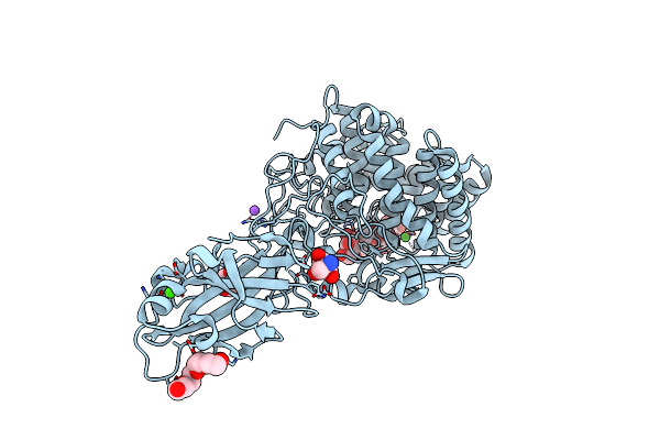
Crystal Structure Of Blcel9A From Glycoside Hydrolase Family 9 In Complex With Cellotriose
Organism: Bacillus licheniformis dsm 13 = atcc 14580
Method: X-RAY DIFFRACTION Resolution:1.97 Å Release Date: 2024-02-14 Classification: HYDROLASE Ligands: TRS, CA, NA, CL |
Organism: Bacillus licheniformis dsm 13 = atcc 14580
Method: X-RAY DIFFRACTION
Release Date: 2024-02-14
Ligands: TRS, CA, NA, CL
 |
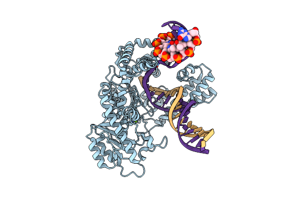
Crystal Structure Of Blcel9A From Glycoside Hydrolase Family 9 In Complex With Cellohexaose
Organism: Bacillus licheniformis dsm 13 = atcc 14580
Method: X-RAY DIFFRACTION Resolution:2.01 Å Release Date: 2024-02-14 Classification: HYDROLASE Ligands: TRS, PG4, BGC, CA, CL, NA |
Organism: Bacillus licheniformis dsm 13 = atcc 14580
Method: X-RAY DIFFRACTION
Release Date: 2024-02-14
Ligands: TRS, PG4, BGC, CA, CL, NA
 |
Pol I Bound To Extended And Displaced Dna Section - Closed Conformation
Organism: Escherichia coli 'bl21-gold(de3)plyss ag', Synthetic construct
Method: ELECTRON MICROSCOPY Release Date: 2023-08-09 Classification: DNA BINDING PROTEIN Ligands: MG |
Organism: Escherichia coli 'bl21-gold(de3)plyss ag', Synthetic construct
Method: ELECTRON MICROSCOPY
Release Date: 2023-08-09
Ligands: MG
 |
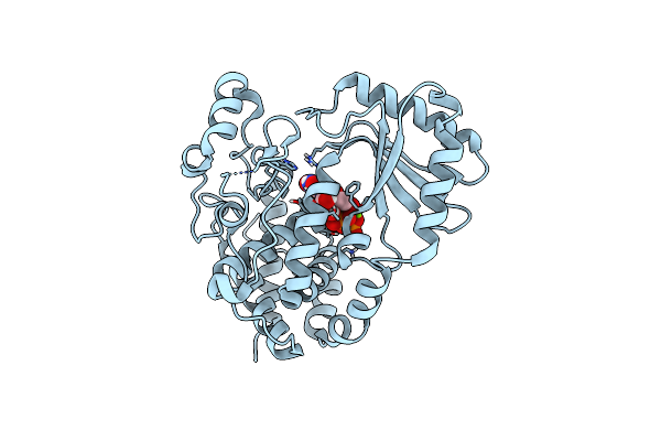
Pol I Bound To Extended And Displaced Dna Section - Open Conformation
Organism: Escherichia coli 'bl21-gold(de3)plyss ag', Synthetic construct
Method: ELECTRON MICROSCOPY Release Date: 2023-08-09 Classification: DNA BINDING PROTEIN Ligands: MG |
Organism: Escherichia coli 'bl21-gold(de3)plyss ag', Synthetic construct
Method: ELECTRON MICROSCOPY
Release Date: 2023-08-09
Ligands: MG
 |
Crystal Structure Of Dmtailor In Complex With Umpnpp
Organism: Drosophila melanogaster
Method: X-RAY DIFFRACTION Resolution:1.90 Å Release Date: 2018-12-05 Classification: TRANSFERASE Ligands: 2KH, MG |
Organism: Drosophila melanogaster
Method: X-RAY DIFFRACTION
Release Date: 2018-12-05
Ligands: 2KH, MG
 |
Crystal Structure Of Dmtailor In Complex With Gpu
Organism: Drosophila melanogaster
Method: X-RAY DIFFRACTION Resolution:2.00 Å Release Date: 2018-12-05 Classification: TRANSFERASE |
Organism: Drosophila melanogaster
Method: X-RAY DIFFRACTION
Release Date: 2018-12-05
 |
Crystal Structure Of Dmtailor In Complex With U6 Rna
Organism: Drosophila melanogaster
Method: X-RAY DIFFRACTION Resolution:2.00 Å Release Date: 2018-12-05 Classification: TRANSFERASE Ligands: MG |
Organism: Drosophila melanogaster
Method: X-RAY DIFFRACTION
Release Date: 2018-12-05
Ligands: MG
 |
Crystal Structure Of Dmtailor In Complex With Cacagu Rna
Organism: Drosophila melanogaster
Method: X-RAY DIFFRACTION Resolution:1.85 Å Release Date: 2018-12-05 Classification: TRANSFERASE Ligands: MG |
Organism: Drosophila melanogaster
Method: X-RAY DIFFRACTION
Release Date: 2018-12-05
Ligands: MG
 |
Crystal Structure Of The Type Ii Dehydroquinase From Pseudomonas Aeruginosa
Organism: Pseudomonas aeruginosa
Method: X-RAY DIFFRACTION Resolution:1.74 Å Release Date: 2014-07-23 Classification: LYASE |
Organism: Pseudomonas aeruginosa
Method: X-RAY DIFFRACTION
Release Date: 2014-07-23
 |
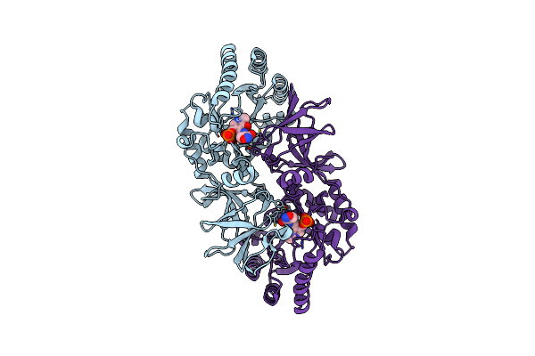
Alanine Racemase [Clostridium Difficile 630]
Organism: Clostridium difficile
Method: X-RAY DIFFRACTION Resolution:2.10 Å Release Date: 2014-06-04 Classification: ISOMERASE Ligands: GOL, TCE |
Organism: Clostridium difficile
Method: X-RAY DIFFRACTION
Release Date: 2014-06-04
Ligands: GOL, TCE
 |
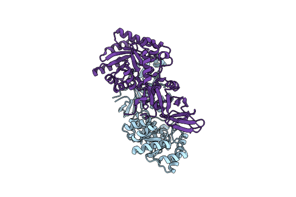
Alanine Racemase [Clostridium Difficile 630] Complex With Cycloserine
Organism: Clostridium difficile
Method: X-RAY DIFFRACTION Resolution:2.26 Å Release Date: 2014-06-04 Classification: ISOMERASE/ISOMERASE INHIBITOR Ligands: DCS |
Organism: Clostridium difficile
Method: X-RAY DIFFRACTION
Release Date: 2014-06-04
Ligands: DCS
 |
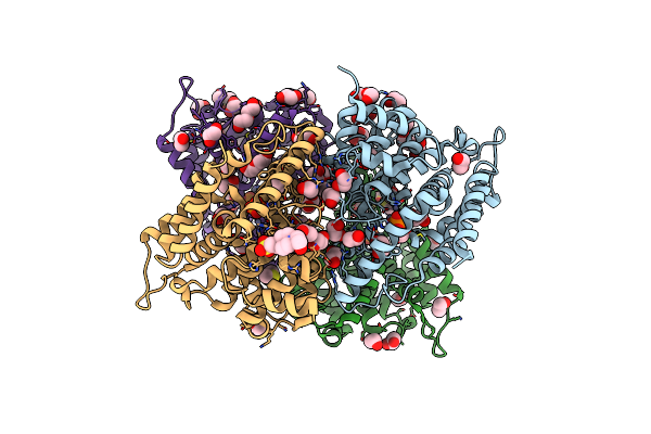
Crystal Structure Of Cdalr Mutant K 271 T
Organism: Clostridium difficile
Method: X-RAY DIFFRACTION Resolution:2.60 Å Release Date: 2014-06-04 Classification: ISOMERASE |
Organism: Clostridium difficile
Method: X-RAY DIFFRACTION
Release Date: 2014-06-04
 |
Crystal Structure Of The Apo Form Of The Catalytic Domain Of Pde4D2
Organism: Homo sapiens
Method: X-RAY DIFFRACTION Resolution:2.10 Å Release Date: 2011-10-26 Classification: HYDROLASE Ligands: EDO, PEG, ZN, PO4, DMS, EPE |
Organism: Homo sapiens
Method: X-RAY DIFFRACTION
Release Date: 2011-10-26
Ligands: EDO, PEG, ZN, PO4, DMS, EPE