

Search Count: 114
 |
Crystal Structure Of A Calcium Bound C2 Domain Containing Protein From Trichomonas Vaginalis
Organism: Trichomonas vaginalis g3
Method: X-RAY DIFFRACTION Resolution:1.40 Å Release Date: 2025-12-17 Classification: LIPID BINDING PROTEIN Ligands: 1PE, CL, PEG, PGE |
Organism: Trichomonas vaginalis g3
Method: X-RAY DIFFRACTION
Release Date: 2025-12-17
Ligands: 1PE, CL, PEG, PGE
 |
Crystal Structure Of A Calcium Bound C2 Domain Containing Protein From Trichomonas Vaginalis (P21 Form)
Organism: Trichomonas vaginalis g3
Method: X-RAY DIFFRACTION Resolution:1.51 Å Release Date: 2025-12-17 Classification: LIPID BINDING PROTEIN Ligands: PG4, CA, MES, 1PE |
Organism: Trichomonas vaginalis g3
Method: X-RAY DIFFRACTION
Release Date: 2025-12-17
Ligands: PG4, CA, MES, 1PE
 |
Crystal Structure Of A Calcium Bound C2 Domain Containing Protein From Trichomonas Vaginalis (Orthorhombic P Form)
Organism: Trichomonas vaginalis g3
Method: X-RAY DIFFRACTION Resolution:1.53 Å Release Date: 2025-12-17 Classification: LIPID BINDING PROTEIN Ligands: CA, 1PE |
Organism: Trichomonas vaginalis g3
Method: X-RAY DIFFRACTION
Release Date: 2025-12-17
Ligands: CA, 1PE
 |
Crystal Structure Of A C2 Domain Containing Protein From Trichomonas Vaginalis
Organism: Trichomonas vaginalis g3
Method: X-RAY DIFFRACTION Resolution:1.35 Å Release Date: 2025-12-17 Classification: LIPID BINDING PROTEIN Ligands: GOL, PGE |
Organism: Trichomonas vaginalis g3
Method: X-RAY DIFFRACTION
Release Date: 2025-12-17
Ligands: GOL, PGE
 |
Crystal Structure Of A Malonate Bound C2 Domain Containing Protein From Trichomonas Vaginalis (P21 Form)
Organism: Trichomonas vaginalis g3
Method: X-RAY DIFFRACTION Resolution:1.30 Å Release Date: 2025-12-17 Classification: LIPID BINDING PROTEIN Ligands: MLI, CL, NA |
Organism: Trichomonas vaginalis g3
Method: X-RAY DIFFRACTION
Release Date: 2025-12-17
Ligands: MLI, CL, NA
 |
Crystal Structure Of A Calcium Bound C2 Domain Containing Protein From Trichomonas Vaginalis (P61 Form)
Organism: Trichomonas vaginalis g3
Method: X-RAY DIFFRACTION Resolution:1.54 Å Release Date: 2025-12-17 Classification: LIPID BINDING PROTEIN Ligands: CA, CL, PG4 |
Organism: Trichomonas vaginalis g3
Method: X-RAY DIFFRACTION
Release Date: 2025-12-17
Ligands: CA, CL, PG4
 |
Crystal Structure Of A C2 Domain Containing Protein From Trichomonas Vaginalis In Complex With Pyrophosphate
Organism: Trichomonas vaginalis g3
Method: X-RAY DIFFRACTION Resolution:1.70 Å Release Date: 2025-12-17 Classification: LIPID BINDING PROTEIN Ligands: PPV |
Organism: Trichomonas vaginalis g3
Method: X-RAY DIFFRACTION
Release Date: 2025-12-17
Ligands: PPV
 |
Crystal Structure Of Phosphoglycerate Mutase From Trichomonas Vaginalis In Complex With 3-Phosphoglyceric Acid
Organism: Trichomonas vaginalis g3
Method: X-RAY DIFFRACTION Resolution:2.10 Å Release Date: 2025-12-03 Classification: ISOMERASE Ligands: CL, 3PG, PG4, SO4 Modified Residue(s): RPI |
Organism: Trichomonas vaginalis g3
Method: X-RAY DIFFRACTION
Release Date: 2025-12-03
Ligands: CL, 3PG, PG4, SO4
 |
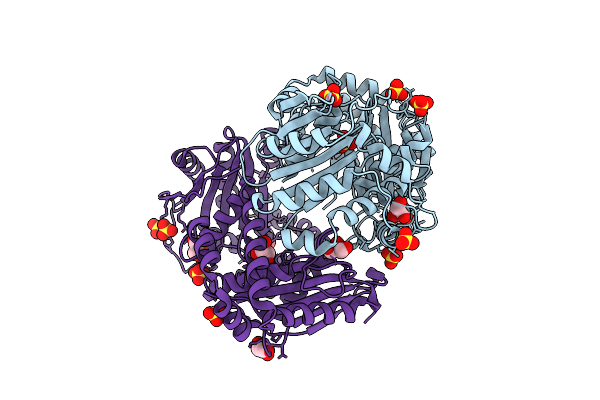
X-Ray Structure Of Trichomonas Vaginalis Inactive Mutant Hydrogenosomal Processing Peptidase Heterodimer (Hppin)
Organism: Trichomonas vaginalis
Method: X-RAY DIFFRACTION Resolution:2.84 Å Release Date: 2025-11-05 Classification: HYDROLASE Ligands: SO4, ZN |
Organism: Trichomonas vaginalis
Method: X-RAY DIFFRACTION
Release Date: 2025-11-05
Ligands: SO4, ZN
 |
X-Ray Structure Of Hydrogenosomal Processing Peptidase (Hpp), E56Q Inactive Mutant, From Trichomonas Vaginalis Co-Crystallized With Presequence Peptide From Adenylate Kinase (Ak) - Not Visible In The Structure Model
Organism: Trichomonas vaginalis
Method: X-RAY DIFFRACTION Resolution:2.00 Å Release Date: 2025-11-05 Classification: HYDROLASE Ligands: SO4, GOL, ZN |
Organism: Trichomonas vaginalis
Method: X-RAY DIFFRACTION
Release Date: 2025-11-05
Ligands: SO4, GOL, ZN
 |
X-Ray Structure Of Hydrogenosomal Processing Peptidase (Hpp), E56Q Inactive Mutant, From Trichomonas Vaginalis Co-Crystallized With Presequence Peptide From Ferredoxin Oxidoreductase (Pfo) - Not Visible In The Structure Model
Organism: Trichomonas vaginalis
Method: X-RAY DIFFRACTION Resolution:2.65 Å Release Date: 2025-11-05 Classification: HYDROLASE Ligands: SO4, GOL, ZN |
Organism: Trichomonas vaginalis
Method: X-RAY DIFFRACTION
Release Date: 2025-11-05
Ligands: SO4, GOL, ZN
 |
48-Nm Doublet Microtubule From Trichomonas Vaginalis Strain G3
Organism: Trichomonas vaginalis g3
Method: ELECTRON MICROSCOPY Release Date: 2025-05-14 Classification: STRUCTURAL PROTEIN Ligands: GDP, GTP, MG |
Organism: Trichomonas vaginalis g3
Method: ELECTRON MICROSCOPY
Release Date: 2025-05-14
Ligands: GDP, GTP, MG
 |
Crystal Structure Of Phosphoglycerate Mutase From Trichomonas Vaginalis (Sulfate Bound)
Organism: Trichomonas vaginalis g3
Method: X-RAY DIFFRACTION Resolution:2.05 Å Release Date: 2025-04-23 Classification: ISOMERASE Ligands: SO4 |
Organism: Trichomonas vaginalis g3
Method: X-RAY DIFFRACTION
Release Date: 2025-04-23
Ligands: SO4
 |
Crystal Structure Of Phosphoglycerate Mutase From Trichomonas Vaginalis
Organism: Trichomonas vaginalis g3
Method: X-RAY DIFFRACTION Resolution:1.95 Å Release Date: 2025-04-02 Classification: ISOMERASE Ligands: SO4, GOL, P33, PG4 |
Organism: Trichomonas vaginalis g3
Method: X-RAY DIFFRACTION
Release Date: 2025-04-02
Ligands: SO4, GOL, P33, PG4
 |
Structure Of Rmld From Trichomonas Vaginalis Is Space Group P1
Organism: Trichomonas vaginalis
Method: X-RAY DIFFRACTION Resolution:2.40 Å Release Date: 2025-03-12 Classification: CYTOSOLIC PROTEIN |
Organism: Trichomonas vaginalis
Method: X-RAY DIFFRACTION
Release Date: 2025-03-12
 |
Structure Of Rmld From Trichomonas Vaginalis Is Space Group P212121
Organism: Trichomonas vaginalis
Method: X-RAY DIFFRACTION Resolution:2.80 Å Release Date: 2025-03-12 Classification: CYTOSOLIC PROTEIN |
Organism: Trichomonas vaginalis
Method: X-RAY DIFFRACTION
Release Date: 2025-03-12
 |
Crystal Structure Of Purine Nucleoside Phosphorylase From Trichomonas Vaginalis (Inosine Bound)
Organism: Trichomonas vaginalis g3
Method: X-RAY DIFFRACTION Resolution:1.20 Å Release Date: 2025-01-22 Classification: TRANSFERASE Ligands: NOS, CL |
Organism: Trichomonas vaginalis g3
Method: X-RAY DIFFRACTION
Release Date: 2025-01-22
Ligands: NOS, CL
 |
Crystal Structure Of Purine Nucleoside Phosphorylase From Trichomonas Vaginalis (C2 Form)
Organism: Trichomonas vaginalis g3
Method: X-RAY DIFFRACTION Resolution:1.80 Å Release Date: 2024-12-25 Classification: TRANSFERASE Ligands: GOL |
Organism: Trichomonas vaginalis g3
Method: X-RAY DIFFRACTION
Release Date: 2024-12-25
Ligands: GOL
 |
Crystal Structure Of Purine Nucleoside Phosphorylase From Trichomonas Vaginalis (P21 Form)
Organism: Trichomonas vaginalis g3
Method: X-RAY DIFFRACTION Resolution:1.59 Å Release Date: 2024-12-25 Classification: TRANSFERASE Ligands: FLC |
Organism: Trichomonas vaginalis g3
Method: X-RAY DIFFRACTION
Release Date: 2024-12-25
Ligands: FLC
 |
Crystal Structure Of Purine Nucleoside Phosphorylase From Trichomonas Vaginalis (Hepes Bound)
Organism: Trichomonas vaginalis g3
Method: X-RAY DIFFRACTION Resolution:1.49 Å Release Date: 2024-12-25 Classification: TRANSFERASE Ligands: GOL, EPE |
Organism: Trichomonas vaginalis g3
Method: X-RAY DIFFRACTION
Release Date: 2024-12-25
Ligands: GOL, EPE