

Search Count: 258
 |
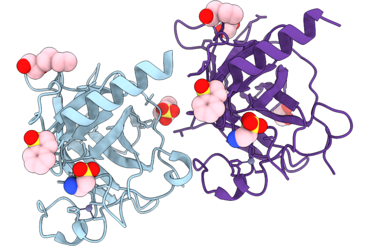
P53-R282W Core Domain Covalently Bound To A Vinyl-Sulfone Fragment
Organism: Homo sapiens
Method: X-RAY DIFFRACTION Resolution:1.56 Å Release Date: 2026-05-27 Classification: CELL CYCLE Ligands: EPE, PEG, A1JQW, GOL, EDO, ZN |
Organism: Homo sapiens
Method: X-RAY DIFFRACTION
Release Date: 2026-05-27
Ligands: EPE, PEG, A1JQW, GOL, EDO, ZN
 |
P53-Y220C Core Domain Covalently Bound To A Vinyl-Sulfone Fragment
Organism: Homo sapiens
Method: X-RAY DIFFRACTION Resolution:1.70 Å Release Date: 2026-05-27 Classification: CELL CYCLE Ligands: A1JQW, EPE, ZN, PGE, PEG, EDO |
Organism: Homo sapiens
Method: X-RAY DIFFRACTION
Release Date: 2026-05-27
Ligands: A1JQW, EPE, ZN, PGE, PEG, EDO
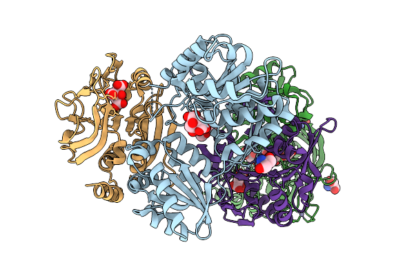 |
Crystal Structure Of The Murt/Gatd Enzyme Complex From Streptococcus Pyogenes
Organism: Streptococcus pyogenes mgas10270
Method: X-RAY DIFFRACTION Resolution:2.05 Å Release Date: 2025-10-08 Classification: LIGASE Ligands: ZN, CIT, TRS, PEG |
Organism: Streptococcus pyogenes mgas10270
Method: X-RAY DIFFRACTION
Release Date: 2025-10-08
Ligands: ZN, CIT, TRS, PEG
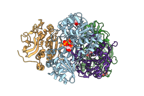 |
Crystal Structure Of The Murt/Gatd Enzyme Complex From Streptococcus Pyogenes With Bound Amppnp
Organism: Streptococcus pyogenes mgas10270
Method: X-RAY DIFFRACTION Resolution:1.98 Å Release Date: 2025-10-08 Classification: LIGASE Ligands: ZN, MG, ANP, GOL |
Organism: Streptococcus pyogenes mgas10270
Method: X-RAY DIFFRACTION
Release Date: 2025-10-08
Ligands: ZN, MG, ANP, GOL
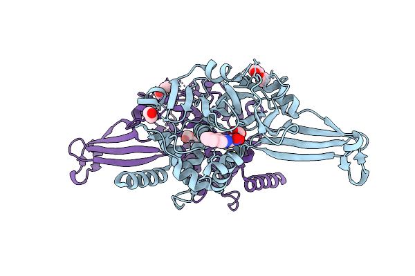 |
Usp7 Covalently Bound To N-(6-Fluoro-3-Nitropyridin-2-Yl)-5-(1-Methyl-1H-Pyrazol-4-Yl)Isoquinolin-3-Amine (Gcl36, 7A) With Partial Occupancy
Organism: Homo sapiens
Method: X-RAY DIFFRACTION Resolution:2.26 Å Release Date: 2025-09-10 Classification: HYDROLASE Ligands: A1I71, EDO, PEG, BR |
Organism: Homo sapiens
Method: X-RAY DIFFRACTION
Release Date: 2025-09-10
Ligands: A1I71, EDO, PEG, BR
 |
P53-Y220C Core Domain Covalently Bound To 2-Chloro-5-Cyanopyrazine Soaked At 5 Mm
Organism: Homo sapiens
Method: X-RAY DIFFRACTION Resolution:1.65 Å Release Date: 2025-06-11 Classification: CELL CYCLE Ligands: A1IIO, EPE, PEG, EDO, ZN, GOL |
Organism: Homo sapiens
Method: X-RAY DIFFRACTION
Release Date: 2025-06-11
Ligands: A1IIO, EPE, PEG, EDO, ZN, GOL
 |
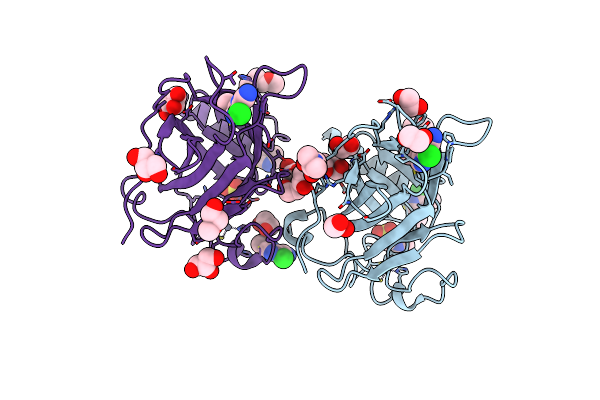
P53-Y220C Core Domain Covalently Bound To 5-Chloro-6-Methylpyrazine-2-Carbonitrile Soaked At 5 Mm
Organism: Homo sapiens
Method: X-RAY DIFFRACTION Resolution:1.56 Å Release Date: 2025-06-11 Classification: CELL CYCLE Ligands: A1II4, EDO, EPE, PEG, GOL, ZN |
Organism: Homo sapiens
Method: X-RAY DIFFRACTION
Release Date: 2025-06-11
Ligands: A1II4, EDO, EPE, PEG, GOL, ZN
 |
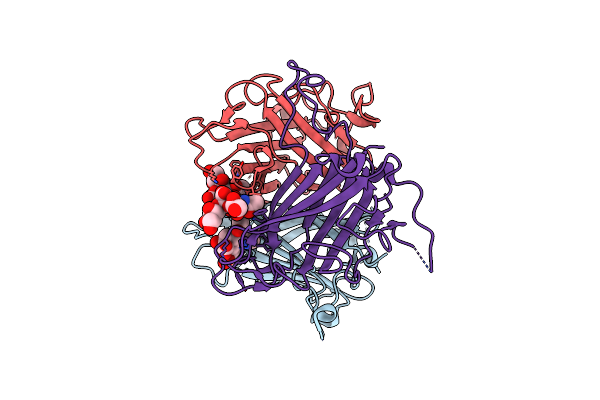
P53-Y220C Core Domain Covalently Bound To 3,5-Dichloro-6-Ethylpyrazine-2-Carbonitirle Soaked At 5 Mm
Organism: Homo sapiens
Method: X-RAY DIFFRACTION Resolution:1.64 Å Release Date: 2025-06-11 Classification: CELL CYCLE Ligands: A1II1, PEG, EDO, EPE, ZN, GOL, PGE |
Organism: Homo sapiens
Method: X-RAY DIFFRACTION
Release Date: 2025-06-11
Ligands: A1II1, PEG, EDO, EPE, ZN, GOL, PGE
 |
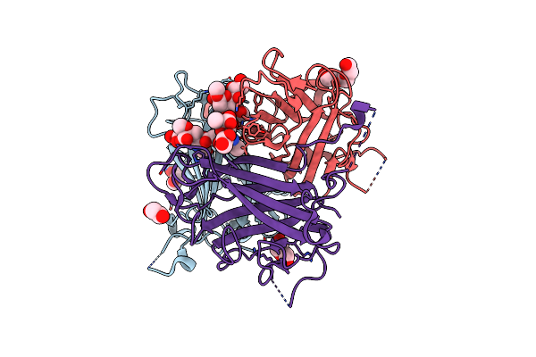
Human Adenovirus Type 36 Fiber Knob In Complex With 4-O-Acetyl-3'-Sialyllactose
Organism: Human adenovirus 36
Method: X-RAY DIFFRACTION Resolution:1.57 Å Release Date: 2025-01-22 Classification: VIRAL PROTEIN Ligands: PKM |
Organism: Human adenovirus 36
Method: X-RAY DIFFRACTION
Release Date: 2025-01-22
Ligands: PKM
 |
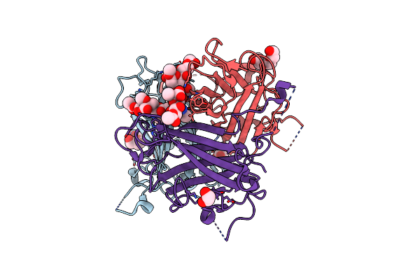
Human Adenovirus Type 36 Fiber Knob In Complex With 4-O,5-N-Diacetylneuraminic Acid
Organism: Human adenovirus 36
Method: X-RAY DIFFRACTION Resolution:1.76 Å Release Date: 2025-01-22 Classification: VIRAL PROTEIN Ligands: ANA, PEG, PGE, EDO |
Organism: Human adenovirus 36
Method: X-RAY DIFFRACTION
Release Date: 2025-01-22
Ligands: ANA, PEG, PGE, EDO
 |
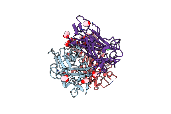
Human Adenovirus Type 36 Fiber Knob In Complex With 4,9-O,5-N-Triacetylneuraminic Acid
Organism: Human adenovirus 36
Method: X-RAY DIFFRACTION Resolution:1.90 Å Release Date: 2025-01-22 Classification: VIRAL PROTEIN Ligands: SIO, PGE, EDO |
Organism: Human adenovirus 36
Method: X-RAY DIFFRACTION
Release Date: 2025-01-22
Ligands: SIO, PGE, EDO
 |
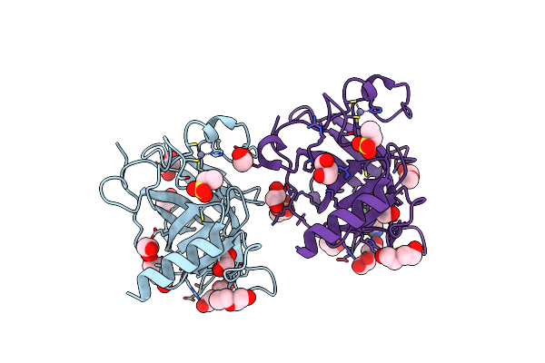
Human Adenovirus Type 37 Fiber Knob In Complex With 4-O,5-N-Diacetylneuraminic Acid
Organism: Human adenovirus d37
Method: X-RAY DIFFRACTION Resolution:1.65 Å Release Date: 2025-01-22 Classification: VIRAL PROTEIN Ligands: ANA, ZN, ACT, CL |
Organism: Human adenovirus d37
Method: X-RAY DIFFRACTION
Release Date: 2025-01-22
Ligands: ANA, ZN, ACT, CL
 |
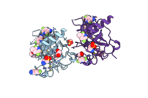
P53-Y220C Core Domain Covalently Bound To 2,5,6-Trifluoropyridine-3-Carbonitrile Soaked At 5 Mm
Organism: Homo sapiens
Method: X-RAY DIFFRACTION Resolution:1.57 Å Release Date: 2024-11-27 Classification: CELL CYCLE Ligands: YLW, PEG, EDO, EPE, ZN, GOL |
Organism: Homo sapiens
Method: X-RAY DIFFRACTION
Release Date: 2024-11-27
Ligands: YLW, PEG, EDO, EPE, ZN, GOL
 |
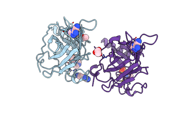
P53-Y220C Core Domain Covalently Bound To 2,5,6-Trifluoropyridine-3-Carbonitrile Soaked At 40 Mm
Organism: Homo sapiens
Method: X-RAY DIFFRACTION Resolution:1.69 Å Release Date: 2024-11-27 Classification: CELL CYCLE Ligands: YLW, GOL, EDO, EPE, ZN |
Organism: Homo sapiens
Method: X-RAY DIFFRACTION
Release Date: 2024-11-27
Ligands: YLW, GOL, EDO, EPE, ZN
 |
P53-Y220C Core Domain Covalently Bound To 3-Amino-5-Chloropyrazine-2,6-Dicarbonitrile Soaked At 5 Mm
Organism: Homo sapiens
Method: X-RAY DIFFRACTION Resolution:2.06 Å Release Date: 2024-11-27 Classification: CELL CYCLE Ligands: YM0, EPE, PEG, ZN |
Organism: Homo sapiens
Method: X-RAY DIFFRACTION
Release Date: 2024-11-27
Ligands: YM0, EPE, PEG, ZN
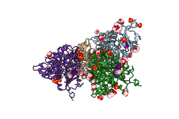 |
Dyrk1A In Complex With 2-Cyclopentyl-7-Iodo-1H-Indole-3-Carbonitrile
Organism: Homo sapiens
Method: X-RAY DIFFRACTION Resolution:2.22 Å Release Date: 2024-09-25 Classification: TRANSFERASE Ligands: YOX, 1PE, PG4, EPE, PEG, GOL, EDO, SO4, PO4 |
Organism: Homo sapiens
Method: X-RAY DIFFRACTION
Release Date: 2024-09-25
Ligands: YOX, 1PE, PG4, EPE, PEG, GOL, EDO, SO4, PO4
 |
Crystal Structure Of Cytochrome P450 Gymb5 From Streptomyces Katrae
Organism: Streptomyces katrae
Method: X-RAY DIFFRACTION Resolution:1.91 Å Release Date: 2024-06-26 Classification: OXIDOREDUCTASE Ligands: HEM |
Organism: Streptomyces katrae
Method: X-RAY DIFFRACTION
Release Date: 2024-06-26
Ligands: HEM
 |
Crystal Structure Of Cytochrome P450 Gymb5 From Streptomyces Katrae In Complex With Cyy And Hypoxanthine
Organism: Streptomyces katrae
Method: X-RAY DIFFRACTION Resolution:1.81 Å Release Date: 2024-06-26 Classification: OXIDOREDUCTASE Ligands: HEM, YTT, HPA, EDO |
Organism: Streptomyces katrae
Method: X-RAY DIFFRACTION
Release Date: 2024-06-26
Ligands: HEM, YTT, HPA, EDO
 |
Crystal Structure Of Cytochrome P450 Gymb1 From Streptomyces Flavidovirens
Organism: Streptomyces flavidovirens dsm 40150
Method: X-RAY DIFFRACTION Resolution:1.96 Å Release Date: 2024-06-26 Classification: OXIDOREDUCTASE Ligands: HEM, GOL |
Organism: Streptomyces flavidovirens dsm 40150
Method: X-RAY DIFFRACTION
Release Date: 2024-06-26
Ligands: HEM, GOL
 |
Tarm(Se)_G117R-Udp-Glucose
Organism: Staphylococcus epidermidis
Method: X-RAY DIFFRACTION Resolution:2.03 Å Release Date: 2023-12-06 Classification: TRANSFERASE Ligands: CL, PO4, UPG, EDO, PG4, PGE |
Organism: Staphylococcus epidermidis
Method: X-RAY DIFFRACTION
Release Date: 2023-12-06
Ligands: CL, PO4, UPG, EDO, PG4, PGE