

Search Count: 33
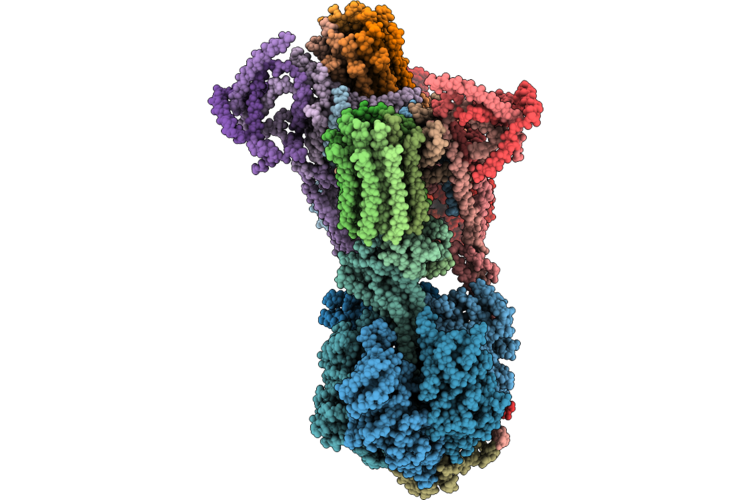 |
Cryo-Em Structure Of The If1 Bound Bovine F-Atp Synthase Planar Dimer
Organism: Bos taurus
Method: ELECTRON MICROSCOPY Release Date: 2026-07-01 Classification: MEMBRANE PROTEIN |
Organism: Bos taurus
Method: ELECTRON MICROSCOPY
Release Date: 2026-07-01
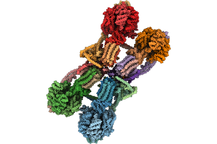 |
Cryo-Em Structure Of The If1 Bound Bovine Atp Synthase Tetramer
Organism: Bos taurus
Method: ELECTRON MICROSCOPY Release Date: 2026-07-01 Classification: MEMBRANE PROTEIN |
Organism: Bos taurus
Method: ELECTRON MICROSCOPY
Release Date: 2026-07-01
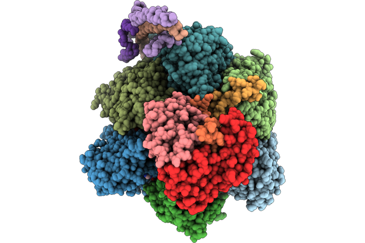 |
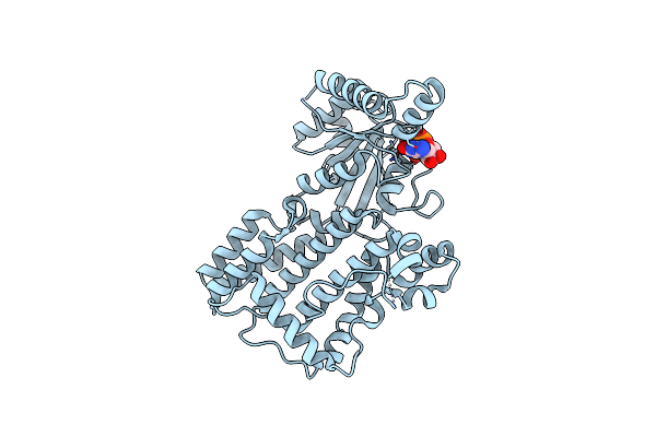
Cryo-Em Structure Of The If1 Bound Bovine Atp Synthase Monomer: Rotary State 1, F1 Focused Map
Organism: Bos taurus
Method: ELECTRON MICROSCOPY Release Date: 2026-07-01 Classification: MEMBRANE PROTEIN Ligands: ATP, MG, ADP |
Organism: Bos taurus
Method: ELECTRON MICROSCOPY
Release Date: 2026-07-01
Ligands: ATP, MG, ADP
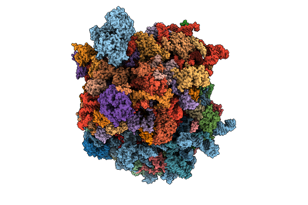 |
Human 80S Ribosome In Complex With Dhx29
Organism: Homo sapiens
Method: ELECTRON MICROSCOPY Release Date: 2026-04-01 Classification: TRANSLATION Ligands: MG, ZN |
Organism: Homo sapiens
Method: ELECTRON MICROSCOPY
Release Date: 2026-04-01
Ligands: MG, ZN
 |
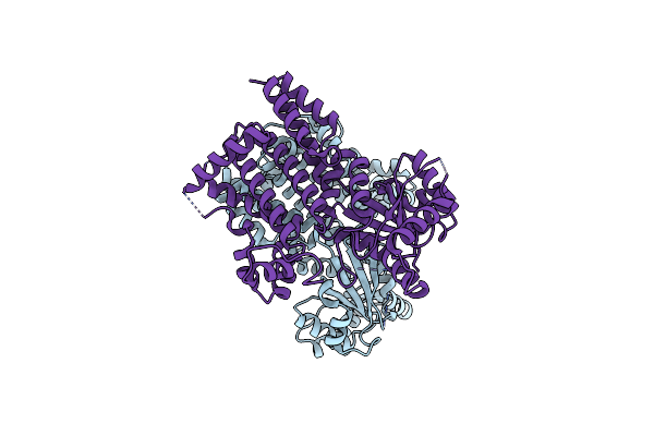
Crystal Structure Of Gtp-Bound Irgb6_T95D Mutant
Organism: Mus musculus
Method: X-RAY DIFFRACTION Resolution:1.68 Å Release Date: 2023-10-18 Classification: IMMUNE SYSTEM Ligands: GTP |
Organism: Mus musculus
Method: X-RAY DIFFRACTION
Release Date: 2023-10-18
Ligands: GTP
 |
Crystal Structure Of Nucleotide-Free Irgb6_T95D Mutant
Organism: Mus musculus
Method: X-RAY DIFFRACTION Resolution:2.05 Å Release Date: 2023-10-18 Classification: IMMUNE SYSTEM |
Organism: Mus musculus
Method: X-RAY DIFFRACTION
Release Date: 2023-10-18
 |
Cryo-Em Structure Of Groel Bound To Unfolded Substrate (Ugt1A) At 2.8 Ang. Resolution (Consensus Refinement)
Organism: Escherichia coli
Method: ELECTRON MICROSCOPY Release Date: 2023-05-03 Classification: CHAPERONE |
Organism: Escherichia coli
Method: ELECTRON MICROSCOPY
Release Date: 2023-05-03
 |
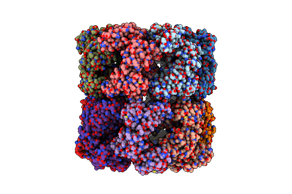
Cryo-Em Structure Of Double Occupied Ring (Dor) Of Groel-Ugt1A Complex At 2.7 Ang. Resolution
Organism: Escherichia coli
Method: ELECTRON MICROSCOPY Release Date: 2023-05-03 Classification: CHAPERONE |
Organism: Escherichia coli
Method: ELECTRON MICROSCOPY
Release Date: 2023-05-03
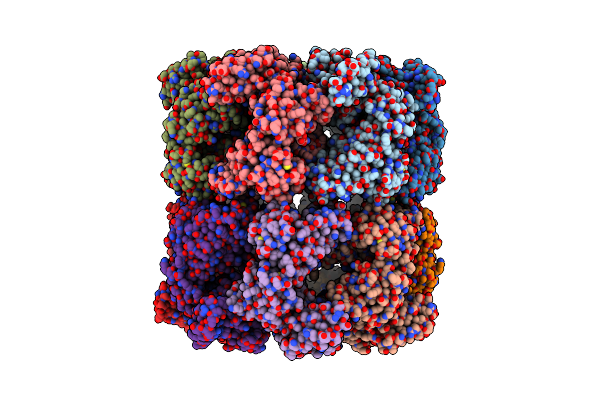 |
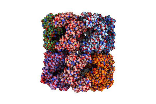
Cryo-Em Structure Of Single Empty Ring 2 (Ser2) Of Groel-Ugt1A Complex At 3.2 Ang. Resolution
Organism: Escherichia coli
Method: ELECTRON MICROSCOPY Release Date: 2023-05-03 Classification: CHAPERONE |
Organism: Escherichia coli
Method: ELECTRON MICROSCOPY
Release Date: 2023-05-03
 |
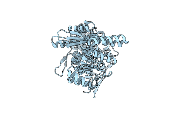
Cryo-Em Structure Of Occupied Ring Subunit 4 (Or4) Of Groel Complexed With Polyalanine Model Of Ugt1A From Groel-Ugt1A Double Occupied Ring Complex
Organism: Escherichia coli, Homo sapiens
Method: ELECTRON MICROSCOPY Release Date: 2023-05-03 Classification: CHAPERONE |
Organism: Escherichia coli, Homo sapiens
Method: ELECTRON MICROSCOPY
Release Date: 2023-05-03
 |
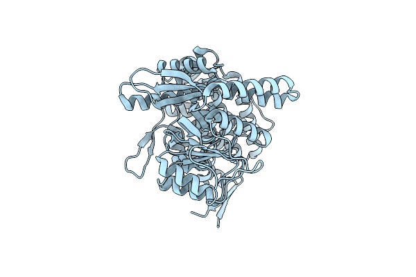
Cryo-Em Structure Of Empty Ring Subunit 1 (Er1) From Single Empty Ring Of Groel-Ugt1A Complex
Organism: Escherichia coli
Method: ELECTRON MICROSCOPY Release Date: 2023-05-03 Classification: CHAPERONE |
Organism: Escherichia coli
Method: ELECTRON MICROSCOPY
Release Date: 2023-05-03
 |
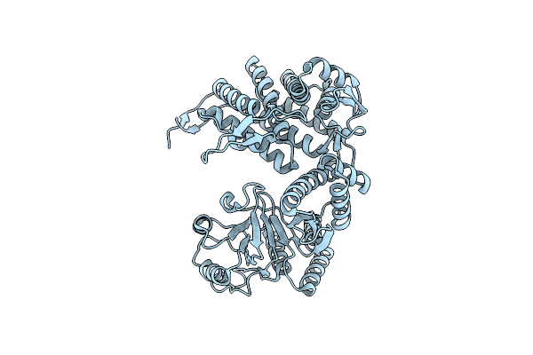
Cryo-Em Structure Of Empty Ring Subunit 2 (Er2) From Groel-Ugt1A Single Empty Ring Complex
Organism: Escherichia coli
Method: ELECTRON MICROSCOPY Release Date: 2023-05-03 Classification: CHAPERONE |
Organism: Escherichia coli
Method: ELECTRON MICROSCOPY
Release Date: 2023-05-03
 |
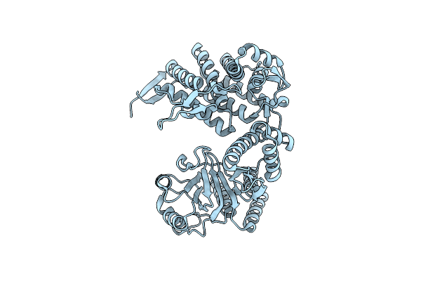
Cryo-Em Structure Of Occupied Ring Subunit 1 (Or1) Of Groel From Groel-Ugt1A Double Occupied Ring Complex
Organism: Escherichia coli
Method: ELECTRON MICROSCOPY Release Date: 2023-05-03 Classification: CHAPERONE |
Organism: Escherichia coli
Method: ELECTRON MICROSCOPY
Release Date: 2023-05-03
 |
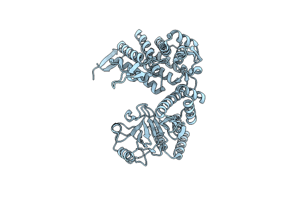
Cryo-Em Structure Of Occupied Ring Subunit 2 (Or2) Of Groel From Groel-Ugt1A Double Occupied Ring Complex
Organism: Escherichia coli
Method: ELECTRON MICROSCOPY Release Date: 2023-05-03 Classification: CHAPERONE |
Organism: Escherichia coli
Method: ELECTRON MICROSCOPY
Release Date: 2023-05-03
 |
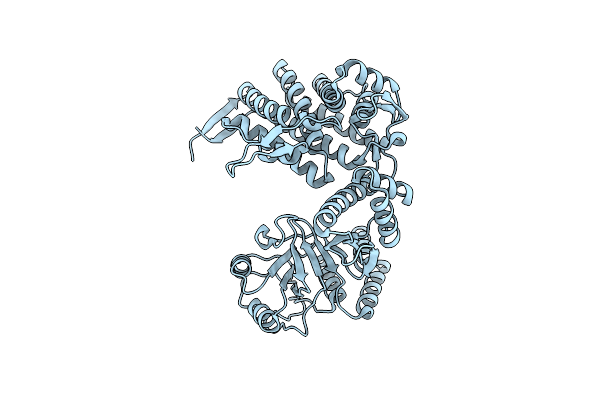
Cryo-Em Structure Of Occupied Ring Subunit 3 (Or3) Of Groel From Groel-Ugt1A Double Occupied Ring Complex
Organism: Escherichia coli
Method: ELECTRON MICROSCOPY Release Date: 2023-05-03 Classification: CHAPERONE |
Organism: Escherichia coli
Method: ELECTRON MICROSCOPY
Release Date: 2023-05-03
 |
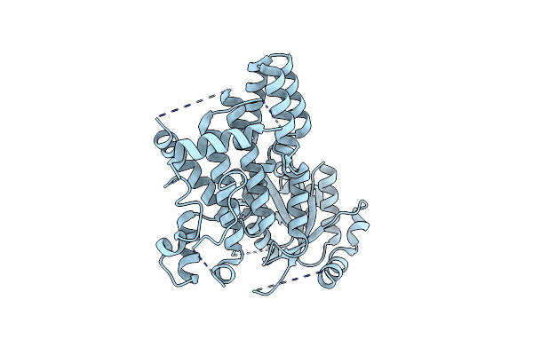
Cryo-Em Structure Of Occupied Ring Subunit 4 (Or4) Of Groel From Groel-Ugt1A Double Occupied Ring Complex
Organism: Escherichia coli
Method: ELECTRON MICROSCOPY Release Date: 2023-05-03 Classification: CHAPERONE |
Organism: Escherichia coli
Method: ELECTRON MICROSCOPY
Release Date: 2023-05-03
 |
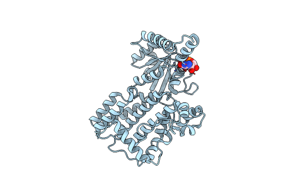
Crystal Structure Of Nucleotide-Free Irgb6
Organism: Mus musculus
Method: X-RAY DIFFRACTION Resolution:2.00 Å Release Date: 2021-11-03 Classification: HYDROLASE |
Organism: Mus musculus
Method: X-RAY DIFFRACTION
Release Date: 2021-11-03
 |
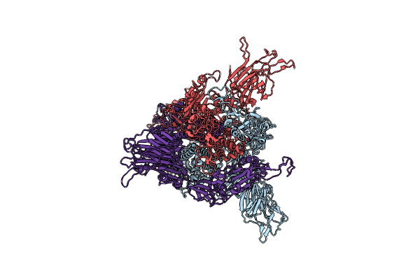
Crystal Structure Of Gtp-Bound Irgb6
Organism: Mus musculus
Method: X-RAY DIFFRACTION Resolution:1.51 Å Release Date: 2021-11-03 Classification: HYDROLASE Ligands: GTP |
Organism: Mus musculus
Method: X-RAY DIFFRACTION
Release Date: 2021-11-03
Ligands: GTP
 |
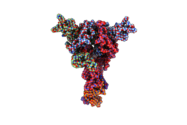
Apo Spike Protein From Sars-Cov2
Organism: Severe acute respiratory syndrome coronavirus 2
Method: ELECTRON MICROSCOPY Release Date: 2021-06-02 Classification: VIRAL PROTEIN |
Organism: Severe acute respiratory syndrome coronavirus 2
Method: ELECTRON MICROSCOPY
Release Date: 2021-06-02
 |
Spike Protein From Sars-Cov2 With Fab Fragment Of Enhancing Antibody 8D2
Organism: Severe acute respiratory syndrome coronavirus 2, Homo sapiens
Method: ELECTRON MICROSCOPY Release Date: 2021-06-02 Classification: VIRAL PROTEIN |
Organism: Severe acute respiratory syndrome coronavirus 2, Homo sapiens
Method: ELECTRON MICROSCOPY
Release Date: 2021-06-02