

Search Count: 2,913
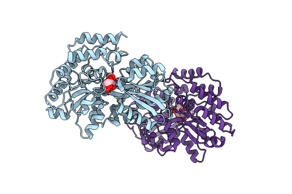 |
Crystal Structure Of Beta-L-Arabinobiose Binding Protein - Native
Organism: Bifidobacterium longum
Method: X-RAY DIFFRACTION Resolution:1.92 Å Release Date: 2020-04-22 Classification: SUGAR BINDING PROTEIN |
Organism: Bifidobacterium longum
Method: X-RAY DIFFRACTION
Release Date: 2020-04-22
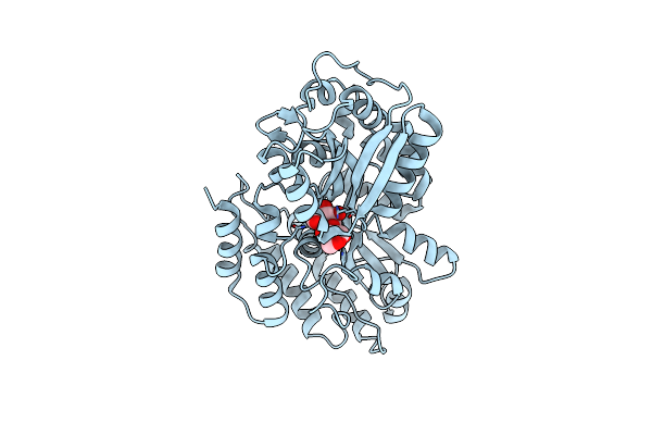 |
Crystal Structure Of Beta-L-Arabinobiose Binding Protein - Selenomethionine Derivative
Organism: Bifidobacterium longum
Method: X-RAY DIFFRACTION Resolution:1.78 Å Release Date: 2020-04-22 Classification: SUGAR BINDING PROTEIN Modified Residue(s): MSE |
Organism: Bifidobacterium longum
Method: X-RAY DIFFRACTION
Release Date: 2020-04-22
 |
Crystal Structure Of Putative Sugar Binding Protein (Yp_001299726.1) From Bacteroides Vulgatus Atcc 8482 At 1.90 A Resolution
Organism: Bacteroides vulgatus atcc 8482
Method: X-RAY DIFFRACTION Resolution:1.90 Å Release Date: 2009-11-10 Classification: SUGAR BINDING PROTEIN Ligands: CA, PO4, EDO Modified Residue(s): MSE |
Organism: Bacteroides vulgatus atcc 8482
Method: X-RAY DIFFRACTION
Release Date: 2009-11-10
Ligands: CA, PO4, EDO
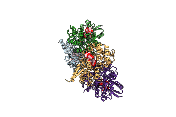 |
Sbp Rafe In Complex With Verbascose
Organism: Streptococcus pneumoniae tigr4
Method: X-RAY DIFFRACTION Resolution:2.40 Å Release Date: 2019-10-02 Classification: SUGAR BINDING PROTEIN Ligands: EDO |
Organism: Streptococcus pneumoniae tigr4
Method: X-RAY DIFFRACTION
Release Date: 2019-10-02
Ligands: EDO
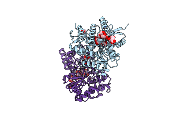 |
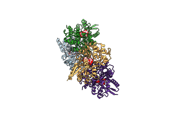
Sbp Rafe In Complex With Stachyose
Organism: Streptococcus pneumoniae tigr4
Method: X-RAY DIFFRACTION Resolution:2.35 Å Release Date: 2019-10-02 Classification: SUGAR BINDING PROTEIN Ligands: CIT, EDO |
Organism: Streptococcus pneumoniae tigr4
Method: X-RAY DIFFRACTION
Release Date: 2019-10-02
Ligands: CIT, EDO
 |
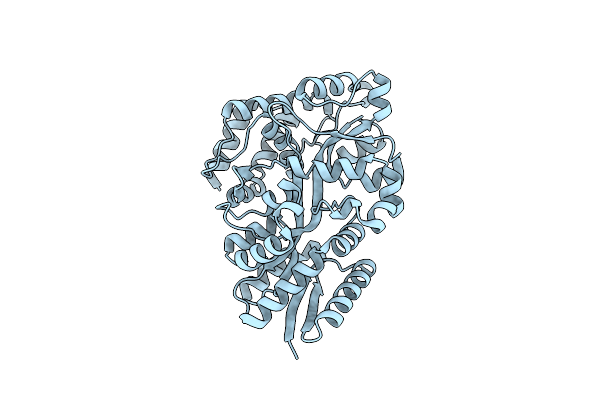
Sbp Rafe In Complex With Raffinose
Organism: Streptococcus pneumoniae tigr4
Method: X-RAY DIFFRACTION Resolution:2.65 Å Release Date: 2019-10-02 Classification: SUGAR BINDING PROTEIN |
Organism: Streptococcus pneumoniae tigr4
Method: X-RAY DIFFRACTION
Release Date: 2019-10-02
 |
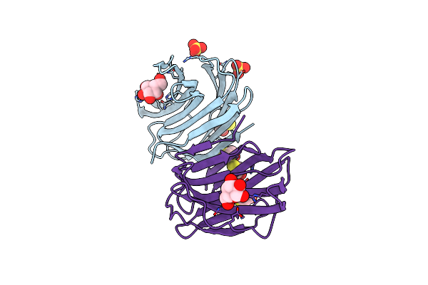
Lmo2839 Abc Transporter Substrate Binding Protein
Organism: Listeria monocytogenes
Method: X-RAY DIFFRACTION Resolution:2.00 Å Release Date: 2025-05-14 Classification: SUGAR BINDING PROTEIN |
Organism: Listeria monocytogenes
Method: X-RAY DIFFRACTION
Release Date: 2025-05-14
 |
X-Ray Crystal Structure Of C2S Human Galectin-1 Complexed With Galactose
Organism: Homo sapiens
Method: X-RAY DIFFRACTION Resolution:2.30 Å Release Date: 2004-10-20 Classification: SUGAR BINDING PROTEIN Ligands: BME, SO4, GAL Modified Residue(s): CSO |
Organism: Homo sapiens
Method: X-RAY DIFFRACTION
Release Date: 2004-10-20
Ligands: BME, SO4, GAL
 |
Zinc-Free Engineered Maltose Binding Protein
Organism: Escherichia coli
Method: X-RAY DIFFRACTION Resolution:2.20 Å Release Date: 2005-12-20 Classification: SUGAR BINDING, METAL BINDING PROTEIN |
Organism: Escherichia coli
Method: X-RAY DIFFRACTION
Release Date: 2005-12-20
 |
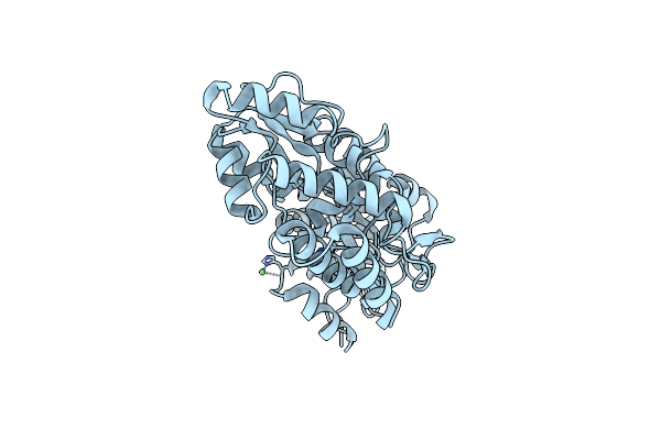
Crystal Structure Of Zinc-Bound Engineered Maltose Binding Protein
Organism: Escherichia coli
Method: X-RAY DIFFRACTION Resolution:2.00 Å Release Date: 2005-12-20 Classification: SUGAR BINDING, METAL BINDING PROTEIN Ligands: ZN |
Organism: Escherichia coli
Method: X-RAY DIFFRACTION
Release Date: 2005-12-20
Ligands: ZN
 |
Crystal Structure Of Nickel-Bound Engineered Maltose Binding Protein
Organism: Escherichia coli
Method: X-RAY DIFFRACTION Resolution:2.00 Å Release Date: 2005-12-20 Classification: SUGAR BINDING, METAL BINDING PROTEIN Ligands: NI |
Organism: Escherichia coli
Method: X-RAY DIFFRACTION
Release Date: 2005-12-20
Ligands: NI
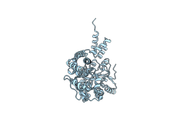 |
Maltose Binding-A1 Homeodomain Protein Chimera, Crystal Form I
Organism: Escherichia coli, Saccharomyces cerevisiae
Method: X-RAY DIFFRACTION Resolution:2.10 Å Release Date: 2002-09-18 Classification: SUGAR BINDING, DNA BINDING PROTEIN |
Organism: Escherichia coli, Saccharomyces cerevisiae
Method: X-RAY DIFFRACTION
Release Date: 2002-09-18
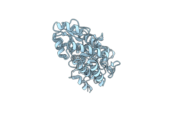 |
Crystal Structure Of Copper-Bound Engineered Maltose Binding Protein
Organism: Escherichia coli
Method: X-RAY DIFFRACTION Resolution:2.50 Å Release Date: 2005-12-20 Classification: SUGAR BINDING, METAL BINDING PROTEIN Ligands: CU |
Organism: Escherichia coli
Method: X-RAY DIFFRACTION
Release Date: 2005-12-20
Ligands: CU
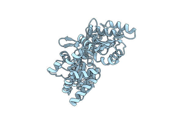 |
Maltose Binding-A1 Homeodomain Protein Chimera, Crystal Form Ii
Organism: Escherichia coli, Saccharomyces cerevisiae
Method: X-RAY DIFFRACTION Resolution:2.30 Å Release Date: 2002-09-18 Classification: SUGAR BINDING, DNA BINDING PROTEIN |
Organism: Escherichia coli, Saccharomyces cerevisiae
Method: X-RAY DIFFRACTION
Release Date: 2002-09-18
 |
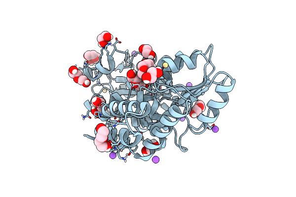
Crystal Structure Of A Novel Alginate-Binding Carbohydrate Binding Module
Organism: Vibrio breoganii
Method: X-RAY DIFFRACTION Resolution:1.50 Å Release Date: 2024-06-12 Classification: SUGAR BINDING PROTEIN |
Organism: Vibrio breoganii
Method: X-RAY DIFFRACTION
Release Date: 2024-06-12
 |
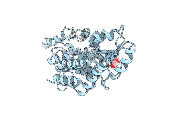
Crystal Structure Of Maltose Binding Protein (Apo)
Organism: Escherichia coli
Method: X-RAY DIFFRACTION Resolution:1.58 Å Release Date: 2024-12-18 Classification: SUGAR BINDING PROTEIN Ligands: PEG, EDO, PGE, NA, CD |
Organism: Escherichia coli
Method: X-RAY DIFFRACTION
Release Date: 2024-12-18
Ligands: PEG, EDO, PGE, NA, CD
 |
Crystal Structure Of The E. Coli Maltodextrin-Binding Protein
Organism: Escherichia coli bl21
Method: X-RAY DIFFRACTION Resolution:1.15 Å Release Date: 2023-02-22 Classification: SUGAR BINDING PROTEIN Ligands: PEG |
Organism: Escherichia coli bl21
Method: X-RAY DIFFRACTION
Release Date: 2023-02-22
Ligands: PEG
 |
Crystal Structure Of The Periplasmic Sugar Binding Protein Chve
Organism: Agrobacterium tumefaciens
Method: X-RAY DIFFRACTION Resolution:1.80 Å Release Date: 2012-11-28 Classification: SUGAR BINDING PROTEIN Ligands: GAL |
Organism: Agrobacterium tumefaciens
Method: X-RAY DIFFRACTION
Release Date: 2012-11-28
Ligands: GAL
 |
Crystal Structure Of The Periplasmic Sugar Binding Protein Chve
Organism: Agrobacterium tumefaciens
Method: X-RAY DIFFRACTION Resolution:1.75 Å Release Date: 2012-11-28 Classification: SUGAR BINDING PROTEIN Ligands: BDP |
Organism: Agrobacterium tumefaciens
Method: X-RAY DIFFRACTION
Release Date: 2012-11-28
Ligands: BDP
 |
The Crystal Structure Of A Solute Binding Protein From Bacillus Anthracis Str. Ames In Complex With Quorum-Sensing Signal Autoinducer-2 (Ai-2)
Organism: Bacillus anthracis
Method: X-RAY DIFFRACTION Resolution:1.25 Å Release Date: 2014-04-09 Classification: SUGAR BINDING PROTEIN Ligands: PAV, EDO, CL Modified Residue(s): MSE |
Organism: Bacillus anthracis
Method: X-RAY DIFFRACTION
Release Date: 2014-04-09
Ligands: PAV, EDO, CL