

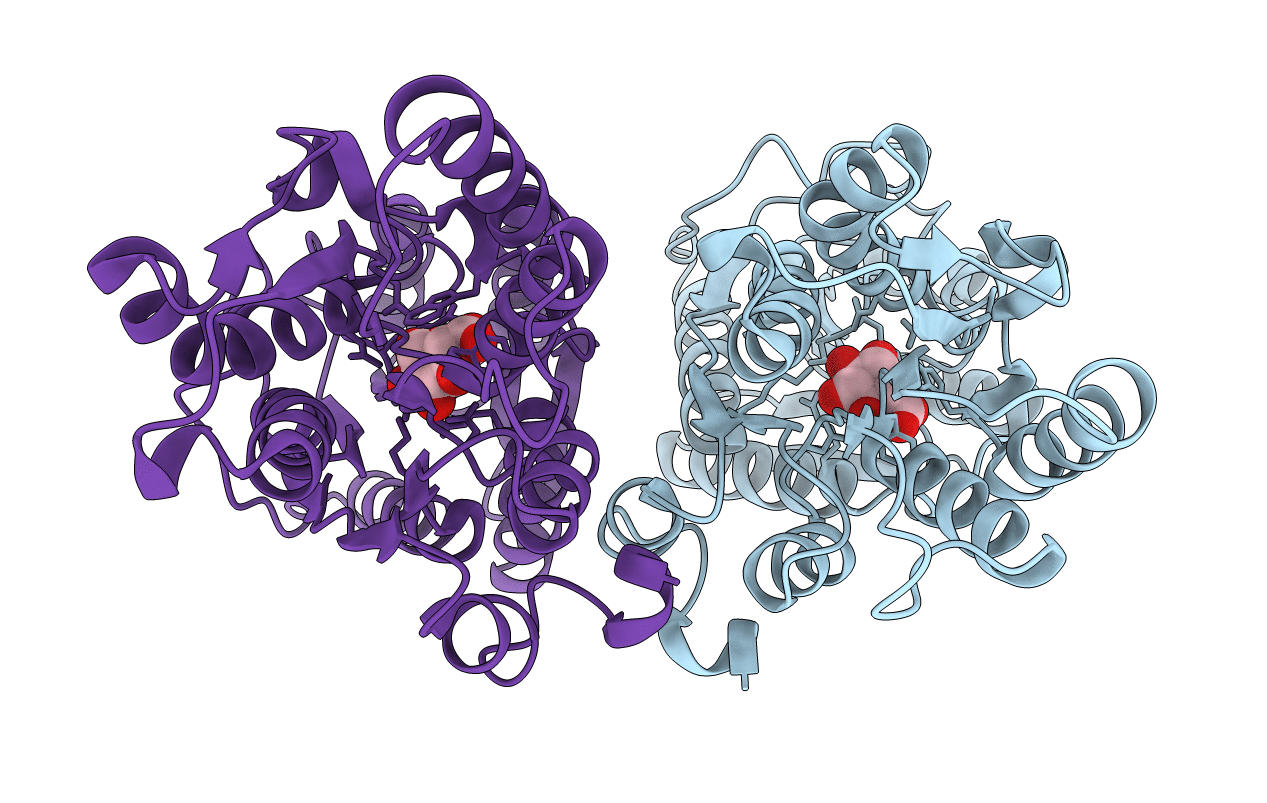
3UUG image
Deposition Date 2011-11-28
Release Date 2012-11-28
Last Version Date 2024-02-28
Entry Detail
PDB ID:
3UUG
Title:
Crystal structure of the periplasmic sugar binding protein ChvE
Biological Source:
Source Organism(s):
Expression System(s):
Method Details:
Experimental Method:
Resolution:
1.75 Å
R-Value Free:
0.19
R-Value Work:
0.15
R-Value Observed:
0.15
Space Group:
P 65
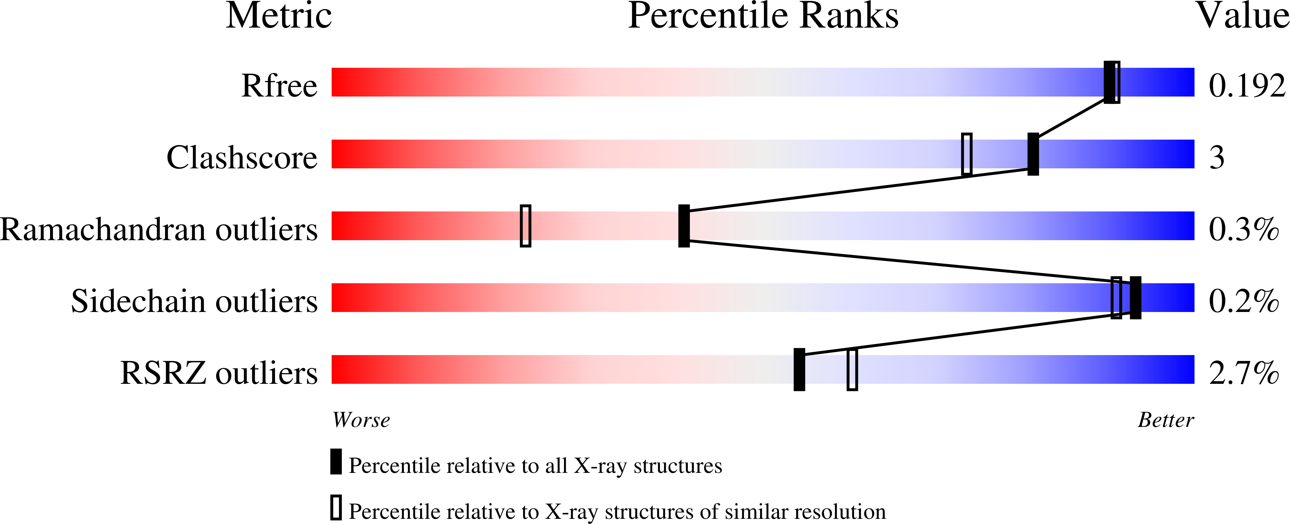
Macromolecular Entities
Polymer Type:polypeptide(L)
Molecule:Multiple sugar-binding peripl
Gene (Uniprot):chvE
Chain IDs:A, B
Chain Length:330
Number of Molecules:2
Biological Source:Agrobacterium tumefaciens
Ligand Molecules
Primary Citation
Agrobacterium tumefaciens recognizes its host environment using ChvE to bind diverse plant sugars as virulence signals.
Proc. Natl. Acad. Sci. U.S.A. 110 678 683 (2013)
PMID: 23267119 DOI: 10.1073/pnas.1215033110

Abstact

Agrobacterium tumefaciens is a broad host range plant pathogen that combinatorially recognizes diverse host molecules including phenolics, low pH, and aldose monosaccharides to activate its pathogenic pathways. Chromosomal virulence gene E (chvE) encodes a periplasmic-binding protein that binds several neutral sugars and sugar acids, and subsequently interacts with the VirA/VirG regulatory system to stimulate virulence (vir) gene expression. Here, a combination of genetics, X-ray crystallography, and isothermal calorimetry reveals how ChvE binds the different monosaccharides and also shows that binding of sugar acids is pH dependent. Moreover, the potency of a sugar for vir gene expression is modulated by a transport system that also relies on ChvE. These two circuits tune the overall system to respond to sugar concentrations encountered in vivo. Finally, using chvE mutants with restricted sugar specificities, we show that there is host variation in regard to the types of sugars that are limiting for vir induction.

Legend

Protein

Chemical

Disease

Primary Citation of related structures
Feedback Form
Name
Email
Institute
Feedback