

Search Count: 28,141
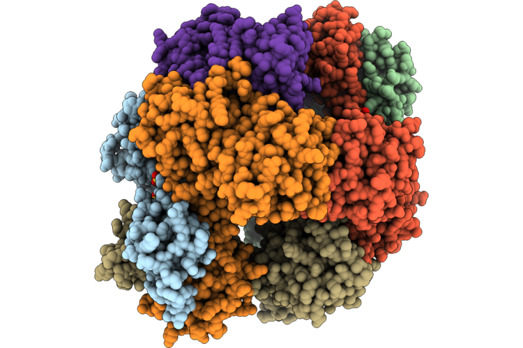 |
Crystal Structure Of Human Nif3L1 Protein With Hexamer In Asu
Organism: Homo sapiens
Method: X-RAY DIFFRACTION Resolution:3.00 Å Release Date: 2026-09-23 Classification: METAL BINDING PROTEIN Ligands: ZN, SO4 |
Organism: Homo sapiens
Method: X-RAY DIFFRACTION
Release Date: 2026-09-23
Ligands: ZN, SO4
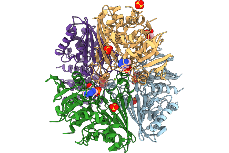 |
Crystal Structure Of The Complex Of Erythrose-4-Phosphate Dehydrogenase From Acinetobacter Baumannii With Adenosine Monophosphate At 2.40 Angstrom Resolution.
Organism: Acinetobacter baumannii
Method: X-RAY DIFFRACTION Resolution:2.40 Å Release Date: 2026-09-23 Classification: OXIDOREDUCTASE Ligands: AMP, SO4, MG, GOL |
Organism: Acinetobacter baumannii
Method: X-RAY DIFFRACTION
Release Date: 2026-09-23
Ligands: AMP, SO4, MG, GOL
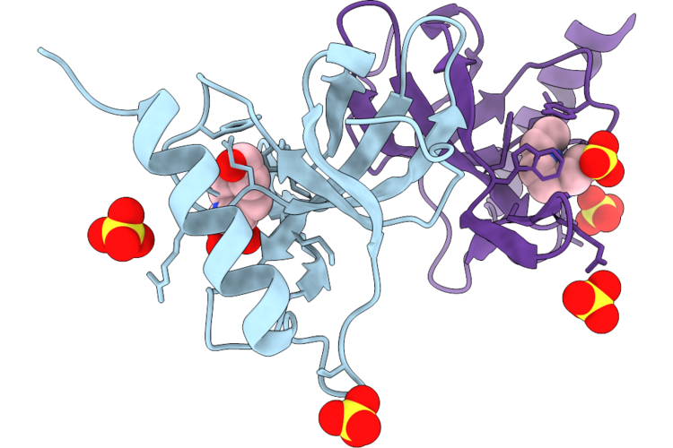 |
Crystal Structure Of Human Nipbl C-Terminal Domain
Organism: Homo sapiens
Method: X-RAY DIFFRACTION Resolution:2.10 Å Release Date: 2026-09-23 Classification: GENE REGULATION Ligands: EPE, SO4 |
Organism: Homo sapiens
Method: X-RAY DIFFRACTION
Release Date: 2026-09-23
Ligands: EPE, SO4
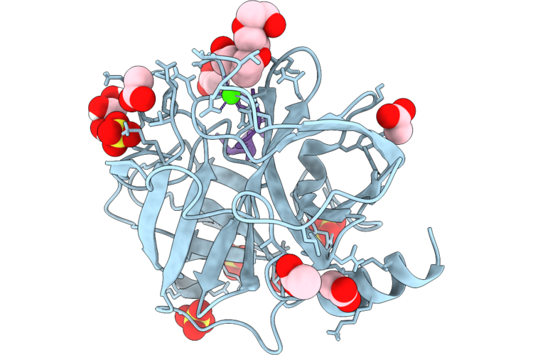 |
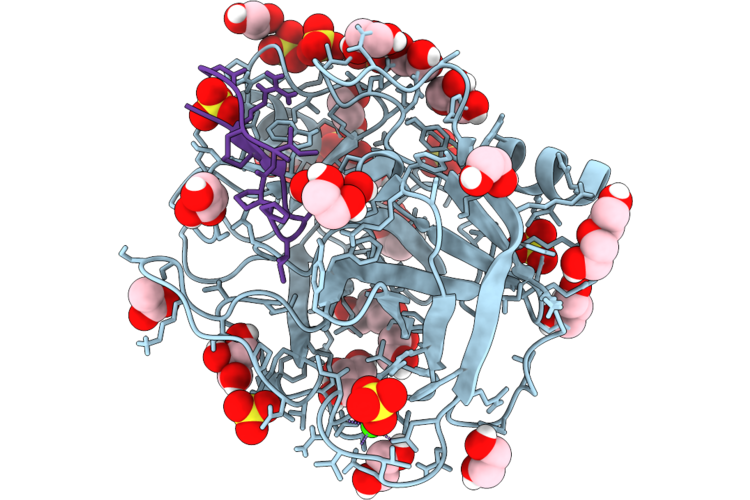
Sfti-1 Containing L-2-Thiaproline At Residue 8 In Complex With Trypsin
Organism: Helianthus annuus, Bos taurus
Method: X-RAY DIFFRACTION Resolution:1.47 Å Release Date: 2026-09-23 Classification: HYDROLASE/HYDROLASE INHIBITOR Ligands: ACT, CA, SO4, PG4, GOL |
Organism: Helianthus annuus, Bos taurus
Method: X-RAY DIFFRACTION
Release Date: 2026-09-23
Ligands: ACT, CA, SO4, PG4, GOL
 |
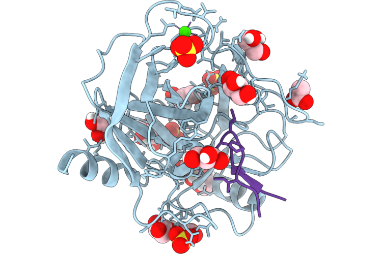
Sfti-1 Containing L-2-Thiaproline At Residue 9 In Complex With Trypsin
Organism: Helianthus annuus, Bos taurus
Method: X-RAY DIFFRACTION Resolution:1.22 Å Release Date: 2026-09-23 Classification: HYDROLASE/HYDROLASE INHIBITOR Ligands: CA, GOL, ACT, EDO, SO4, PEG |
Organism: Helianthus annuus, Bos taurus
Method: X-RAY DIFFRACTION
Release Date: 2026-09-23
Ligands: CA, GOL, ACT, EDO, SO4, PEG
 |
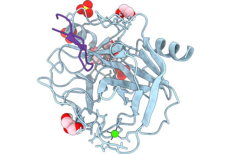
Sfti-1 Containing L-2-Thiaproline At Residue 13 In Complex With Trypsin
Organism: Helianthus annuus, Bos taurus
Method: X-RAY DIFFRACTION Resolution:1.13 Å Release Date: 2026-09-23 Classification: HYDROLASE/HYDROLASE INHIBITOR Ligands: SO4, CA, GOL, EDO, ACT |
Organism: Helianthus annuus, Bos taurus
Method: X-RAY DIFFRACTION
Release Date: 2026-09-23
Ligands: SO4, CA, GOL, EDO, ACT
 |
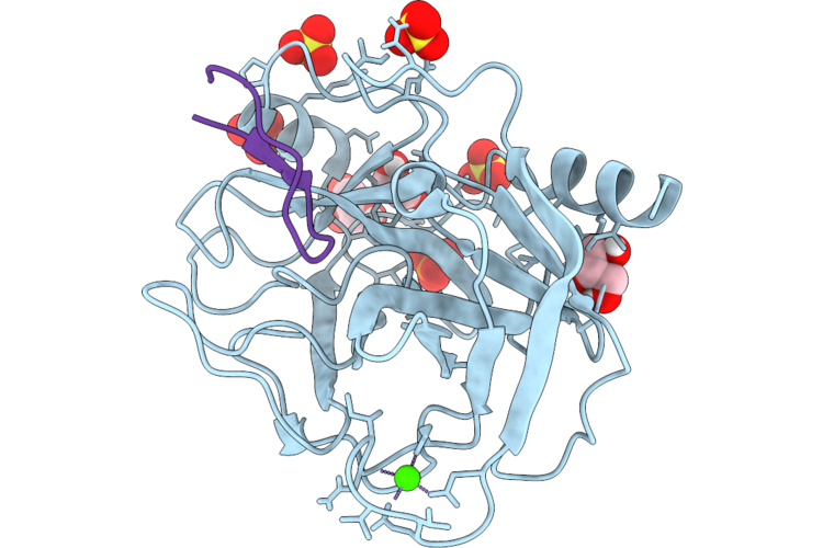
Sfti-1 Containing L-2-Selenaproline At Residue 8 In Complex With Trypsin
Organism: Helianthus annuus, Bos taurus
Method: X-RAY DIFFRACTION Resolution:1.17 Å Release Date: 2026-09-23 Classification: HYDROLASE/HYDROLASE INHIBITOR Ligands: CA, EDO, GOL, SO4 |
Organism: Helianthus annuus, Bos taurus
Method: X-RAY DIFFRACTION
Release Date: 2026-09-23
Ligands: CA, EDO, GOL, SO4
 |
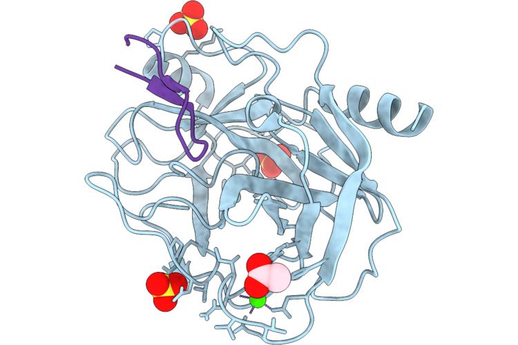
Sfti-1 Containing L-2-Selenaproline At Residue 9 In Complex With Trypsin
Organism: Helianthus annuus, Bos taurus
Method: X-RAY DIFFRACTION Resolution:1.45 Å Release Date: 2026-09-23 Classification: HYDROLASE/HYDROLASE INHIBITOR Ligands: CA, SO4, PO4, GOL, EDO |
Organism: Helianthus annuus, Bos taurus
Method: X-RAY DIFFRACTION
Release Date: 2026-09-23
Ligands: CA, SO4, PO4, GOL, EDO
 |
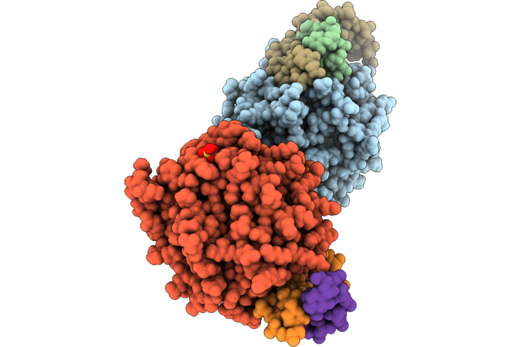
Sfti-1 Containing L-2-Selenaproline At Residue 13 In Complex With Trypsin
Organism: Helianthus annuus, Bos taurus
Method: X-RAY DIFFRACTION Resolution:1.90 Å Release Date: 2026-09-23 Classification: HYDROLASE/HYDROLASE INHIBITOR Ligands: CA, SO4, ACT |
Organism: Helianthus annuus, Bos taurus
Method: X-RAY DIFFRACTION
Release Date: 2026-09-23
Ligands: CA, SO4, ACT
 |
Human Ape1 D210N With Mg And Substrate Tetrahydrofuran-Containing Dna
Organism: Homo sapiens, Synthetic construct
Method: X-RAY DIFFRACTION Resolution:2.05 Å Release Date: 2026-09-23 Classification: DNA BINDING PROTEIN/DNA Ligands: MG, SO4 |
Organism: Homo sapiens, Synthetic construct
Method: X-RAY DIFFRACTION
Release Date: 2026-09-23
Ligands: MG, SO4
 |
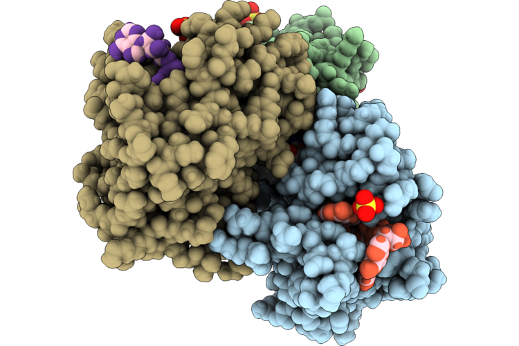
Crystal Structure Of The N4Bp2 Polynucleotide Kinase (Pnk) Domain In Complex With Adp And Mg.
Organism: Homo sapiens
Method: X-RAY DIFFRACTION Resolution:1.95 Å Release Date: 2026-09-23 Classification: TRANSFERASE Ligands: MG, ADP, SO4, EDO, PO4, IPA |
Organism: Homo sapiens
Method: X-RAY DIFFRACTION
Release Date: 2026-09-23
Ligands: MG, ADP, SO4, EDO, PO4, IPA
 |
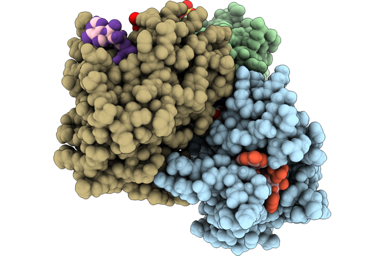
Crystal Structure Of The N4Bp2 Polynucleotide Kinase Domain Complexed With Atp Or Adp, Mg, And The Trinucleotide Substrate Aaa Or Its 5'-Phosphorylated Product Paaa.
Organism: Homo sapiens, Synthetic construct
Method: X-RAY DIFFRACTION Resolution:2.30 Å Release Date: 2026-09-23 Classification: TRANSFERASE Ligands: MG, ATP, SO4, ADP |
Organism: Homo sapiens, Synthetic construct
Method: X-RAY DIFFRACTION
Release Date: 2026-09-23
Ligands: MG, ATP, SO4, ADP
 |
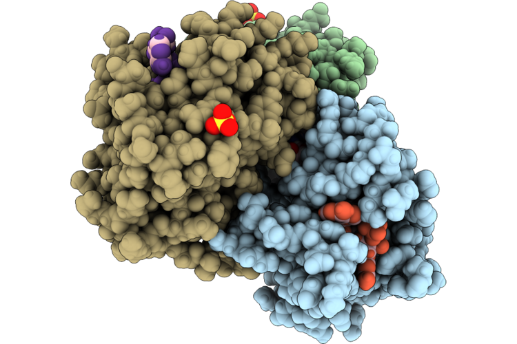
Crystal Structure Of The N4Bp2 Polynucleotide Kinase Domain Complexed With Atp Or Adp, Mg, And The Trinucleotide Substrate Aca Or Its 5'-Phosphorylated Product Paca.
Organism: Homo sapiens, Synthetic construct
Method: X-RAY DIFFRACTION Resolution:2.00 Å Release Date: 2026-09-23 Classification: TRANSFERASE Ligands: MG, ATP, SO4, ADP |
Organism: Homo sapiens, Synthetic construct
Method: X-RAY DIFFRACTION
Release Date: 2026-09-23
Ligands: MG, ATP, SO4, ADP
 |
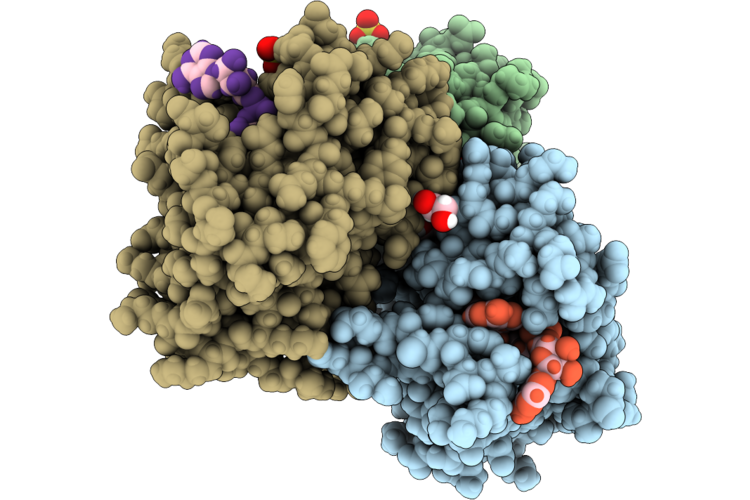
Crystal Structure Of The N4Bp2 Polynucleotide Kinase Domain Complexed With Atp Or Adp, Mg, And The Trinucleotide Substrate Aga Or Its 5'-Phosphorylated Product Paga.
Organism: Homo sapiens, Synthetic construct
Method: X-RAY DIFFRACTION Resolution:2.72 Å Release Date: 2026-09-23 Classification: TRANSFERASE Ligands: MG, ATP, SO4, ADP |
Organism: Homo sapiens, Synthetic construct
Method: X-RAY DIFFRACTION
Release Date: 2026-09-23
Ligands: MG, ATP, SO4, ADP
 |
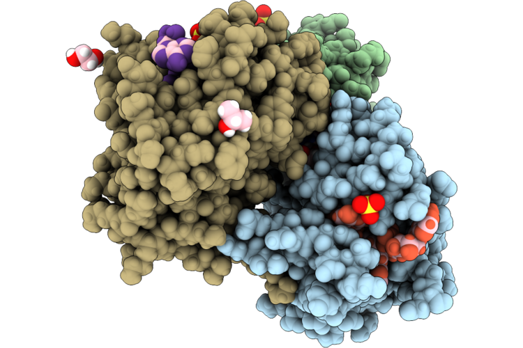
Crystal Structure Of The N4Bp2 Polynucleotide Kinase Domain Complexed With Atp Or Adp, Mg, And The Trinucleotide Substrate Aua Or Its 5'-Phosphorylated Product Paua.
Organism: Homo sapiens, Synthetic construct
Method: X-RAY DIFFRACTION Resolution:2.23 Å Release Date: 2026-09-23 Classification: TRANSFERASE Ligands: MG, ATP, SO4, ADP, EDO |
Organism: Homo sapiens, Synthetic construct
Method: X-RAY DIFFRACTION
Release Date: 2026-09-23
Ligands: MG, ATP, SO4, ADP, EDO
 |
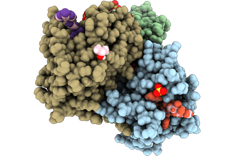
Crystal Structure Of The N4Bp2 Polynucleotide Kinase Domain Complexed With Atp, Mg, And The Trinucleotide Substrate Caa.
Organism: Homo sapiens, Synthetic construct
Method: X-RAY DIFFRACTION Resolution:2.26 Å Release Date: 2026-09-23 Classification: TRANSFERASE Ligands: MG, ATP, SO4, IPA, EDO |
Organism: Homo sapiens, Synthetic construct
Method: X-RAY DIFFRACTION
Release Date: 2026-09-23
Ligands: MG, ATP, SO4, IPA, EDO
 |
Crystal Structure Of The N4Bp2 Polynucleotide Kinase Domain Complexed With Atp, Mg, And The Trinucleotide Substrate Cca.
Organism: Homo sapiens, Synthetic construct
Method: X-RAY DIFFRACTION Resolution:1.85 Å Release Date: 2026-09-23 Classification: TRANSFERASE Ligands: MG, ATP, SO4, EDO, IPA |
Organism: Homo sapiens, Synthetic construct
Method: X-RAY DIFFRACTION
Release Date: 2026-09-23
Ligands: MG, ATP, SO4, EDO, IPA
 |
Crystal Structure Of The N4Bp2 Polynucleotide Kinase Domain Complexed With Atp, Mg, And The Trinucleotide Substrate Cga.
Organism: Homo sapiens, Synthetic construct
Method: X-RAY DIFFRACTION Resolution:2.12 Å Release Date: 2026-09-23 Classification: TRANSFERASE Ligands: MG, ATP, SO4 |
Organism: Homo sapiens, Synthetic construct
Method: X-RAY DIFFRACTION
Release Date: 2026-09-23
Ligands: MG, ATP, SO4
 |
Crystal Structure Of The N4Bp2 Polynucleotide Kinase Domain Complexed With Atp, Mg, And The Trinucleotide Substrate Cua.
Organism: Homo sapiens, Synthetic construct
Method: X-RAY DIFFRACTION Resolution:2.10 Å Release Date: 2026-09-23 Classification: TRANSFERASE Ligands: MG, ATP, SO4, EDO |
Organism: Homo sapiens, Synthetic construct
Method: X-RAY DIFFRACTION
Release Date: 2026-09-23
Ligands: MG, ATP, SO4, EDO
 |
Crystal Structure Of The N4Bp2 Polynucleotide Kinase Domain Complexed With Atp Or Adp, Mg, And The Trinucleotide Substrate Gaa Or Its 5'-Phosphorylated Product Pgaa.
Organism: Homo sapiens, Synthetic construct
Method: X-RAY DIFFRACTION Resolution:2.16 Å Release Date: 2026-09-23 Classification: TRANSFERASE Ligands: MG, ATP, ADP, SO4 |
Organism: Homo sapiens, Synthetic construct
Method: X-RAY DIFFRACTION
Release Date: 2026-09-23
Ligands: MG, ATP, ADP, SO4