

Search Count: 28
 |
Crystal Structure Of Wild-Type Trypanosoma Brucei Dhodh In Fmn-Reduced, Ligand-Free Form
Organism: Trypanosoma brucei brucei treu927
Method: X-RAY DIFFRACTION Resolution:1.75 Å Release Date: 2025-11-26 Classification: FLAVOPROTEIN Ligands: FNR, MLI, SO2, SO3 |
Organism: Trypanosoma brucei brucei treu927
Method: X-RAY DIFFRACTION
Release Date: 2025-11-26
Ligands: FNR, MLI, SO2, SO3
 |
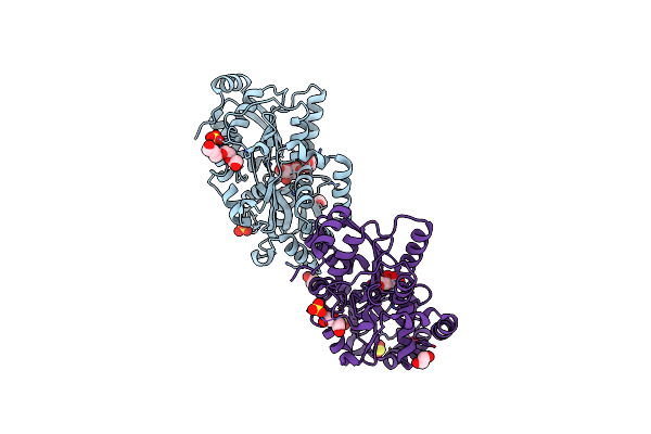
W-Formate Dehydrogenase U192C From Desulfovibrio Vulgaris - Soaked With Dithionite
Organism: Nitratidesulfovibrio vulgaris str. hildenborough
Method: X-RAY DIFFRACTION Resolution:2.00 Å Release Date: 2025-07-16 Classification: OXIDOREDUCTASE Ligands: GOL, EDO, PEG, SO2, H2S, W, MGD, SF4 |
Organism: Nitratidesulfovibrio vulgaris str. hildenborough
Method: X-RAY DIFFRACTION
Release Date: 2025-07-16
Ligands: GOL, EDO, PEG, SO2, H2S, W, MGD, SF4
 |
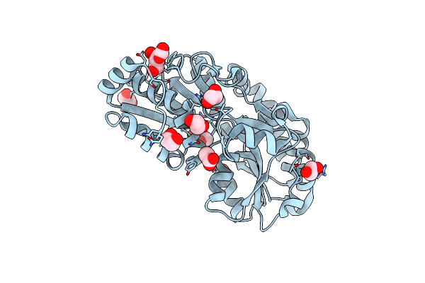
Crystal Structure Of Metal-Citrate-Binding Mutant (S26A) Protein (Mcta) Of Abc Transporter Endogenously Bound To Mn2+-Citrate Complex
Organism: Thermus thermophilus (strain atcc 27634 / dsm 579 / hb8)
Method: X-RAY DIFFRACTION Resolution:2.50 Å Release Date: 2021-12-01 Classification: TRANSPORT PROTEIN Ligands: MN, CIT, SO4, SO3, EDO, GOL, PG4, SO2, PEG |
Organism: Thermus thermophilus (strain atcc 27634 / dsm 579 / hb8)
Method: X-RAY DIFFRACTION
Release Date: 2021-12-01
Ligands: MN, CIT, SO4, SO3, EDO, GOL, PG4, SO2, PEG
 |
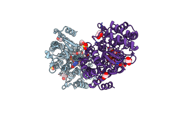
Crystal Structure Of Metal-Citrate-Binding Mutant (S79A) Protein (Mcta) Of Abc Transporter In Apo State
Organism: Thermus thermophilus (strain atcc 27634 / dsm 579 / hb8)
Method: X-RAY DIFFRACTION Resolution:1.63 Å Release Date: 2021-12-01 Classification: TRANSPORT PROTEIN Ligands: CL, SO2, CO2, ACT, EDO, PDO, PEG |
Organism: Thermus thermophilus (strain atcc 27634 / dsm 579 / hb8)
Method: X-RAY DIFFRACTION
Release Date: 2021-12-01
Ligands: CL, SO2, CO2, ACT, EDO, PDO, PEG
 |
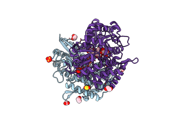
Crystal Structure Of A Dinucleotide-Binding Protein Of Abc Transporter Endogenously Bound To Uridylyl-3'-5'-Phospho-Guanosine (Form Iii)
Organism: Thermus thermophilus hb8
Method: X-RAY DIFFRACTION Resolution:1.85 Å Release Date: 2021-03-10 Classification: TRANSPORT PROTEIN Ligands: CL, SO3, EDO, GOL, FGO, CO2, SO2 |
Organism: Thermus thermophilus hb8
Method: X-RAY DIFFRACTION
Release Date: 2021-03-10
Ligands: CL, SO3, EDO, GOL, FGO, CO2, SO2
 |
Crystal Structure Of Beta-Glycosides-Binding Protein Of Abc Transporter In A Closed State Bound To Cellotriose
Organism: Thermus thermophilus (strain hb8 / atcc 27634 / dsm 579)
Method: X-RAY DIFFRACTION Resolution:2.00 Å Release Date: 2020-10-21 Classification: SUGAR BINDING PROTEIN Ligands: CL, NA, SO2, SO4, EDO, CO3 |
Organism: Thermus thermophilus (strain hb8 / atcc 27634 / dsm 579)
Method: X-RAY DIFFRACTION
Release Date: 2020-10-21
Ligands: CL, NA, SO2, SO4, EDO, CO3
 |
Crystal Structure Of Beta-Glycosides-Binding Protein Of Abc Transporter In A Closed State Bound To Cellobiose
Organism: Thermus thermophilus (strain hb8 / atcc 27634 / dsm 579)
Method: X-RAY DIFFRACTION Resolution:2.30 Å Release Date: 2020-09-16 Classification: SUGAR BINDING PROTEIN Ligands: CL, SO3, EDO, PEG, SO2 |
Organism: Thermus thermophilus (strain hb8 / atcc 27634 / dsm 579)
Method: X-RAY DIFFRACTION
Release Date: 2020-09-16
Ligands: CL, SO3, EDO, PEG, SO2
 |
Crystal Structure Of Beta-Glycosides-Binding Protein Of Abc Transporter In A Closed State Bound To Cellotetraose
Organism: Thermus thermophilus (strain hb8 / atcc 27634 / dsm 579)
Method: X-RAY DIFFRACTION Resolution:2.05 Å Release Date: 2020-09-16 Classification: SUGAR BINDING PROTEIN Ligands: CL, SO2, SO3, SO4, CO2, EDO |
Organism: Thermus thermophilus (strain hb8 / atcc 27634 / dsm 579)
Method: X-RAY DIFFRACTION
Release Date: 2020-09-16
Ligands: CL, SO2, SO3, SO4, CO2, EDO
 |
Crystal Structure Of Beta-Glycosides-Binding Protein (W177X) Of Abc Transporter In An Open-Liganded State Bound To Sophorose
Organism: Thermus thermophilus (strain hb8 / atcc 27634 / dsm 579)
Method: X-RAY DIFFRACTION Resolution:1.70 Å Release Date: 2020-09-16 Classification: SUGAR BINDING PROTEIN Ligands: SO2, CO2, ACT, SO4, EDO, BU2 |
Organism: Thermus thermophilus (strain hb8 / atcc 27634 / dsm 579)
Method: X-RAY DIFFRACTION
Release Date: 2020-09-16
Ligands: SO2, CO2, ACT, SO4, EDO, BU2
 |
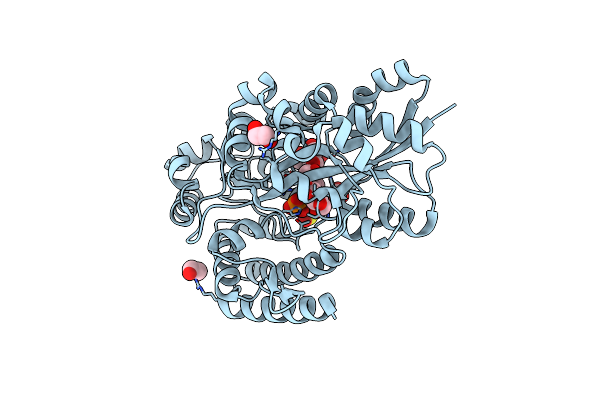
Crystal Structure Of Beta-Glycosides-Binding Protein (W177X) Of Abc Transporter In A Closed State Bound To Cellobiose
Organism: Thermus thermophilus (strain hb8 / atcc 27634 / dsm 579)
Method: X-RAY DIFFRACTION Resolution:2.10 Å Release Date: 2020-09-16 Classification: SUGAR BINDING PROTEIN Ligands: CL, SO2, ACT, FJO |
Organism: Thermus thermophilus (strain hb8 / atcc 27634 / dsm 579)
Method: X-RAY DIFFRACTION
Release Date: 2020-09-16
Ligands: CL, SO2, ACT, FJO
 |
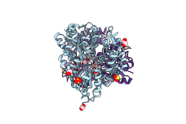
Crystal Structure Of Beta-Glycosides-Binding Protein (W177X) Of Abc Transporter In A Closed State Bound To Laminaritetraose
Organism: Thermus thermophilus hb8
Method: X-RAY DIFFRACTION Resolution:1.88 Å Release Date: 2020-09-16 Classification: SUGAR BINDING PROTEIN Ligands: CL, SO2, CO3, EDO, PO4 |
Organism: Thermus thermophilus hb8
Method: X-RAY DIFFRACTION
Release Date: 2020-09-16
Ligands: CL, SO2, CO3, EDO, PO4
 |
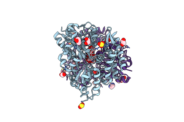
Crystal Structure Of Beta-Glycosides-Binding Protein (W67A) Of Abc Transporter In An Open State
Organism: Thermus thermophilus hb8
Method: X-RAY DIFFRACTION Resolution:1.63 Å Release Date: 2020-09-16 Classification: SUGAR BINDING PROTEIN Ligands: CL, CO2, SO3, EDO, PEG, PGE, SO2, PO4 |
Organism: Thermus thermophilus hb8
Method: X-RAY DIFFRACTION
Release Date: 2020-09-16
Ligands: CL, CO2, SO3, EDO, PEG, PGE, SO2, PO4
 |
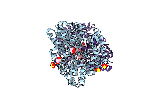
Crystal Structure Of Beta-Glycosides-Binding Protein (W67A) Of Abc Transporter In An Open-Liganded State Bound To Gentiobiose
Organism: Thermus thermophilus hb8
Method: X-RAY DIFFRACTION Resolution:1.63 Å Release Date: 2020-09-16 Classification: SUGAR BINDING PROTEIN Ligands: SO2, SO3, CO2, PGO, EDO, PEG, SO4, PO4, IMD |
Organism: Thermus thermophilus hb8
Method: X-RAY DIFFRACTION
Release Date: 2020-09-16
Ligands: SO2, SO3, CO2, PGO, EDO, PEG, SO4, PO4, IMD
 |
Crystal Structure Of Beta-Glycosides-Binding Protein (E117A) Of Abc Transporter In An Open State
Organism: Thermus thermophilus hb8
Method: X-RAY DIFFRACTION Resolution:2.10 Å Release Date: 2020-09-16 Classification: SUGAR BINDING PROTEIN Ligands: CL, SO2, SO3, EDO, PGE |
Organism: Thermus thermophilus hb8
Method: X-RAY DIFFRACTION
Release Date: 2020-09-16
Ligands: CL, SO2, SO3, EDO, PGE
 |
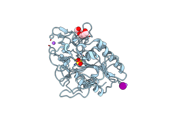
Wolinella Succinogenes Octaheme Sulfite Reductase Mcca, Form I
Organism: Wolinella succinogenes dsm 1740
Method: X-RAY DIFFRACTION Resolution:2.20 Å Release Date: 2015-02-04 Classification: UNKNOWN FUNCTION Ligands: CU1, SX, BU3, ACT, HEM, SO2 |
Organism: Wolinella succinogenes dsm 1740
Method: X-RAY DIFFRACTION
Release Date: 2015-02-04
Ligands: CU1, SX, BU3, ACT, HEM, SO2
 |
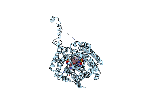
The Crystal Structure Of Phaz7 At Atomic (1.2 Angstrom) Resolution Reveals Details Of The Active Site And Suggests A Substrate Binding Mode
Organism: Paucimonas lemoignei
Method: X-RAY DIFFRACTION Resolution:1.20 Å Release Date: 2010-06-09 Classification: HYDROLASE Ligands: IOD, SO2, PG4, CL, NA |
Organism: Paucimonas lemoignei
Method: X-RAY DIFFRACTION
Release Date: 2010-06-09
Ligands: IOD, SO2, PG4, CL, NA
 |
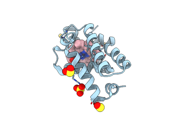
Structure Of The E. Coli Puta Proline Dehydrogenase Domain Reduced By Dithionite And Complexed With So2
Organism: Escherichia coli
Method: X-RAY DIFFRACTION Resolution:2.30 Å Release Date: 2007-04-03 Classification: OXIDOREDUCTASE Ligands: FAD, SO2 |
Organism: Escherichia coli
Method: X-RAY DIFFRACTION
Release Date: 2007-04-03
Ligands: FAD, SO2
 |
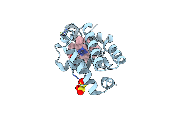
The X-Ray Crystal Structure Of Ferrous Synechocystis Hemoglobin With A Covalent Linkage
Organism: Synechocystis sp.
Method: X-RAY DIFFRACTION Resolution:1.80 Å Release Date: 2006-08-29 Classification: OXYGEN STORAGE/TRANSPORT Ligands: CD, SO3, HEM, SO2 |
Organism: Synechocystis sp.
Method: X-RAY DIFFRACTION
Release Date: 2006-08-29
Ligands: CD, SO3, HEM, SO2
 |
The X-Ray Crystal Structure Of Ferrous Synechocystis Hemoglobin H117A Mutant With A Covalent Linkage
Organism: Synechocystis sp.
Method: X-RAY DIFFRACTION Resolution:1.90 Å Release Date: 2006-08-29 Classification: oxygen storage/transport Ligands: CD, SO2, HEM |
Organism: Synechocystis sp.
Method: X-RAY DIFFRACTION
Release Date: 2006-08-29
Ligands: CD, SO2, HEM
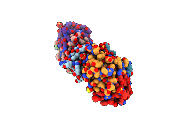 |
Conserved Dimerization Of The Ib1 Src-Homology 3 Domain
Organism: Rattus norvegicus
Method: X-RAY DIFFRACTION Resolution:1.75 Å Release Date: 2006-02-28 Classification: SIGNALING PROTEIN Ligands: SO4, P6G, SO2 Modified Residue(s): MSE |
Organism: Rattus norvegicus
Method: X-RAY DIFFRACTION
Release Date: 2006-02-28
Ligands: SO4, P6G, SO2