
Search Count: 44
All
Selected
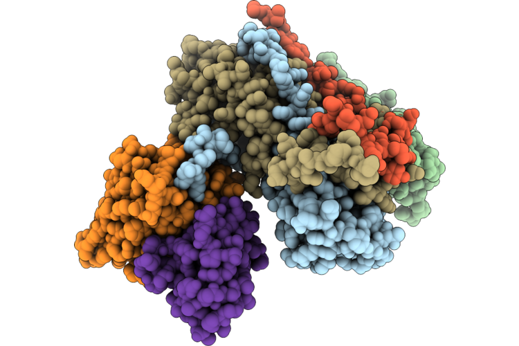 |
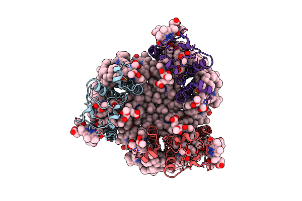
Complex Of Fmdv O/18074 And Porcine-Derived Neutralizing Monoclonal Antibody Po18-10
Organism: Sus scrofa, Foot-and-mouth disease virus o
Method: ELECTRON MICROSCOPY Resolution:2.27 Å Release Date: 2026-04-22 Classification: VIRUS |
 |
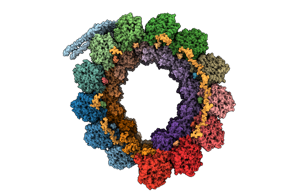
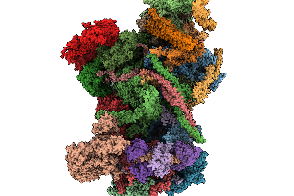
Cryo-Em Structure Of The Chromera Velia Psi Supercomplex At 1.84 Angstrom Resolution
Organism: Chromera velia
Method: ELECTRON MICROSCOPY Resolution:1.84 Å Release Date: 2026-01-28 Classification: PHOTOSYNTHESIS Ligands: FE, CLA, CL0, SF4, PQN, XAT, LMG, DGD, LMT, BCR, AV0, A1L6D, SQD, A1I05 |
 |
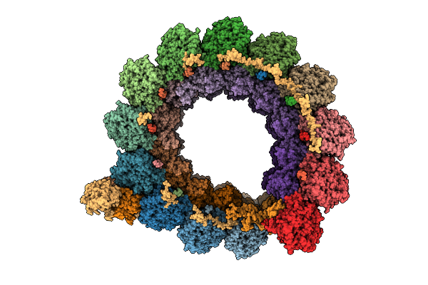
Cryo Em Structure Of Rc-Dlh Complex Model Ii From Gemmatimonas Groenlandica
Organism: Gemmatimonas groenlandica
Method: ELECTRON MICROSCOPY Release Date: 2025-12-03 Classification: PHOTOSYNTHESIS Ligands: BCL, BPH, LMT, MQ8, FE, CD4, CRT, PEX, HEC, V7N |
 |
Organism: Toxoplasma gondii
Method: ELECTRON MICROSCOPY Release Date: 2025-12-03 Classification: STRUCTURAL PROTEIN Ligands: GTP, MG, GDP |
 |
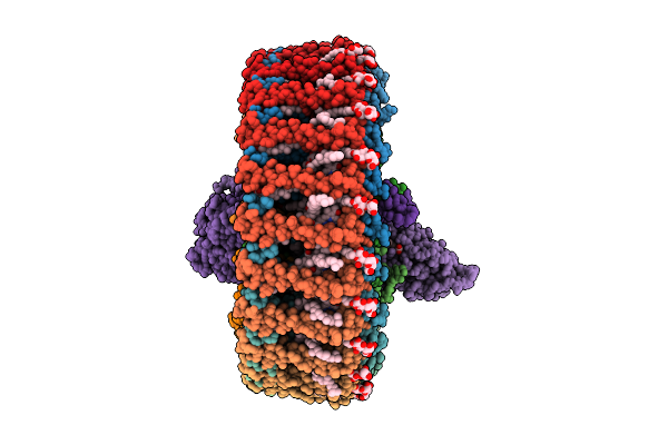
Cryo-Em Structure Of Intraconoidal Microtubule 2 (Icmt2) From Toxoplasma Gondii (8-Nm Repeat)
Organism: Toxoplasma gondii
Method: ELECTRON MICROSCOPY Release Date: 2025-12-03 Classification: STRUCTURAL PROTEIN Ligands: GTP, MG, GDP |
 |
Cryo-Em Structure Of Intraconoidal Microtubule 1 (Icmt1) From Toxoplasma Gondii (8-Nm Repeat)
Organism: Toxoplasma gondii
Method: ELECTRON MICROSCOPY Release Date: 2025-12-03 Classification: STRUCTURAL PROTEIN Ligands: GTP, MG, GDP |
 |
Cryo-Em Structure Of The Apical Region Of Subpellicular Microtubule (Spmt) From Toxoplasma Gondii (8-Nm Repeat)
Organism: Toxoplasma gondii
Method: ELECTRON MICROSCOPY Release Date: 2025-12-03 Classification: STRUCTURAL PROTEIN Ligands: GDP, GTP, MG |
 |
Organism: Toxoplasma gondii
Method: ELECTRON MICROSCOPY Release Date: 2025-11-19 Classification: STRUCTURAL PROTEIN |
 |
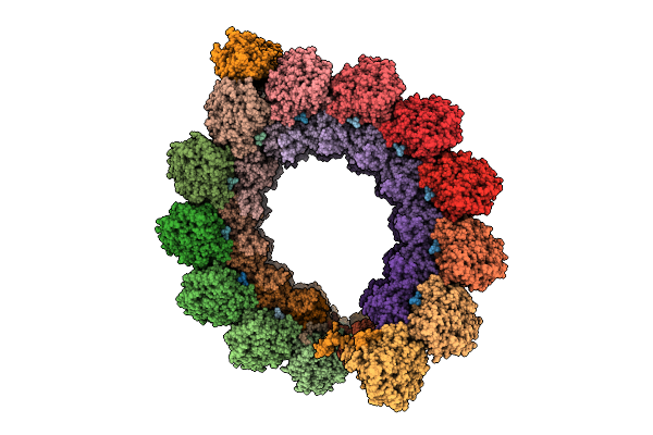
Cryo-Em Structure Of Rc-Dlh Complex Model I From Gem. Groenlandica Strain Tet16
Organism: Gemmatimonas groenlandica
Method: ELECTRON MICROSCOPY Release Date: 2025-10-29 Classification: PHOTOSYNTHESIS Ligands: V7N, BCL, LMT, PEX, MQ8, CD4, BPH, FE, CRT, HEC |
 |
The Cryo-Em Structure Of Siphonaxanthin Chlorophyll A/B Type Light-Harvesting Complex Ii
Organism: Codium fragile
Method: ELECTRON MICROSCOPY Release Date: 2022-11-23 Classification: PHOTOSYNTHESIS Ligands: CHL, CLA, NEX, 0UR, 0IE, LHG |
 |
Organism: Rhodopseudomonas palustris atcc 17001
Method: ELECTRON MICROSCOPY Release Date: 2022-10-12 Classification: PHOTOSYNTHESIS Ligands: BCL, IRM |
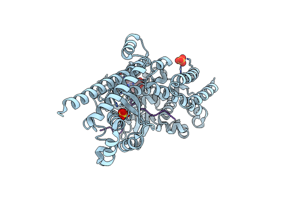 |
Organism: Homo sapiens
Method: X-RAY DIFFRACTION Resolution:1.79 Å Release Date: 2022-10-05 Classification: STRUCTURAL PROTEIN Ligands: SAH, SO4 |
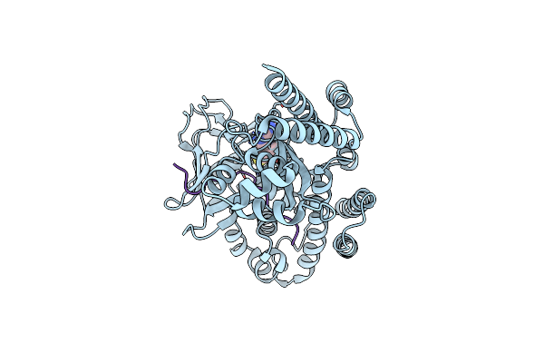 |
Organism: Homo sapiens
Method: X-RAY DIFFRACTION Resolution:2.90 Å Release Date: 2022-10-05 Classification: STRUCTURAL PROTEIN Ligands: SAH |
 |
Organism: Rhodopseudomonas palustris
Method: ELECTRON MICROSCOPY Release Date: 2022-10-05 Classification: PHOTOSYNTHESIS Ligands: BCL, IRM |
 |
Organism: Rhodopseudomonas palustris
Method: ELECTRON MICROSCOPY Release Date: 2022-10-05 Classification: PHOTOSYNTHESIS Ligands: BCL, ZE0 |
 |
Organism: Rhodopseudomonas palustris
Method: ELECTRON MICROSCOPY Release Date: 2022-10-05 Classification: PHOTOSYNTHESIS Ligands: BCL, IRM |
 |
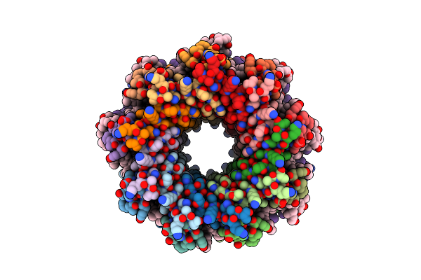
Cryo-Em Structure (Model_1A) Of The Rc-Dlh Complex From Gemmatimonas Phototrophica At 2.4 A
Organism: Gemmatimonas phototrophica
Method: ELECTRON MICROSCOPY Release Date: 2022-03-02 Classification: MEMBRANE PROTEIN Ligands: BCL, LMT, V7N, 0V9, HEC, NDG, V75, CD4, PGW, MQ8, BPH, FE, CRT, V7B, UYH |
 |
Cryo-Em Structure (Model_2A) Of The Rc-Dlh Complex From Gemmatimonas Phototrophica At 2.5 A
Organism: Gemmatimonas phototrophica
Method: ELECTRON MICROSCOPY Release Date: 2022-03-02 Classification: MEMBRANE PROTEIN Ligands: BCL, LMT, V7N, HEC, V75, NDG, 0V9, CD4, PGW, MQ8, V7B, BPH, FE, CRT, UYH |
 |
Cryo-Em Structure Of The Rc-Dlh Complex (Model_1B) From Gemmatimonas Phototrophica At 2.47 A
Organism: Gemmatimonas phototrophica
Method: ELECTRON MICROSCOPY Release Date: 2022-03-02 Classification: MEMBRANE PROTEIN Ligands: BCL, LMT, V7N, 0V9, HEC, V75, NDG, PGW, CD4, BPH, MQ8, FE, CRT, V7B, UYH |
 |
Cryo-Em Structure (Model_2B) Of The Rc-Dlh Complex From Gemmatimonas Phototrophica At 2.44 A
Organism: Gemmatimonas phototrophica
Method: ELECTRON MICROSCOPY Release Date: 2022-03-02 Classification: MEMBRANE PROTEIN Ligands: BCL, LMT, V7N, HEC, NDG, V75, PGW, 0V9, CD4, BPH, MQ8, FE, CRT, V7B, UYH |