

Search Count: 272
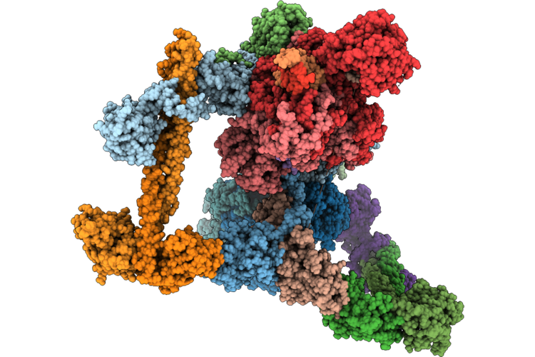 |
Nhej Short-Range Complex With Polymerase Lambda
Organism: Homo sapiens
Method: ELECTRON MICROSCOPY Release Date: 2026-07-01 Classification: DNA BINDING PROTEIN/DNA |
Organism: Homo sapiens
Method: ELECTRON MICROSCOPY
Release Date: 2026-07-01
 |
Crystal Structure Of A P. Aeruginosa Gyrase Chimera In Complex With 20Mer Dna And Ciprofloxacin
Organism: Pseudomonas aeruginosa, Escherichia coli
Method: X-RAY DIFFRACTION Resolution:2.85 Å Release Date: 2026-04-22 Classification: DNA BINDING PROTEIN Ligands: CIT, CPF, SO4, MN, CL |
Organism: Pseudomonas aeruginosa, Escherichia coli
Method: X-RAY DIFFRACTION
Release Date: 2026-04-22
Ligands: CIT, CPF, SO4, MN, CL
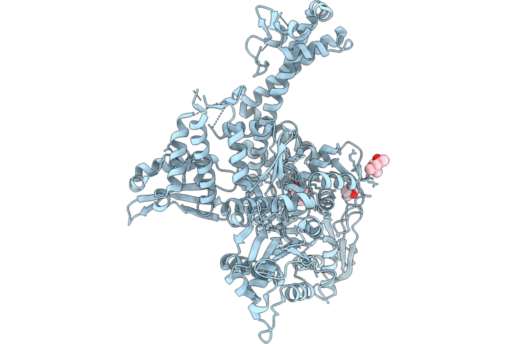 |
Crystal Structure Of A P. Aeruginosa Gyrase
Organism: Pseudomonas aeruginosa
Method: X-RAY DIFFRACTION Resolution:2.40 Å Release Date: 2026-04-22 Classification: DNA BINDING PROTEIN |
Organism: Pseudomonas aeruginosa
Method: X-RAY DIFFRACTION
Release Date: 2026-04-22
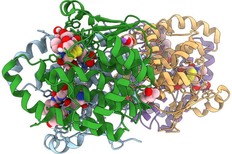 |
Crystal Structure Of Glutathione S-Transferase Per A 23 (Sigma Class)
Organism: Periplaneta americana
Method: X-RAY DIFFRACTION Resolution:1.50 Å Release Date: 2026-04-22 Classification: TRANSFERASE Ligands: GDS, EDO, MG, MPD, NA |
Organism: Periplaneta americana
Method: X-RAY DIFFRACTION
Release Date: 2026-04-22
Ligands: GDS, EDO, MG, MPD, NA
 |
Crystal Structure Of Glutathione S-Transferase Per A 5 (Delta Class)
Organism: Periplaneta americana
Method: X-RAY DIFFRACTION Resolution:1.30 Å Release Date: 2026-04-22 Classification: TRANSFERASE Ligands: GSH, EDO, MG, CL |
Organism: Periplaneta americana
Method: X-RAY DIFFRACTION
Release Date: 2026-04-22
Ligands: GSH, EDO, MG, CL
 |
Crystal Structure Of Glutathione S-Transferase Bla G 5 (Sigma Class)
Organism: Blattella germanica
Method: X-RAY DIFFRACTION Resolution:1.35 Å Release Date: 2026-04-22 Classification: TRANSFERASE Ligands: GDS, GOL, GSH |
Organism: Blattella germanica
Method: X-RAY DIFFRACTION
Release Date: 2026-04-22
Ligands: GDS, GOL, GSH
 |
Crystal Structure Of Glutathione S-Transferase Bla G 24 (Delta Class)
Organism: Blattella germanica
Method: X-RAY DIFFRACTION Resolution:1.33 Å Release Date: 2026-04-22 Classification: TRANSFERASE Ligands: GSH, EDO, CA |
Organism: Blattella germanica
Method: X-RAY DIFFRACTION
Release Date: 2026-04-22
Ligands: GSH, EDO, CA
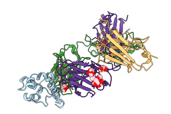 |
Crystal Structure Of The Peanut Allergen Ara H 9 With Bound Fab Igx-3103 And Antifab Nanobody
Organism: Homo sapiens, Lama glama, Arachis hypogaea
Method: X-RAY DIFFRACTION Resolution:1.60 Å Release Date: 2026-02-18 Classification: ALLERGEN Ligands: EDO, CL |
Organism: Homo sapiens, Lama glama, Arachis hypogaea
Method: X-RAY DIFFRACTION
Release Date: 2026-02-18
Ligands: EDO, CL
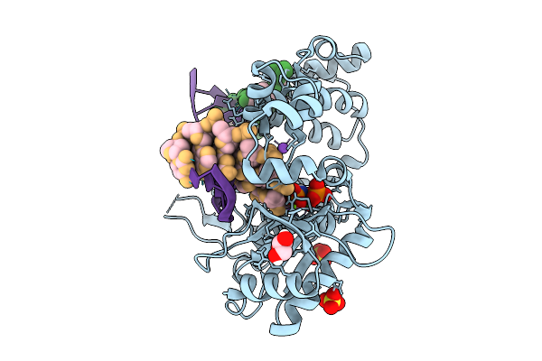 |
Pre-Catalytic Ternary Complex Of Dna Polymerase Lambda With Bound 1-Nt Gapped Ssb Substrate Containing Template Ribonucleotide Opposite Primer Terminus, And Incoming Dumpnpp
Organism: Homo sapiens, Synthetic construct
Method: X-RAY DIFFRACTION Resolution:1.63 Å Release Date: 2026-01-21 Classification: TRANSFERASE Ligands: SO4, GOL, DUP, MG, NA, CL |
Organism: Homo sapiens, Synthetic construct
Method: X-RAY DIFFRACTION
Release Date: 2026-01-21
Ligands: SO4, GOL, DUP, MG, NA, CL
 |
Crystal Structure Of The Walnut Allergen Jug R 2 Bound To The Human-Derived Fab 6D12
Organism: Homo sapiens, Juglans regia
Method: X-RAY DIFFRACTION Resolution:1.56 Å Release Date: 2026-01-21 Classification: ALLERGEN/IMMUNE SYSTEM Ligands: EDO, CL |
Organism: Homo sapiens, Juglans regia
Method: X-RAY DIFFRACTION
Release Date: 2026-01-21
Ligands: EDO, CL
 |
Co-Crystal Structure Of Maltose Binding Protein (Mbp)-Human Senp3 Fusion Protein In Complex With Pelp1 Peptide
Organism: Homo sapiens
Method: X-RAY DIFFRACTION Resolution:2.93 Å Release Date: 2025-08-06 Classification: HYDROLASE |
Organism: Homo sapiens
Method: X-RAY DIFFRACTION
Release Date: 2025-08-06
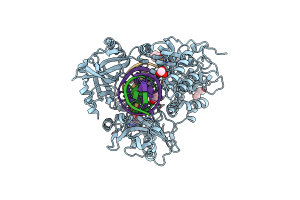 |
Human Dna Ligase I F872A Bound To Adenylated Nicked Dna With A 5' Terminal Ribonucleotide
Organism: Homo sapiens, Synthetic construct
Method: X-RAY DIFFRACTION Resolution:2.00 Å Release Date: 2025-07-02 Classification: LIGASE/DNA Ligands: PEG, MES, CL, GOL, NA, AMP |
Organism: Homo sapiens, Synthetic construct
Method: X-RAY DIFFRACTION
Release Date: 2025-07-02
Ligands: PEG, MES, CL, GOL, NA, AMP
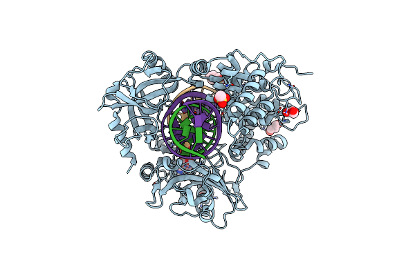 |
Human Dna Ligase I F872L Bound To Adenylated Nicked Dna
Organism: Homo sapiens, Synthetic construct
Method: X-RAY DIFFRACTION Resolution:2.30 Å Release Date: 2025-07-02 Classification: LIGASE/DNA Ligands: GOL, MES, AMP, NA |
Organism: Homo sapiens, Synthetic construct
Method: X-RAY DIFFRACTION
Release Date: 2025-07-02
Ligands: GOL, MES, AMP, NA
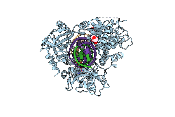 |
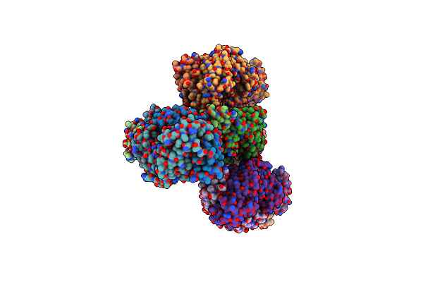
Human Dna Ligase I F872A Bound To Adenylated Nicked Dna
Organism: Homo sapiens, Synthetic construct
Method: X-RAY DIFFRACTION Resolution:2.20 Å Release Date: 2025-07-02 Classification: LIGASE/DNA Ligands: MES, AMP |
Organism: Homo sapiens, Synthetic construct
Method: X-RAY DIFFRACTION
Release Date: 2025-07-02
Ligands: MES, AMP
 |
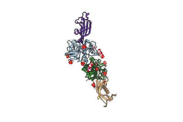
Crystal Structure Of The Peanut Allergen Ara H 2 With Two Human Derived Fab Antibodies 22S1 And 23P34
Organism: Homo sapiens, Arachis hypogaea
Method: X-RAY DIFFRACTION Resolution:2.95 Å Release Date: 2025-06-11 Classification: ALLERGEN |
Organism: Homo sapiens, Arachis hypogaea
Method: X-RAY DIFFRACTION
Release Date: 2025-06-11
 |
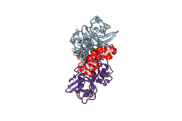
Complex Of Murine Cathepsin K With Bound Cystatin C Inhibitor
Organism: Mus musculus
Method: X-RAY DIFFRACTION Resolution:2.75 Å Release Date: 2024-10-09 Classification: HYDROLASE Ligands: EDO, SO4 |
Organism: Mus musculus
Method: X-RAY DIFFRACTION
Release Date: 2024-10-09
Ligands: EDO, SO4
 |
Complex Of Murine Cathepsin K With Bound Heparan Sulfate 12Mer
Organism: Mus musculus
Method: X-RAY DIFFRACTION Resolution:3.10 Å Release Date: 2024-10-09 Classification: HYDROLASE Ligands: NAG, EDO |
Organism: Mus musculus
Method: X-RAY DIFFRACTION
Release Date: 2024-10-09
Ligands: NAG, EDO
 |
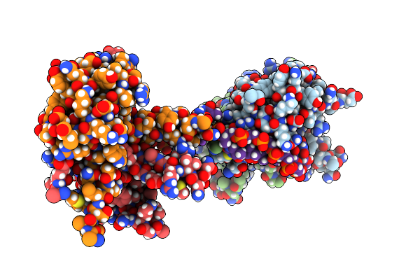
Crystal Structure Of Heparosan Synthase 2 From Pasteurella Multocida At 2.85 A
Organism: Pasteurella multocida
Method: X-RAY DIFFRACTION Resolution:2.85 Å Release Date: 2024-09-04 Classification: TRANSFERASE Ligands: MN, UDP, NA, CL, EDO, CA |
Organism: Pasteurella multocida
Method: X-RAY DIFFRACTION
Release Date: 2024-09-04
Ligands: MN, UDP, NA, CL, EDO, CA
 |
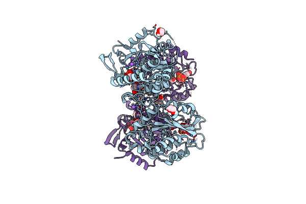
Mitochondrial Single-Stranded Binding Protein Bound To Dna
Organism: Homo sapiens, Synthetic construct
Method: X-RAY DIFFRACTION Resolution:1.90 Å Release Date: 2024-08-21 Classification: DNA BINDING PROTEIN/DNA Ligands: GOL, NA |
Organism: Homo sapiens, Synthetic construct
Method: X-RAY DIFFRACTION
Release Date: 2024-08-21
Ligands: GOL, NA
 |
Crystal Structure Of Heparosan Synthase 2 From Pasteurella Multocida At 1.98 A
Organism: Pasteurella multocida
Method: X-RAY DIFFRACTION Resolution:1.98 Å Release Date: 2024-07-24 Classification: TRANSFERASE Ligands: MN, UDP, EDO, NA |
Organism: Pasteurella multocida
Method: X-RAY DIFFRACTION
Release Date: 2024-07-24
Ligands: MN, UDP, EDO, NA