
Search Count: 20
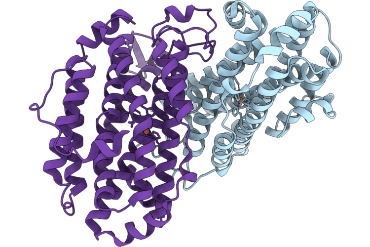 |
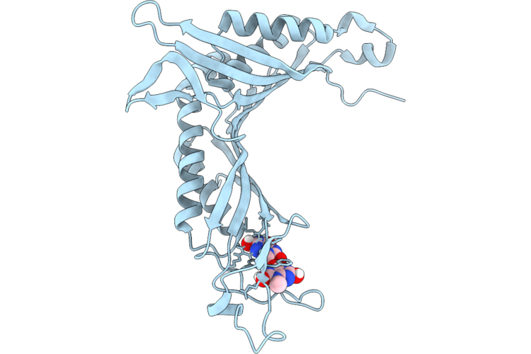
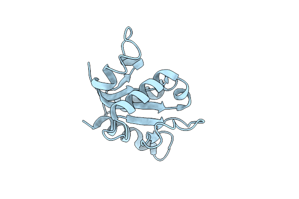
Organism: Escherichia coli
Method: X-RAY DIFFRACTION Resolution:1.90 Å Release Date: 2026-05-13 Classification: OXIDOREDUCTASE Ligands: FE |
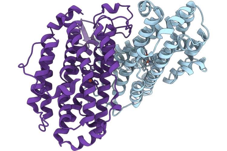 |
Organism: Escherichia coli
Method: X-RAY DIFFRACTION Resolution:1.70 Å Release Date: 2026-05-13 Classification: OXIDOREDUCTASE Ligands: FE2 |
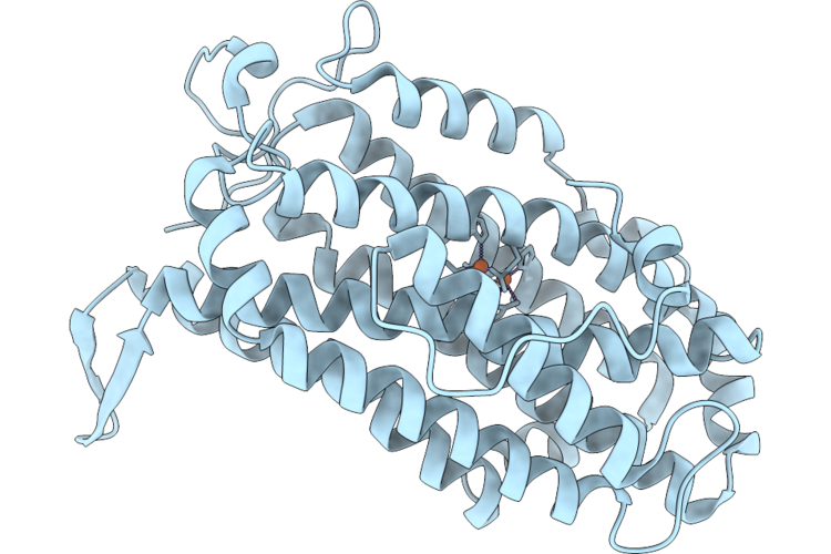 |
Organism: Escherichia coli
Method: X-RAY DIFFRACTION Resolution:2.70 Å Release Date: 2026-05-13 Classification: OXIDOREDUCTASE Ligands: FE |
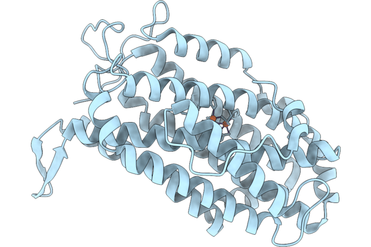 |
Organism: Escherichia coli
Method: X-RAY DIFFRACTION Resolution:2.70 Å Release Date: 2026-05-13 Classification: OXIDOREDUCTASE Ligands: FE2 |
 |
Organism: Aspergillus flavus
Method: ELECTRON CRYSTALLOGRAPHY Resolution:1.25 Å Release Date: 2026-04-29 Classification: OXIDOREDUCTASE Ligands: MUA |
 |
Organism: Aspergillus flavus
Method: ELECTRON CRYSTALLOGRAPHY Resolution:1.75 Å Release Date: 2026-04-29 Classification: OXIDOREDUCTASE Ligands: OXY, URC |
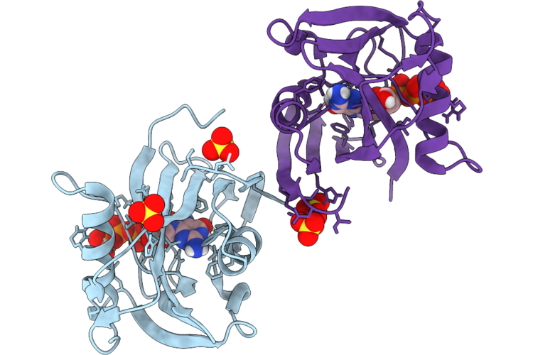 |
Organism: Homo sapiens
Method: ELECTRON CRYSTALLOGRAPHY Resolution:1.66 Å Release Date: 2026-04-22 Classification: HYDROLASE Ligands: SO4, 8DG |
 |
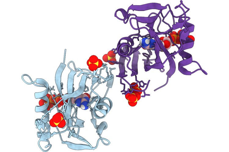
Organism: Homo sapiens
Method: ELECTRON CRYSTALLOGRAPHY Resolution:2.32 Å Release Date: 2026-04-22 Classification: HYDROLASE Ligands: 8DG, SO4 |
 |
Organism: Homo sapiens
Method: ELECTRON CRYSTALLOGRAPHY Resolution:2.86 Å Release Date: 2026-04-22 Classification: HYDROLASE Ligands: 8DG, SO4 |
 |
Organism: Gallus gallus
Method: ELECTRON CRYSTALLOGRAPHY Resolution:0.83 Å Release Date: 2026-04-22 Classification: HYDROLASE Ligands: ACT, CL |
 |
Organism: Escherichia coli
Method: ELECTRON CRYSTALLOGRAPHY Resolution:1.80 Å Release Date: 2026-04-22 Classification: OXIDOREDUCTASE Ligands: FE |
 |
Organism: Escherichia coli
Method: ELECTRON CRYSTALLOGRAPHY Resolution:2.00 Å Release Date: 2026-04-22 Classification: OXIDOREDUCTASE Ligands: FE |
 |
Organism: Escherichia coli
Method: ELECTRON CRYSTALLOGRAPHY Resolution:1.80 Å Release Date: 2026-04-22 Classification: OXIDOREDUCTASE Ligands: FE |
 |
Organism: Escherichia coli
Method: ELECTRON CRYSTALLOGRAPHY Resolution:1.70 Å Release Date: 2026-04-22 Classification: OXIDOREDUCTASE Ligands: FE |
 |
Organism: Escherichia coli
Method: ELECTRON CRYSTALLOGRAPHY Resolution:1.80 Å Release Date: 2026-04-22 Classification: OXIDOREDUCTASE Ligands: FE2 |
 |
Organism: Escherichia coli
Method: ELECTRON CRYSTALLOGRAPHY Resolution:1.80 Å Release Date: 2026-04-22 Classification: OXIDOREDUCTASE Ligands: FE2 |
 |
Organism: Streptomyces lividans
Method: ELECTRON CRYSTALLOGRAPHY Resolution:1.10 Å Release Date: 2025-07-16 Classification: OXIDOREDUCTASE Ligands: AZI, HEM |
 |
Organism: Streptomyces lividans
Method: ELECTRON CRYSTALLOGRAPHY Resolution:2.40 Å Release Date: 2025-07-16 Classification: OXIDOREDUCTASE Ligands: HEM |
 |
Organism: Streptomyces lividans
Method: ELECTRON CRYSTALLOGRAPHY Resolution:1.30 Å Release Date: 2025-07-16 Classification: OXIDOREDUCTASE Ligands: HEM |
 |
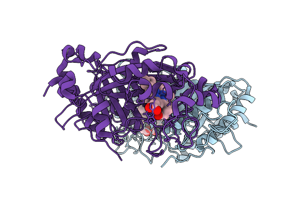
Organism: Escherichia coli bl21(de3)
Method: ELECTRON CRYSTALLOGRAPHY Resolution:2.85 Å Release Date: 2025-02-05 Classification: IMMUNE SYSTEM |