9SIH image
Deposition Date 2025-08-28
Release Date 2026-05-13
Last Version Date 2026-06-17
Entry Detail
PDB ID:
9SIH
Keywords:
Title:
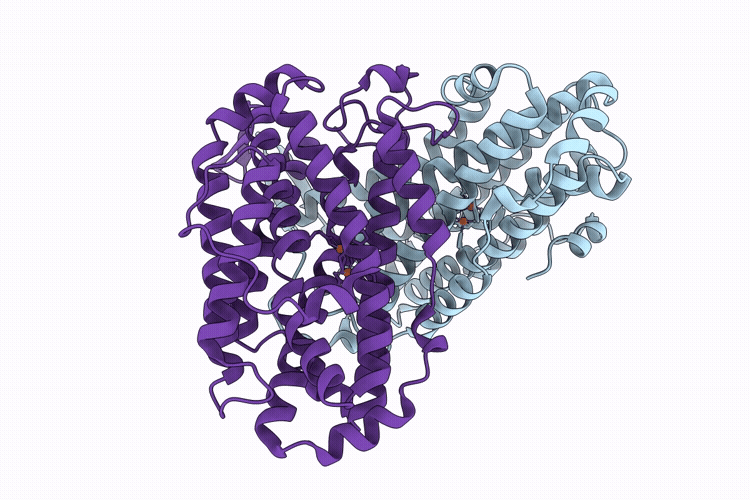
XFEL structure of Ribonucleotide reductase R2a Y122F mutant from E. coli,reduced form
Biological Source:
Source Organism(s):
Escherichia coli (Taxon ID: 562)
Method Details:
Experimental Method:
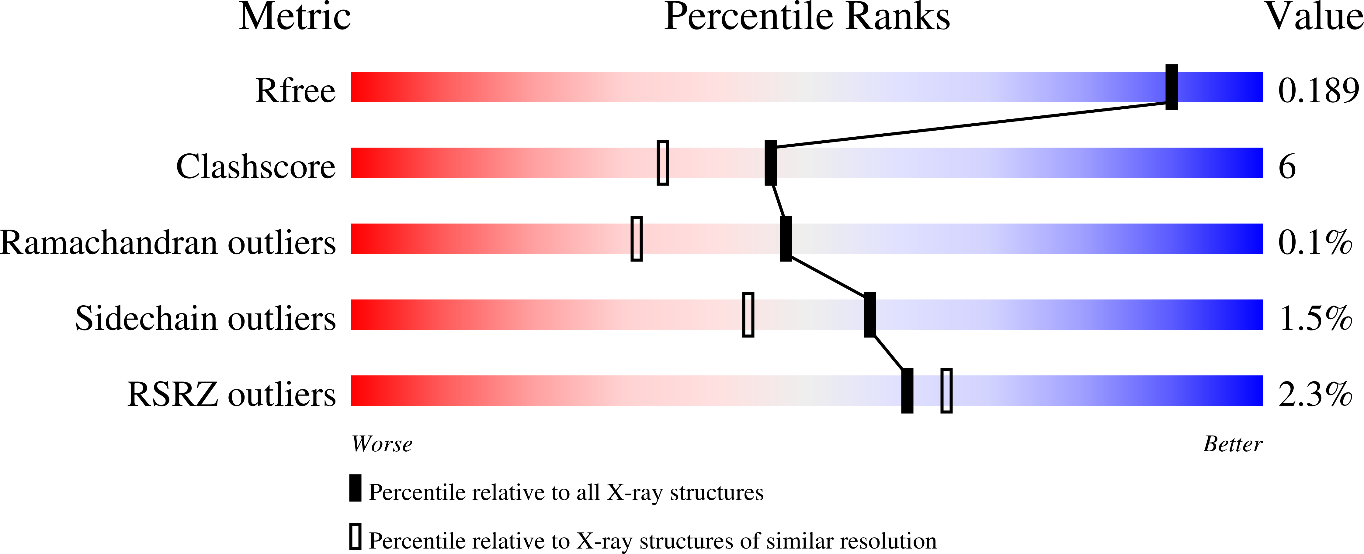
Resolution:
1.70 Å
R-Value Free:
0.19
R-Value Work:
0.15
R-Value Observed:
0.15
Space Group:
P 21 21 21
Macromolecular Entities
Structures with similar UniProt ID
Protein Blast
Polymer Type:polypeptide(L)
Molecule:Ribonucleoside-diphosphate re
Gene (Uniprot):nrdB
Mutagens:Y122F
Chain IDs:A, B
Chain Length:375
Number of Molecules:2
Biological Source:Escherichia coli
Ligand Molecules
Primary Citation

Abstact

Serial femtosecond crystallography (SFX) and continuous serial electron diffraction (c-SerialED) both enable high-resolution structure determination from protein microcrystals with minimal radiation damage, making it ideal for studying redox-active metalloenzymes. Here, c-SerialED and SFX were used to solve structures of the class Ia ribonucleotide reductase R2 subunit in oxidized (Fe(III)-Fe(III)), reduced (Fe(II)-Fe(II)), and re-oxidized states at approximately 1.8 A resolution, capturing three points in a redox reaction. These results demonstrate that c-SerialED can track reversible changes at the redox-site, enabling future time-resolved studies. Comparison between c-SerialED structures and SFX diffraction and emission data confirmed minimal radiation damage. Furthermore, previously reported structures use mercury in the crystallization condition and show mercury-induced conformational changes. Here, we use mercury-free crystallization conditions and reveal a water molecule in the redox center of the reduced state, absent in the previous structures, making these structures more representative of the physiological state.

Legend

Protein

Chemical

Disease

Primary Citation of related structures
Feedback Form
Name
Email
Institute
Feedback