
Search Count: 4,088
 |
Organism: Homo sapiens
Method: ELECTRON MICROSCOPY Release Date: 2026-05-27 Classification: MEMBRANE PROTEIN |
 |
Organism: Homo sapiens
Method: ELECTRON MICROSCOPY Resolution:2.60 Å Release Date: 2026-05-27 Classification: MEMBRANE PROTEIN |
 |
Organism: Homo sapiens
Method: ELECTRON MICROSCOPY Release Date: 2021-10-06 Classification: MEMBRANE PROTEIN |
 |
Organism: Homo sapiens
Method: ELECTRON MICROSCOPY Release Date: 2021-10-06 Classification: MEMBRANE PROTEIN |
 |
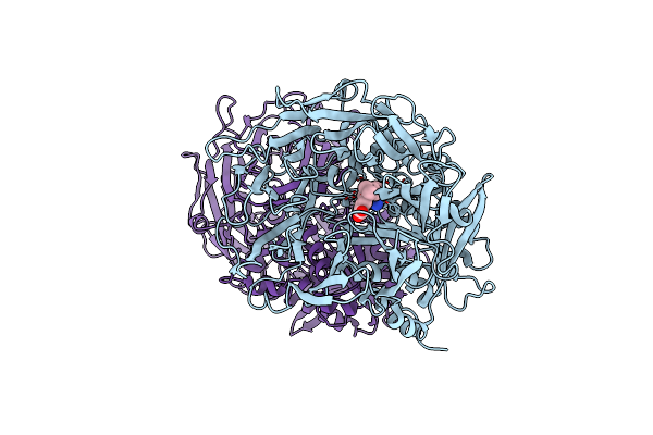
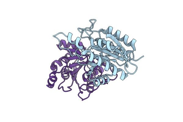
Human Dpp1 In Complex With (2S)-2-Amino-N-((1S)-1-Cyano-2-(4- Phenylphenyl)Ethyl)Butanamide
Organism: Homo sapiens
Method: X-RAY DIFFRACTION Resolution:2.40 Å Release Date: 2014-03-19 Classification: HYDROLASE Ligands: NAG, 6AO, CL, GOL |
 |
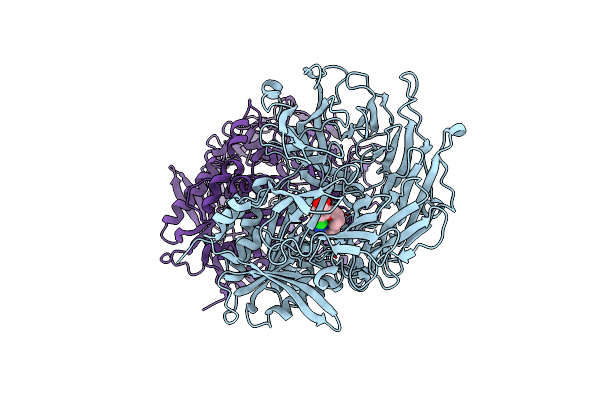
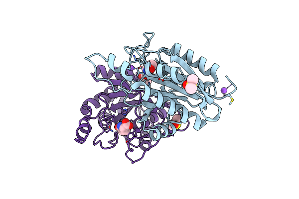
Human Dpp1 In Complex With (2S)-N-((1S)-1-Cyano-2-(4-(4-Cyanophenyl) Phenyl)Ethyl)Piperidine-2-Carboxamide
Organism: Homo sapiens
Method: X-RAY DIFFRACTION Resolution:2.35 Å Release Date: 2014-03-19 Classification: HYDROLASE Ligands: NAG, GDI, CL |
 |
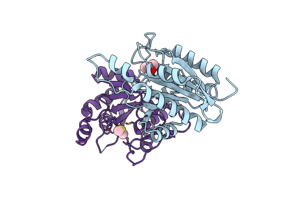
Human Dpp1 In Complex With 4-Amino-N-((1S)-1-Cyano-2-(4-(4- Cyanophenyl)Phenyl)Ethyl)Tetrahydropyran-4-Carboxamide
Organism: Homo sapiens
Method: X-RAY DIFFRACTION Resolution:2.40 Å Release Date: 2014-03-19 Classification: HYDROLASE Ligands: NAG, U6B, CL |
 |
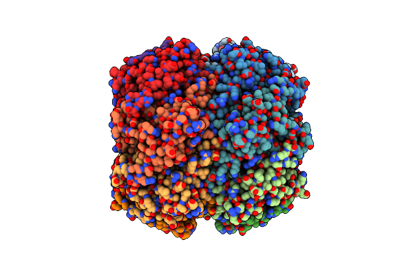
Human Dpp1 In Complex With (2S,4S)-N-((1S)-1-Cyano-2-(4-(4- Cyanophenyl)Phenyl)Ethyl)-4-Hydroxy-Piperidine-2-Carboxamide
Organism: Homo sapiens
Method: X-RAY DIFFRACTION Resolution:2.20 Å Release Date: 2014-03-19 Classification: HYDROLASE Ligands: NAG, W2C, CL |
 |
Crystal Structure Of A Mutant Pyrrolidone Carboxyl Peptidase (A199P) From P. Furiosus
Organism: Pyrococcus furiosus
Method: X-RAY DIFFRACTION Resolution:2.30 Å Release Date: 2007-10-02 Classification: HYDROLASE |
 |
Crystal Structure Of Human Dipeptidyl Peptidase Iv (Dppiv) Complexed With Cyanopyrrolidine (C5-Pro-Pro) Inhibitor 21Ac
Organism: Homo sapiens
Method: X-RAY DIFFRACTION Resolution:2.40 Å Release Date: 2006-07-04 Classification: HYDROLASE Ligands: ADF |
 |
Crystal Structure Of Human Dipeptidyl Peptidase Iv (Dppiv) Complexed With Cyanopyrrolidine (C5-Pro-Pro) Inhibitor 21Ag
Organism: Homo sapiens
Method: X-RAY DIFFRACTION Resolution:2.30 Å Release Date: 2006-07-04 Classification: HYDROLASE Ligands: ACF |
 |
Crystal Structure Of Human Dipeptidyl Peptidase Iv (Dppiv) Complexed With Cyanopyrrolidine (C5-Pro-Pro) Inhibitor 24B
Organism: Homo sapiens
Method: X-RAY DIFFRACTION Resolution:2.00 Å Release Date: 2006-07-04 Classification: HYDROLASE Ligands: AAF |
 |
Organism: Thermococcus litoralis
Method: X-RAY DIFFRACTION Resolution:1.73 Å Release Date: 1998-07-15 Classification: PEPTIDASE Ligands: SO4 |
 |
Crystal Structure Of Pyrrolidone Carboxylate Peptidase I From Deionococcus Radiodurans R1
Organism: Deinococcus radiodurans (strain atcc 13939 / dsm 20539 / jcm 16871 / lmg 4051 / nbrc 15346 / ncimb 9279 / r1 / vkm b-1422)
Method: X-RAY DIFFRACTION Resolution:1.84 Å Release Date: 2019-01-16 Classification: HYDROLASE |
 |
Crystal Structure Of Pyrrolidone Carboxylate Peptidase I From Deinococcus Radiodurans R1 Bound To Pyroglutamate
Organism: Deinococcus radiodurans r1
Method: X-RAY DIFFRACTION Resolution:1.55 Å Release Date: 2019-01-16 Classification: HYDROLASE |
 |
Crystal Structure Of Pyrrolidone Carboxylate Peptidase I With Disordered Loop A From Deinococcus Radiodurans R1
Organism: Deinococcus radiodurans r1
Method: X-RAY DIFFRACTION Resolution:1.70 Å Release Date: 2019-01-16 Classification: HYDROLASE Ligands: DMS |
 |
Structure Of E.Coli Class 2 L-Asparaginase Ecaiii, Mutant Rdm1-38 (R207C, D210S, S211V)
Organism: Escherichia coli
Method: X-RAY DIFFRACTION Resolution:1.95 Å Release Date: 2022-07-13 Classification: HYDROLASE |
 |
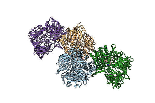
X-Ray Structure Of Hydrogenosomal Processing Peptidase (Hpp), E56Q Inactive Mutant, From Trichomonas Vaginalis Co-Crystallized With Presequence Peptide From Ferredoxin Oxidoreductase (Pfo) - Not Visible In The Structure Model
Organism: Trichomonas vaginalis
Method: X-RAY DIFFRACTION Resolution:2.65 Å Release Date: 2025-11-05 Classification: HYDROLASE Ligands: SO4, GOL, ZN |
 |
The Structure Of Clpp At 2.3 Angstrom Resolution Suggests A Model For Atp-Dependent Proteolysis
Organism: Escherichia coli
Method: X-RAY DIFFRACTION Resolution:2.30 Å Release Date: 1998-06-17 Classification: PEPTIDASE |
 |
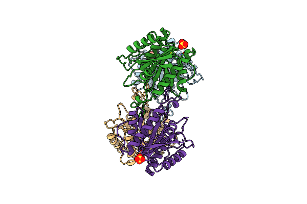
X-Ray Structure Of Hydrogenosomal Processing Peptidase (Hpp), E56Q Inactive Mutant, From Trichomonas Vaginalis Co-Crystallized With Presequence Peptide From Adenylate Kinase (Ak) - Not Visible In The Structure Model
Organism: Trichomonas vaginalis
Method: X-RAY DIFFRACTION Resolution:2.00 Å Release Date: 2025-11-05 Classification: HYDROLASE Ligands: SO4, GOL, ZN |