5Z47 image
Deposition Date 2018-01-10
Release Date 2019-01-16
Last Version Date 2023-11-22
Entry Detail
PDB ID:
5Z47
Keywords:
Title:
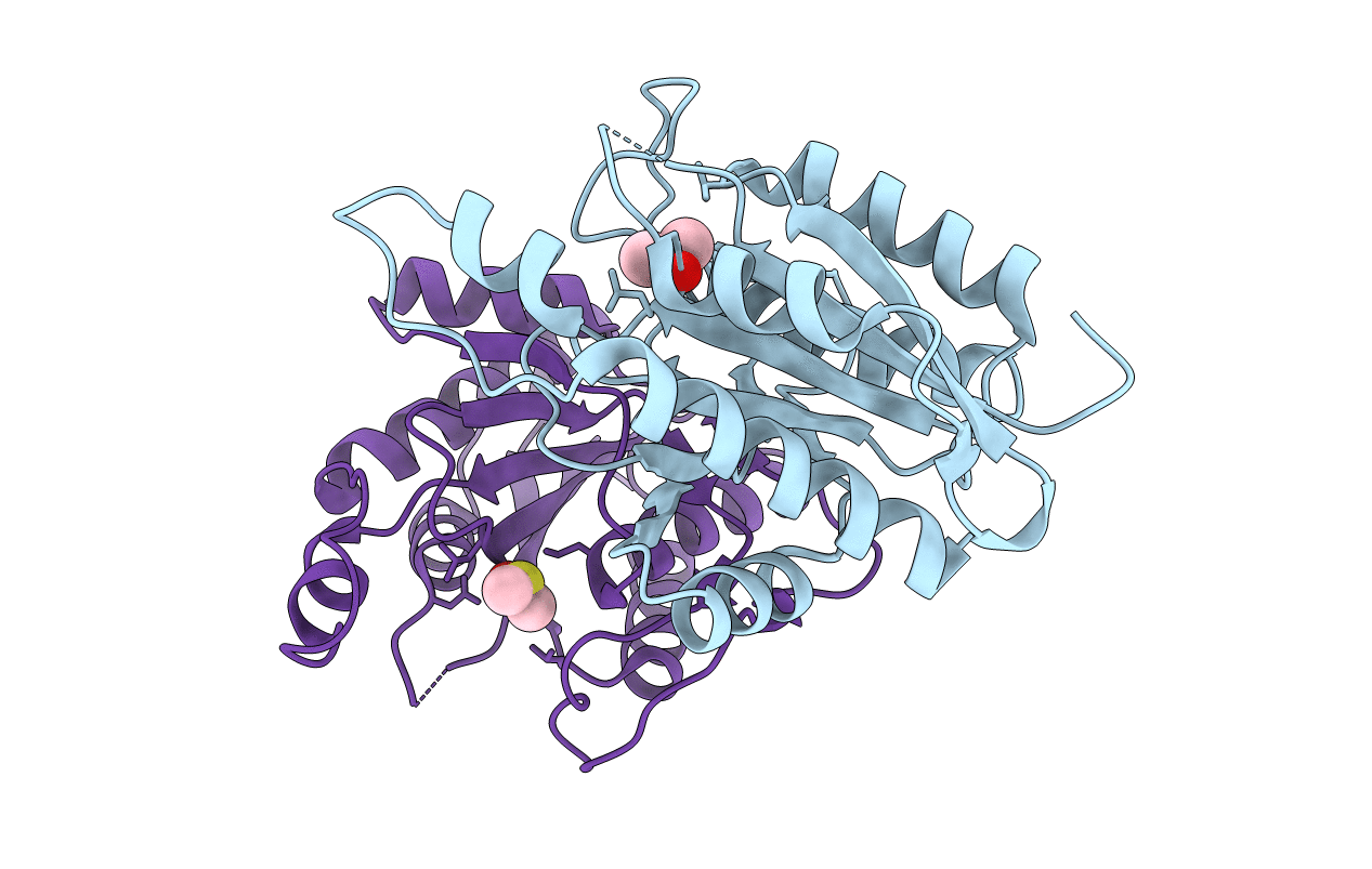
Crystal structure of pyrrolidone carboxylate peptidase I with disordered loop A from Deinococcus radiodurans R1
Biological Source:
Source Organism(s):
Expression System(s):
Method Details:
Experimental Method:
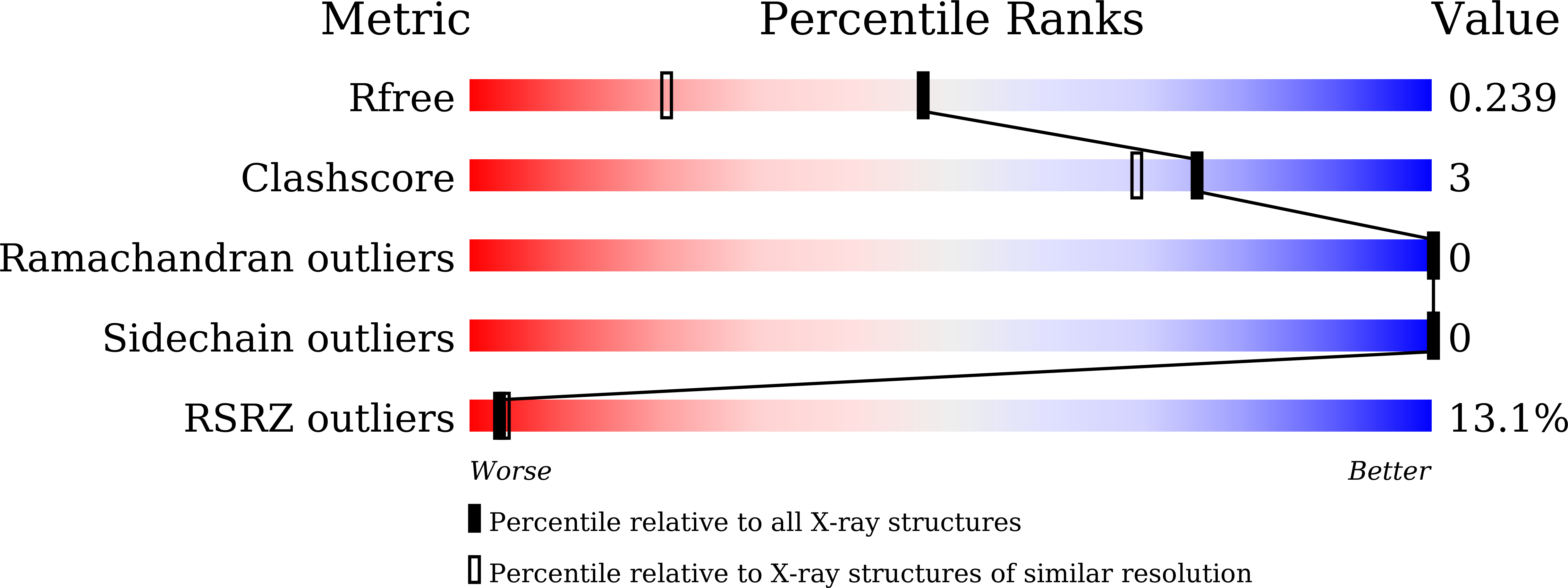
Resolution:
1.70 Å
R-Value Free:
0.23
R-Value Work:
0.20
R-Value Observed:
0.20
Space Group:
I 1 2 1
Macromolecular Entities
Structures with similar UniProt ID
Protein Blast
Polymer Type:polypeptide(L)
Molecule:Pyrrolidone-carboxylate pepti
Gene (Uniprot):pcp
Chain IDs:A, B
Chain Length:220
Number of Molecules:2
Biological Source:Deinococcus radiodurans R1
Modified Residue
Compound ID Chain ID Parent Comp ID Details 2D Image
CSX A CYS modified residue
Ligand Molecules
Primary Citation
Crystal structures of pyrrolidone-carboxylate peptidase I from Deinococcus radiodurans reveal the mechanism of L-pyroglutamate recognition.
Acta Crystallogr D Struct Biol 75 308 316 (2019)
PMID: 30950401 DOI: 10.1107/S2059798319000676

Abstact

Pyrrolidone-carboxylate peptidase (PCP) catalyzes the removal of an unusual amino acid, L-pyroglutamate (pG), from the N-termini of peptides and proteins. It has implications in the functional regulation of different peptides in both prokaryotes and eukaryotes. However, the pG-recognition mechanism of the PCP enzyme remains largely unknown. Here, crystal structures of PCP I from Deinococcus radiodurans (PCPdr) are reported in pG-free and pG-bound forms at resolutions of 1.73 and 1.55 Å, respectively. Four protomers in PCPdr form a tetrameric structure. The residues responsible for recognizing the pG residue are mostly contributed by a flexible loop (loop A) that is present near the active site. These residues are conserved in all known PCPs I, including those from mammals. Phe9 and Phe12 of loop A form stacking interactions with the pyrrolidone ring of pG, while Asn18 forms a hydrogen bond to OE of pG. The main chain of a nonconserved residue, Leu71, forms two hydrogen bonds to NH and OE of pG. Thus, pG is recognized in the S1 substrate subsite of the enzyme by both van der Waals and polar interactions, which provide specificity for the pG residue of the peptide. In contrast to previously reported PCP I structures, the PCPdr tetramer is in a closed conformation with an inaccessible active site. The structures show that the active site can be accessed by the substrates via disordering of loop A. This disordering could also prevent product inhibition by releasing the bound pG product from the S1 subsite, thus allowing the enzyme to engage a fresh substrate.

Legend

Protein

Chemical

Disease

Primary Citation of related structures
Feedback Form
Name
Email
Institute
Feedback