

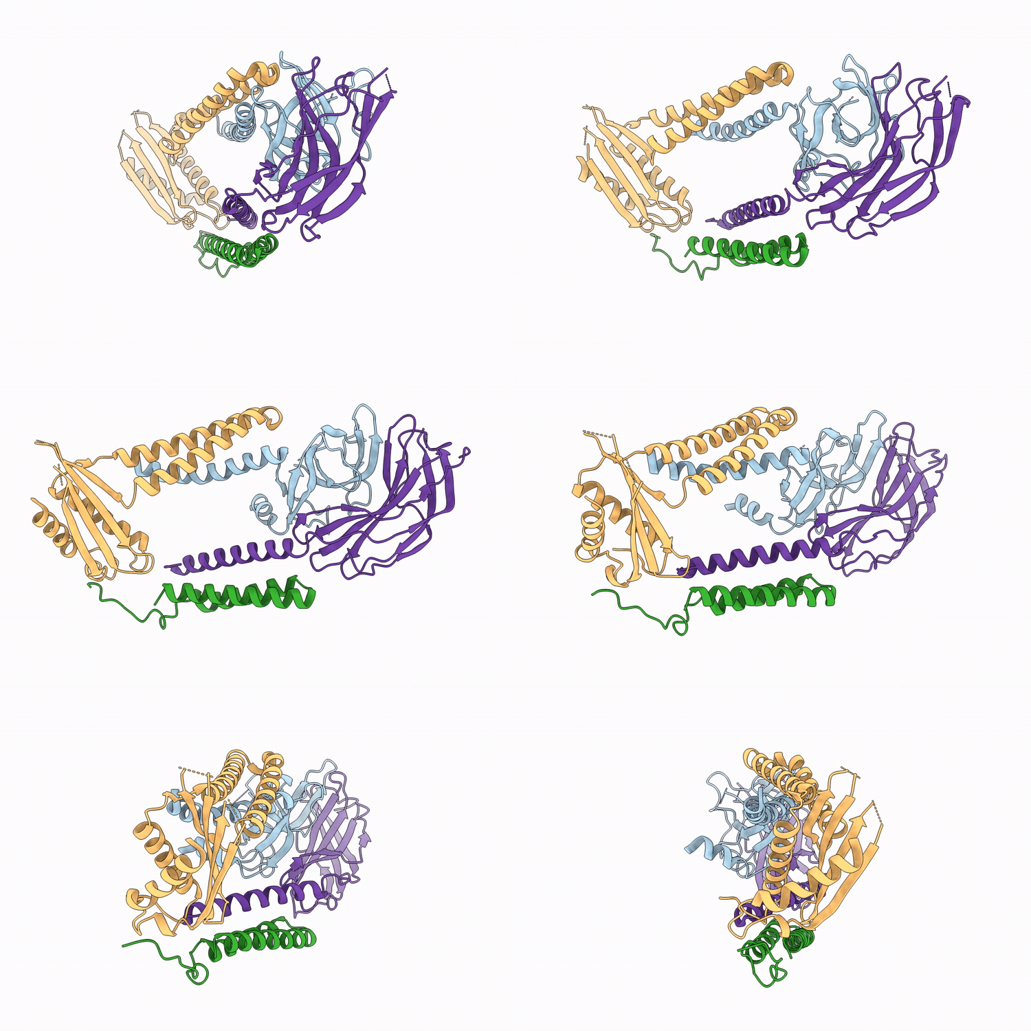
9RJC image
Deposition Date 2025-06-12
Release Date 2026-05-27
Last Version Date 2026-06-03
Entry Detail
PDB ID:
9RJC
Title:
Apo Structure of the Human Signal Peptidase
Biological Source:
Source Organism(s):
Homo sapiens (Taxon ID: 9606)
Expression System(s):
Method Details:
Experimental Method:
Resolution:
4.20 Å
Aggregation State:
PARTICLE
Reconstruction Method:
SINGLE PARTICLE
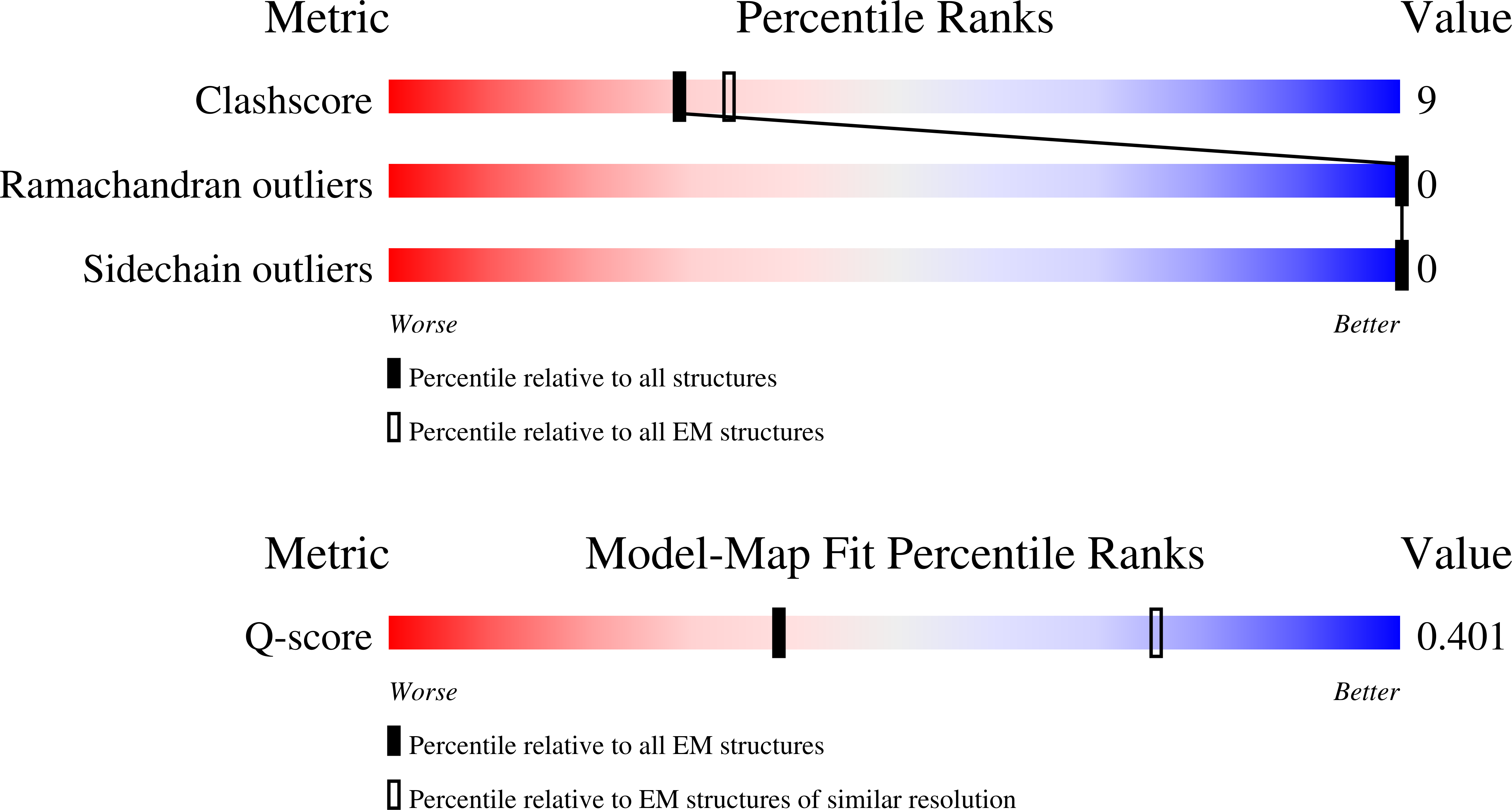
Macromolecular Entities
Polymer Type:polypeptide(L)
Molecule:Signal peptidase complex cata
Gene (Uniprot):SEC11A
Chain IDs:A
Chain Length:177
Number of Molecules:1
Biological Source:Homo sapiens
Polymer Type:polypeptide(L)
Molecule:Signal peptidase complex subu
Gene (Uniprot):SPCS1
Chain IDs:B
Chain Length:64
Number of Molecules:1
Biological Source:Homo sapiens
Polymer Type:polypeptide(L)
Molecule:Signal peptidase complex subu
Gene (Uniprot):SPCS2
Chain IDs:C
Chain Length:177
Number of Molecules:1
Biological Source:Homo sapiens
Polymer Type:polypeptide(L)
Molecule:Signal peptidase complex subu
Gene (Uniprot):SPCS3
Chain IDs:D
Chain Length:174
Number of Molecules:1
Biological Source:Homo sapiens
Ligand Molecules
Primary Citation
Structural basis of signal peptide recognition by the signal peptidase complex.
Nat Commun ? ? ? (2026)
PMID: 42173868 DOI: 10.1038/s41467-026-73423-3

Abstact

The signal peptidase complex (SPC) is responsible for cleaving signal peptides (SPs) from approximately 10% of the human proteome. SPs are characterized by a tripartite structure, consisting of an N-terminal n-region, a central helical h-region and a C-terminal c-region, each defined by rather general chemical properties rather than strict sequence conservation. Despite their sequence diversity, SPC recognizes and processes SPs with exquisite specificity. Here, we present a 2.6 A cryo-EM map of the human SPC-A, one of two SPC paralogs, bound to a model SP. The c-region binds to a hydrophobic binding groove near the active site, a narrow gate marks the transition from c- to h-region, and the h-region localizes in a transmembrane (TM) window. Substrate engagement stabilizes N- and C-terminal helices of Sec11A, which frame the SP and are unresolved in the apo structure. Molecular dynamics (MD) simulations confirm a stable hydrogen-bonding network at the c-region and indicate dynamic interactions within a thinned lipid environment at the TM window. AlphaFold modeling supports this binding mode across physiological SPs. Collectively, our structural and computational analyses explain how the SPC achieves its specificity by combining the selectivity of the luminal binding groove and of the transmembrane window.

Legend

Protein

Chemical

Disease

Primary Citation of related structures
Feedback Form
Name
Email
Institute
Feedback