

Search Count: 1,279
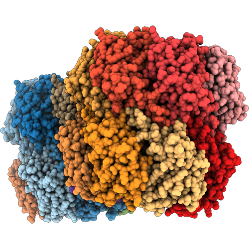 |
Structure Of Pseudomonas Aeruginosa Mutant Clpp2-R140A
Organism: Pseudomonas aeruginosa pao1
Method: X-RAY DIFFRACTION Resolution:3.00 Å Release Date: 2026-10-21 Classification: HYDROLASE |
Organism: Pseudomonas aeruginosa pao1
Method: X-RAY DIFFRACTION
Release Date: 2026-10-21
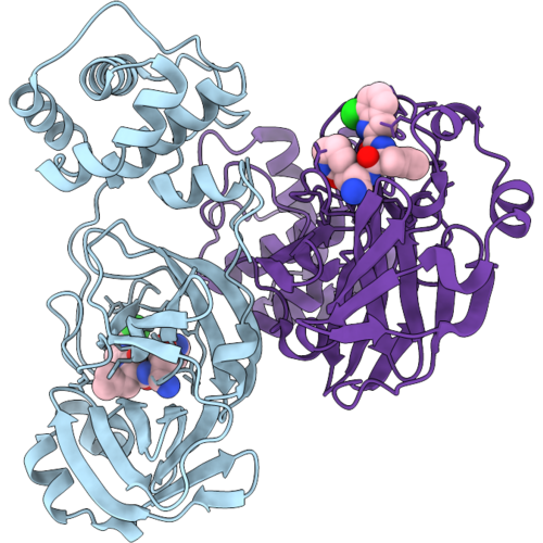 |
Crystal Structure Of Sars-Cov-2 Main Protease In Complex With Pbi0451
Organism: Severe acute respiratory syndrome coronavirus 2
Method: X-RAY DIFFRACTION Resolution:1.95 Å Release Date: 2026-10-07 Classification: VIRAL PROTEIN Ligands: ZQB |
Organism: Severe acute respiratory syndrome coronavirus 2
Method: X-RAY DIFFRACTION
Release Date: 2026-10-07
Ligands: ZQB
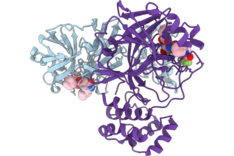 |
Crystal Structure Of Sars-Cov-2 Main Protease In Complex With Leritrelvir
Organism: Severe acute respiratory syndrome coronavirus 2
Method: X-RAY DIFFRACTION Resolution:1.76 Å Release Date: 2026-09-23 Classification: VIRAL PROTEIN Ligands: A1EN0 |
Organism: Severe acute respiratory syndrome coronavirus 2
Method: X-RAY DIFFRACTION
Release Date: 2026-09-23
Ligands: A1EN0
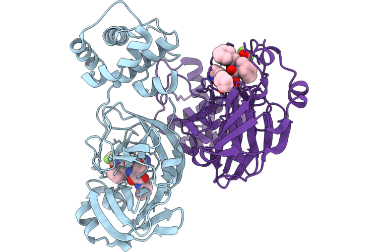 |
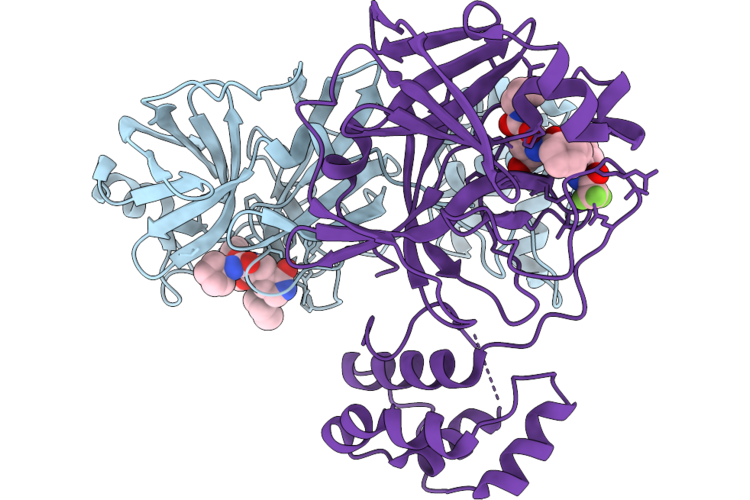
Crystal Structure Of Sars-Cov-2 Main Protease M49I Mutant In Complex With Leritrelvir
Organism: Severe acute respiratory syndrome coronavirus 2
Method: X-RAY DIFFRACTION Resolution:1.82 Å Release Date: 2026-09-23 Classification: VIRAL PROTEIN Ligands: 7ON |
Organism: Severe acute respiratory syndrome coronavirus 2
Method: X-RAY DIFFRACTION
Release Date: 2026-09-23
Ligands: 7ON
 |
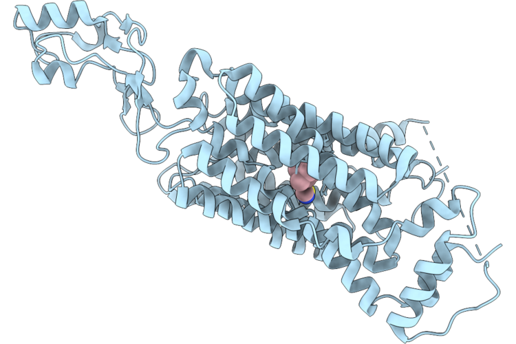
Crystal Structure Of Sars-Cov-2 Main Protease P132H Mutant In Complex With Leritrelvir
Organism: Severe acute respiratory syndrome coronavirus 2
Method: X-RAY DIFFRACTION Resolution:1.96 Å Release Date: 2026-09-23 Classification: VIRAL PROTEIN Ligands: A1EN0 |
Organism: Severe acute respiratory syndrome coronavirus 2
Method: X-RAY DIFFRACTION
Release Date: 2026-09-23
Ligands: A1EN0
 |
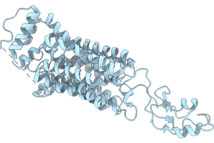
Structure Of Hoctn1-Ergothioneine Complex
Organism: Homo sapiens
Method: ELECTRON MICROSCOPY Release Date: 2026-09-23 Classification: MEMBRANE PROTEIN Ligands: LW8 |
Organism: Homo sapiens
Method: ELECTRON MICROSCOPY
Release Date: 2026-09-23
Ligands: LW8
 |
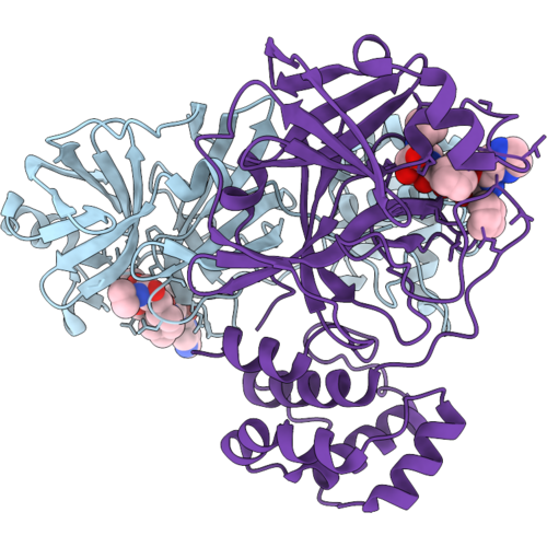
Structure Of Hoctn1 In Apo State
Organism: Homo sapiens
Method: ELECTRON MICROSCOPY Resolution:3.11 Å Release Date: 2026-09-23 Classification: MEMBRANE PROTEIN |
Organism: Homo sapiens
Method: ELECTRON MICROSCOPY
Release Date: 2026-09-23
 |
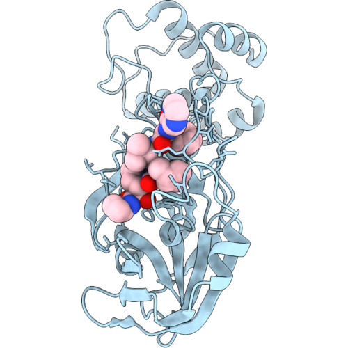
Crystal Structure Of Sars-Cov-2 Main Protease In Complex With Telaprevir
Organism: Severe acute respiratory syndrome coronavirus 2
Method: X-RAY DIFFRACTION Resolution:2.27 Å Release Date: 2026-09-09 Classification: VIRAL PROTEIN Ligands: SV6 |
Organism: Severe acute respiratory syndrome coronavirus 2
Method: X-RAY DIFFRACTION
Release Date: 2026-09-09
Ligands: SV6
 |
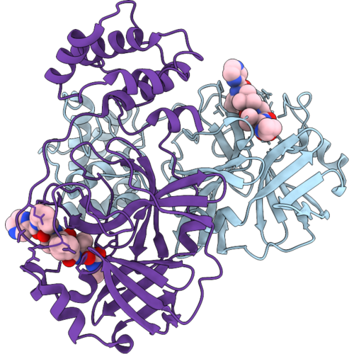
Crystal Structure Of Sars-Cov-2 Main Protease M49I In Complex With Telaprevir
Organism: Severe acute respiratory syndrome coronavirus 2
Method: X-RAY DIFFRACTION Resolution:1.96 Å Release Date: 2026-09-09 Classification: VIRAL PROTEIN Ligands: SV6 |
Organism: Severe acute respiratory syndrome coronavirus 2
Method: X-RAY DIFFRACTION
Release Date: 2026-09-09
Ligands: SV6
 |
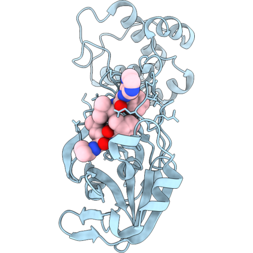
Crystal Structure Of Sars-Cov-2 Main Protease K90R Mutant In Complex With Telaprevir
Organism: Severe acute respiratory syndrome coronavirus 2
Method: X-RAY DIFFRACTION Resolution:2.09 Å Release Date: 2026-09-09 Classification: VIRAL PROTEIN Ligands: SV6 |
Organism: Severe acute respiratory syndrome coronavirus 2
Method: X-RAY DIFFRACTION
Release Date: 2026-09-09
Ligands: SV6
 |
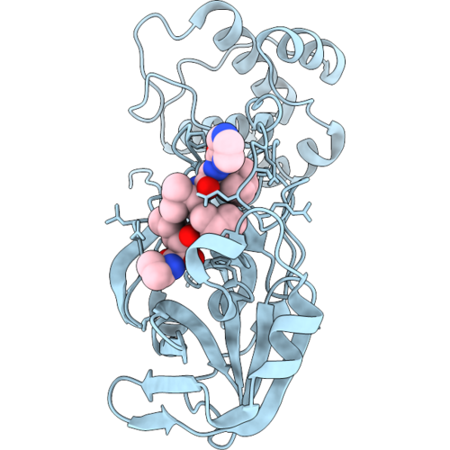
Crystal Structure Of Sars-Cov-2 Main Protease V186F Mutant In Complex With Telaprevir
Organism: Severe acute respiratory syndrome coronavirus 2
Method: X-RAY DIFFRACTION Resolution:2.47 Å Release Date: 2026-09-09 Classification: VIRAL PROTEIN Ligands: SV6 |
Organism: Severe acute respiratory syndrome coronavirus 2
Method: X-RAY DIFFRACTION
Release Date: 2026-09-09
Ligands: SV6
 |
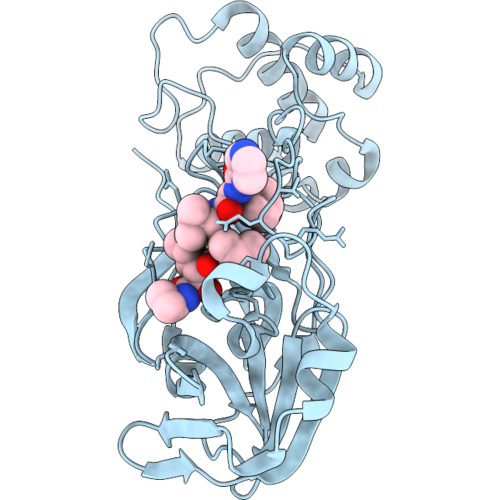
Crystal Structure Of Sars-Cov-2 Main Protease E166N Mutant In Complex With Telaprevir
Organism: Severe acute respiratory syndrome coronavirus 2
Method: X-RAY DIFFRACTION Resolution:2.45 Å Release Date: 2026-09-09 Classification: VIRAL PROTEIN Ligands: SV6 |
Organism: Severe acute respiratory syndrome coronavirus 2
Method: X-RAY DIFFRACTION
Release Date: 2026-09-09
Ligands: SV6
 |
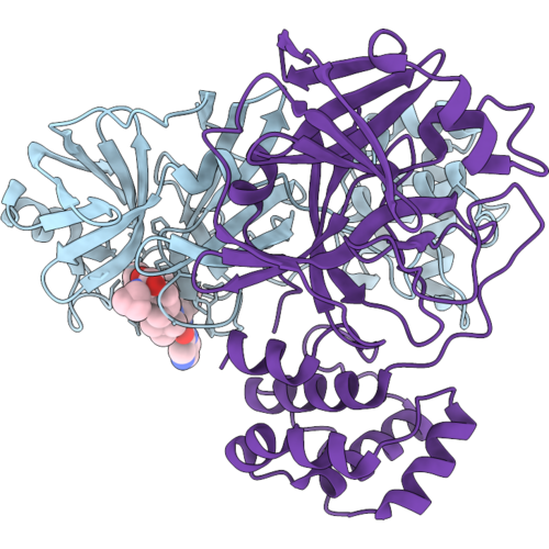
Crystal Structure Of Sars-Cov-2 Main Protease E166R Mutant In Complex With Telaprevir
Organism: Severe acute respiratory syndrome coronavirus 2
Method: X-RAY DIFFRACTION Resolution:1.91 Å Release Date: 2026-09-09 Classification: VIRAL PROTEIN Ligands: SV6 |
Organism: Severe acute respiratory syndrome coronavirus 2
Method: X-RAY DIFFRACTION
Release Date: 2026-09-09
Ligands: SV6
 |
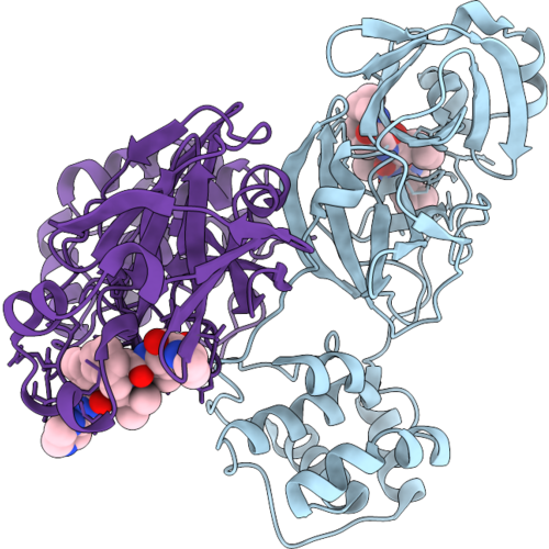
Crystal Structure Of Sars-Cov-2 Main Protease E166V Mutant In Complex With Telaprevir
Organism: Severe acute respiratory syndrome coronavirus 2
Method: X-RAY DIFFRACTION Resolution:2.08 Å Release Date: 2026-09-09 Classification: VIRAL PROTEIN Ligands: SV6 |
Organism: Severe acute respiratory syndrome coronavirus 2
Method: X-RAY DIFFRACTION
Release Date: 2026-09-09
Ligands: SV6
 |
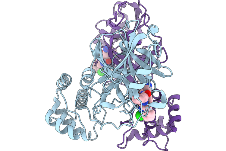
Crystal Structure Of Sars-Cov Main Protease In Complex With Telaprevir
Organism: Severe acute respiratory syndrome coronavirus
Method: X-RAY DIFFRACTION Resolution:2.02 Å Release Date: 2026-09-09 Classification: VIRAL PROTEIN Ligands: SV6 |
Organism: Severe acute respiratory syndrome coronavirus
Method: X-RAY DIFFRACTION
Release Date: 2026-09-09
Ligands: SV6
 |
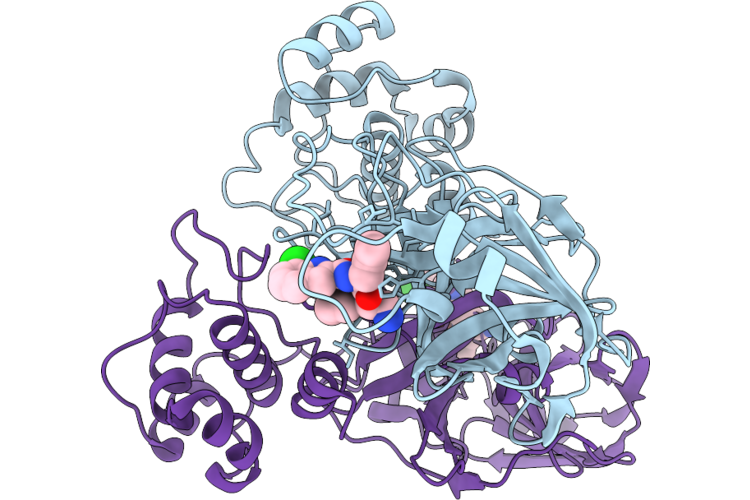
Crystal Structure Of Mers Main Protease In Complex With Pomotrelvir
Organism: Middle east respiratory syndrome-related coronavirus (isolate united kingdom/h123990006/2012)
Method: X-RAY DIFFRACTION Resolution:2.14 Å Release Date: 2026-08-26 Classification: VIRAL PROTEIN Ligands: ZQB |
Organism: Middle east respiratory syndrome-related coronavirus (isolate united kingdom/h123990006/2012)
Method: X-RAY DIFFRACTION
Release Date: 2026-08-26
Ligands: ZQB
 |
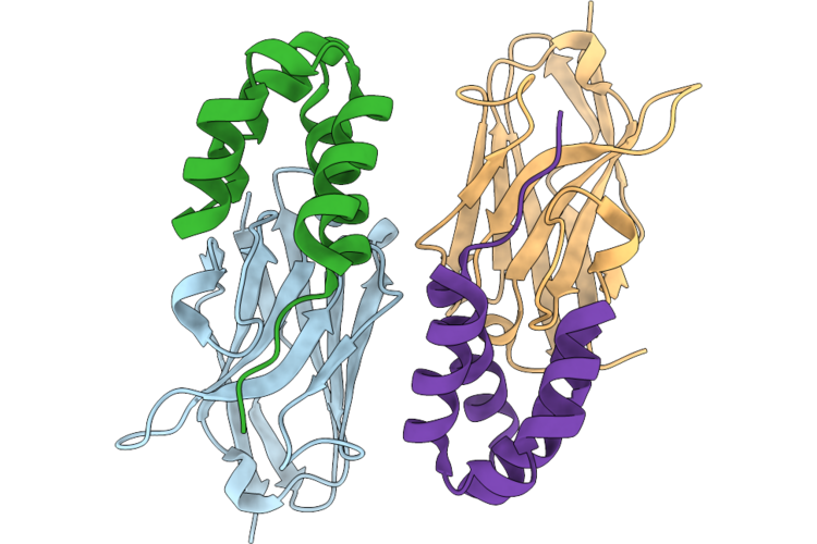
Crystal Structure Of Hcov-Nl63 Main Protease With Pomotrelvir
Organism: Human coronavirus nl63
Method: X-RAY DIFFRACTION Resolution:1.83 Å Release Date: 2026-08-26 Classification: VIRAL PROTEIN Ligands: ZQB |
Organism: Human coronavirus nl63
Method: X-RAY DIFFRACTION
Release Date: 2026-08-26
Ligands: ZQB
 |
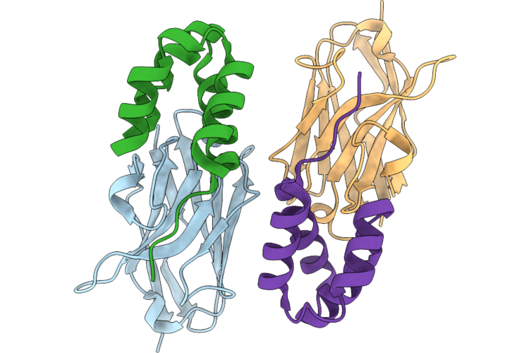
Genetically Encoded Fluorescent Nanobody H11 With 4-(7-Nitro-2,1,3-Benzoxadiazolyl)-Alanine (Nbdo) At The W37 Position Complexed With Arc-Nl
Organism: Vicugna pacos, Homo sapiens
Method: X-RAY DIFFRACTION Resolution:1.56 Å Release Date: 2026-08-26 Classification: PROTEIN BINDING |
Organism: Vicugna pacos, Homo sapiens
Method: X-RAY DIFFRACTION
Release Date: 2026-08-26
 |
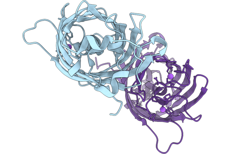
Genetically Encoded Fluorescent Nanobody H11 With 4-(7-Nitro-2,1,3-Benzoselenadiazolyl)-Alanine (Nbdse) At The W37 Position Complexed With Arc-Nl
Organism: Vicugna pacos, Homo sapiens
Method: X-RAY DIFFRACTION Resolution:2.84 Å Release Date: 2026-08-26 Classification: PROTEIN BINDING |
Organism: Vicugna pacos, Homo sapiens
Method: X-RAY DIFFRACTION
Release Date: 2026-08-26
 |
Nowgfp Mutant With Non-Canonical Amino Acid 4-(7-Nitro-2,1,3-Benzoxadiazolyl)-Alanine (Nbdo) In The Chromophore Site
Organism: Synthetic construct
Method: X-RAY DIFFRACTION Resolution:1.36 Å Release Date: 2026-08-26 Classification: FLUORESCENT PROTEIN |
Organism: Synthetic construct
Method: X-RAY DIFFRACTION
Release Date: 2026-08-26