

Search Count: 539
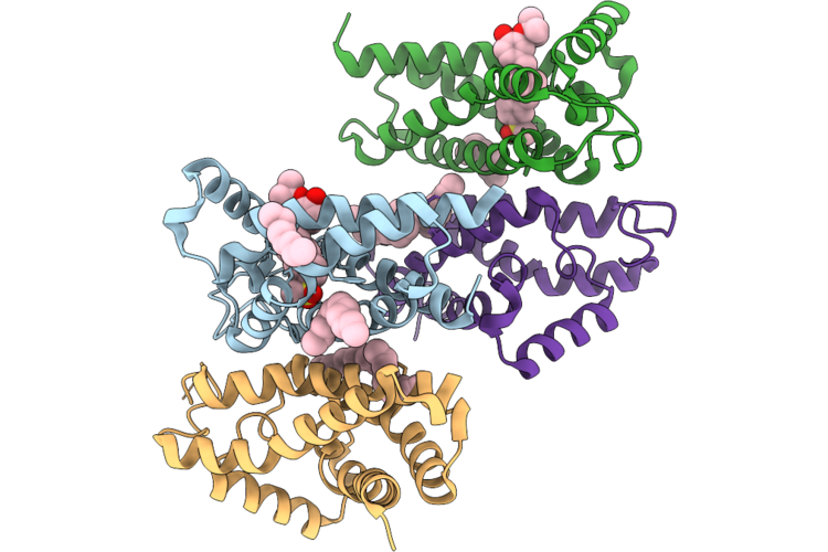 |
Crystal Structure Of Sigma-2 Receptor With Zervimesine (Ct1812)
Organism: Bos taurus
Method: X-RAY DIFFRACTION Resolution:2.74 Å Release Date: 2026-08-19 Classification: MEMBRANE PROTEIN Ligands: A1C2J, OLC |
Organism: Bos taurus
Method: X-RAY DIFFRACTION
Release Date: 2026-08-19
Ligands: A1C2J, OLC
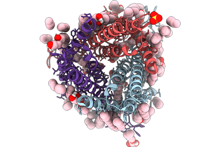 |
Crystal Structure Of Sphingobium Lactosutens Styrene-Oxide Isomerase With Unknown Ligand
Organism: Sphingobium lactosutens
Method: X-RAY DIFFRACTION Resolution:1.70 Å Release Date: 2026-07-22 Classification: MEMBRANE PROTEIN Ligands: LFA, OLC, OLB, HEM, SO4, CL |
Organism: Sphingobium lactosutens
Method: X-RAY DIFFRACTION
Release Date: 2026-07-22
Ligands: LFA, OLC, OLB, HEM, SO4, CL
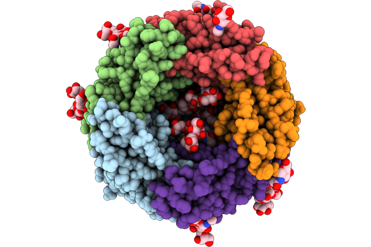 |
Cryoem Structure Of Human Alpha1Beta3Gamma2L Gaba(A)R In Cl47A
Organism: Homo sapiens
Method: ELECTRON MICROSCOPY Release Date: 2026-06-17 Classification: MEMBRANE PROTEIN Ligands: D10, PT5, R16, EPE, PX2, OLC, PLM, HEX, NAG, PGW, CL |
Organism: Homo sapiens
Method: ELECTRON MICROSCOPY
Release Date: 2026-06-17
Ligands: D10, PT5, R16, EPE, PX2, OLC, PLM, HEX, NAG, PGW, CL
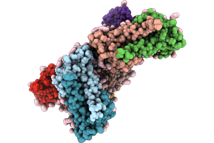 |
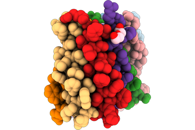
Crystal Structure Of Eukaryotic Heliorhodopsin Cv06 From Chromera Velia Strain Ccmp2878
Organism: Chromera velia ccmp2878
Method: X-RAY DIFFRACTION Resolution:2.60 Å Release Date: 2026-06-17 Classification: MEMBRANE PROTEIN Ligands: LFA, RET, OLC |
Organism: Chromera velia ccmp2878
Method: X-RAY DIFFRACTION
Release Date: 2026-06-17
Ligands: LFA, RET, OLC
 |
Designed Pentameric Proton Channel Llql I13S
Organism: Synthetic construct
Method: X-RAY DIFFRACTION Resolution:2.80 Å Release Date: 2026-05-06 Classification: MEMBRANE PROTEIN Ligands: OLC, PEG, ZN |
Organism: Synthetic construct
Method: X-RAY DIFFRACTION
Release Date: 2026-05-06
Ligands: OLC, PEG, ZN
 |
The Lbd-Tmd Structure Of Native Mouse Ampar With 4 Tarps
Organism: Mus musculus
Method: ELECTRON MICROSCOPY Resolution:3.13 Å Release Date: 2026-04-29 Classification: SIGNALING PROTEIN Ligands: PLM, OLC, POV |
Organism: Mus musculus
Method: ELECTRON MICROSCOPY
Release Date: 2026-04-29
Ligands: PLM, OLC, POV
 |
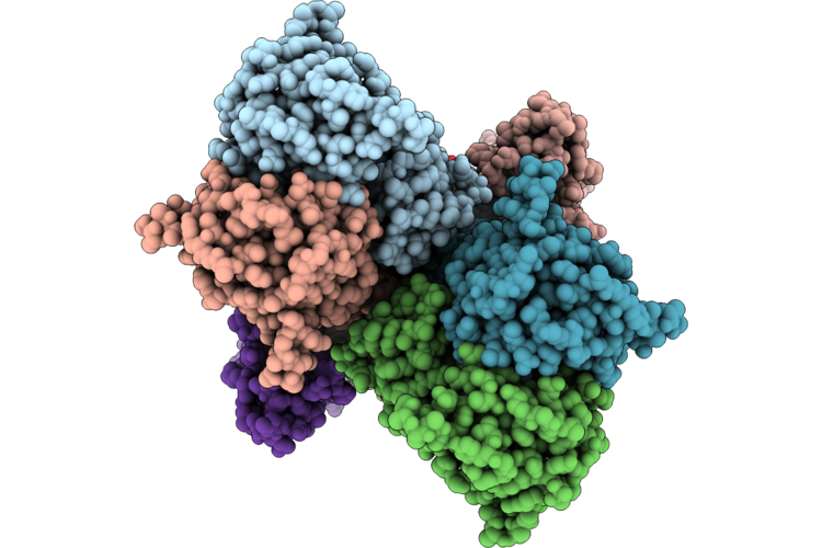
The Tmd Structure Of Native Mouse Ampar With 4 Tarps
Organism: Mus musculus
Method: ELECTRON MICROSCOPY Resolution:2.80 Å Release Date: 2026-04-29 Classification: SIGNALING PROTEIN Ligands: PLM, OLC, POV |
Organism: Mus musculus
Method: ELECTRON MICROSCOPY
Release Date: 2026-04-29
Ligands: PLM, OLC, POV
 |
The Lbd-Tmd Structure Of Native Mouse Ampar With 2 Tarps 2 Cnihs
Organism: Mus musculus
Method: ELECTRON MICROSCOPY Resolution:3.04 Å Release Date: 2026-04-29 Classification: SIGNALING PROTEIN Ligands: PLM, OLC, POV |
Organism: Mus musculus
Method: ELECTRON MICROSCOPY
Release Date: 2026-04-29
Ligands: PLM, OLC, POV
 |
The Tmd Structure Of Native Mouse Ampar With 2 Tarps 2 Cnihs
Organism: Mus musculus
Method: ELECTRON MICROSCOPY Resolution:2.87 Å Release Date: 2026-04-29 Classification: SIGNALING PROTEIN Ligands: PLM, OLC, POV |
Organism: Mus musculus
Method: ELECTRON MICROSCOPY
Release Date: 2026-04-29
Ligands: PLM, OLC, POV
 |
The Lbd-Tmd Structure Of Native Mouse Ampar With 3 Tarps 1 Cnih
Organism: Mus musculus
Method: ELECTRON MICROSCOPY Resolution:3.36 Å Release Date: 2026-04-29 Classification: SIGNALING PROTEIN Ligands: PLM, OLC, POV |
Organism: Mus musculus
Method: ELECTRON MICROSCOPY
Release Date: 2026-04-29
Ligands: PLM, OLC, POV
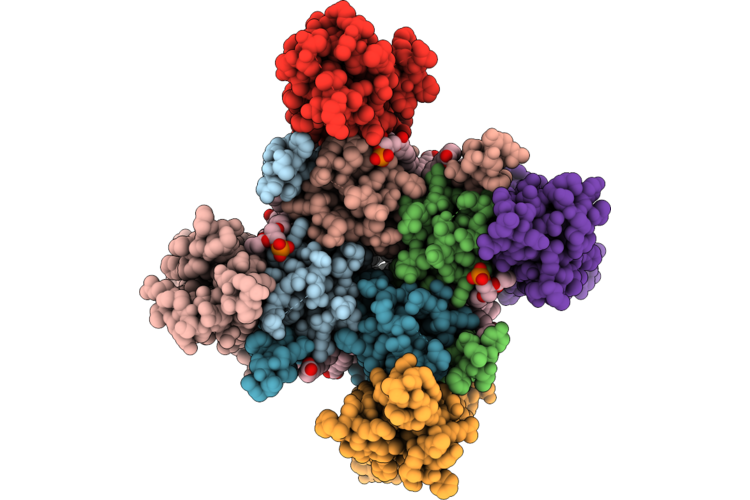 |
The Tmd Structure Of Native Mouse Ampar With 3 Tarps 1 Cnih
Organism: Mus musculus
Method: ELECTRON MICROSCOPY Resolution:3.08 Å Release Date: 2026-04-29 Classification: SIGNALING PROTEIN Ligands: PLM, OLC, POV |
Organism: Mus musculus
Method: ELECTRON MICROSCOPY
Release Date: 2026-04-29
Ligands: PLM, OLC, POV
 |
The Lbd-Tmd Structure Of Native Mouse Ampar With 2 Tarps 2 Cnihs And Prrt1/Syndig4
Organism: Mus musculus
Method: ELECTRON MICROSCOPY Resolution:3.36 Å Release Date: 2026-04-29 Classification: SIGNALING PROTEIN Ligands: PLM, OLC, POV |
Organism: Mus musculus
Method: ELECTRON MICROSCOPY
Release Date: 2026-04-29
Ligands: PLM, OLC, POV
 |
The Tmd Structure Of Native Mouse Ampar With 2 Tarps 2 Cnihs And Prrt1/Syndig4
Organism: Mus musculus
Method: ELECTRON MICROSCOPY Resolution:3.04 Å Release Date: 2026-04-29 Classification: SIGNALING PROTEIN Ligands: PLM, OLC, POV |
Organism: Mus musculus
Method: ELECTRON MICROSCOPY
Release Date: 2026-04-29
Ligands: PLM, OLC, POV
 |
The Lbd-Tmd Structure Of Native Mouse Ampar With 3 Tarps 1 Cnih And Prrt1/Syndig4
Organism: Mus musculus
Method: ELECTRON MICROSCOPY Resolution:3.75 Å Release Date: 2026-04-29 Classification: SIGNALING PROTEIN Ligands: PLM, OLC, POV |
Organism: Mus musculus
Method: ELECTRON MICROSCOPY
Release Date: 2026-04-29
Ligands: PLM, OLC, POV
 |
The Lbd-Tmd Structure Of Native Mouse Ampar With 2 Tarps
Organism: Mus musculus
Method: ELECTRON MICROSCOPY Resolution:3.45 Å Release Date: 2026-04-29 Classification: SIGNALING PROTEIN Ligands: PLM, OLC, POV |
Organism: Mus musculus
Method: ELECTRON MICROSCOPY
Release Date: 2026-04-29
Ligands: PLM, OLC, POV
 |
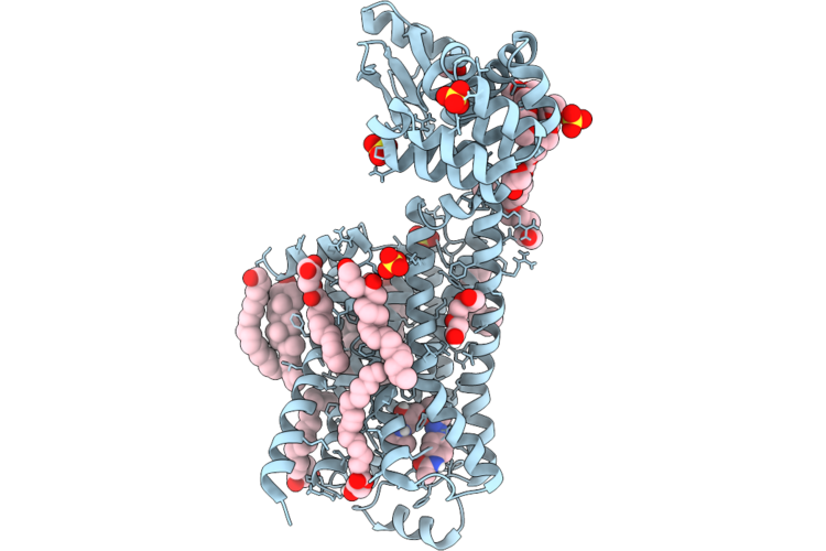
The Lbd-Tmd Structure Of Native Mouse Ampar With 3 Tarps
Organism: Mus musculus
Method: ELECTRON MICROSCOPY Resolution:3.33 Å Release Date: 2026-04-29 Classification: SIGNALING PROTEIN Ligands: PLM, OLC, POV |
Organism: Mus musculus
Method: ELECTRON MICROSCOPY
Release Date: 2026-04-29
Ligands: PLM, OLC, POV
 |
Dark Structure Of Beta-2 Adrenergic Receptor With Photoazolol In Dark State Recorded At Swissfel
Organism: Homo sapiens, Enterobacteria phage t4
Method: X-RAY DIFFRACTION Resolution:2.45 Å Release Date: 2026-04-22 Classification: MEMBRANE PROTEIN Ligands: SO4, CLR, 12P, OLC, 1PE, EDO, GOL, A1JHU, PLM |
Organism: Homo sapiens, Enterobacteria phage t4
Method: X-RAY DIFFRACTION
Release Date: 2026-04-22
Ligands: SO4, CLR, 12P, OLC, 1PE, EDO, GOL, A1JHU, PLM
 |
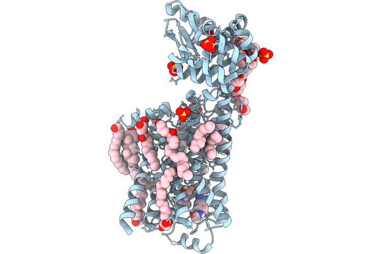
Mixed Model Refinement Of Beta-2 Adrenergic Receptor With Photoazolol In Dark State And Light State, 10 Seconds After Light Activation, Recorded At Swissfel
Organism: Homo sapiens, Tequatrovirus t4
Method: X-RAY DIFFRACTION Resolution:2.45 Å Release Date: 2026-04-22 Classification: MEMBRANE PROTEIN Ligands: SO4, ACM, CLR, PLM, 12P, OLC, 1PE, EDO, GOL, A1JHU |
Organism: Homo sapiens, Tequatrovirus t4
Method: X-RAY DIFFRACTION
Release Date: 2026-04-22
Ligands: SO4, ACM, CLR, PLM, 12P, OLC, 1PE, EDO, GOL, A1JHU
 |
Dark Structure Of Beta-2 Adrenergic Receptor With Photoazolol In Dark State Recorded At Lcls
Organism: Homo sapiens, Enterobacteria phage t4
Method: X-RAY DIFFRACTION Resolution:2.50 Å Release Date: 2026-04-22 Classification: MEMBRANE PROTEIN Ligands: SO4, CLR, PLM, 12P, A1JHU, OLC, EDO, GOL |
Organism: Homo sapiens, Enterobacteria phage t4
Method: X-RAY DIFFRACTION
Release Date: 2026-04-22
Ligands: SO4, CLR, PLM, 12P, A1JHU, OLC, EDO, GOL
 |
Mixed Model Refinement Of Beta-2 Adrenergic Receptor With Photoazolol In Dark State And Light State, 17 Nanoseconds After Light Activation, Recorded At Lcls
Organism: Homo sapiens, Tequatrovirus t4
Method: X-RAY DIFFRACTION Resolution:2.60 Å Release Date: 2026-04-22 Classification: MEMBRANE PROTEIN Ligands: SO4, ACM, CLR, PLM, 12P, OLC, EDO, GOL, 1PE, A1JHU |
Organism: Homo sapiens, Tequatrovirus t4
Method: X-RAY DIFFRACTION
Release Date: 2026-04-22
Ligands: SO4, ACM, CLR, PLM, 12P, OLC, EDO, GOL, 1PE, A1JHU