

Search Count: 50
 |
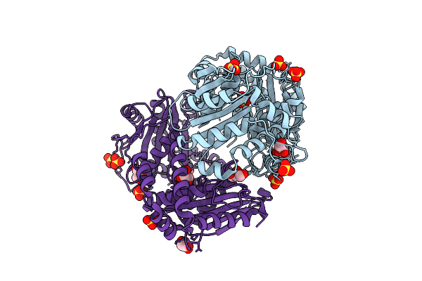
X-Ray Structure Of Trichomonas Vaginalis Inactive Mutant Hydrogenosomal Processing Peptidase Heterodimer (Hppin)
Organism: Trichomonas vaginalis
Method: X-RAY DIFFRACTION Resolution:2.84 Å Release Date: 2025-11-05 Classification: HYDROLASE Ligands: SO4, ZN |
Organism: Trichomonas vaginalis
Method: X-RAY DIFFRACTION
Release Date: 2025-11-05
Ligands: SO4, ZN
 |
X-Ray Structure Of Hydrogenosomal Processing Peptidase (Hpp), E56Q Inactive Mutant, From Trichomonas Vaginalis Co-Crystallized With Presequence Peptide From Adenylate Kinase (Ak) - Not Visible In The Structure Model
Organism: Trichomonas vaginalis
Method: X-RAY DIFFRACTION Resolution:2.00 Å Release Date: 2025-11-05 Classification: HYDROLASE Ligands: SO4, GOL, ZN |
Organism: Trichomonas vaginalis
Method: X-RAY DIFFRACTION
Release Date: 2025-11-05
Ligands: SO4, GOL, ZN
 |
X-Ray Structure Of Hydrogenosomal Processing Peptidase (Hpp), E56Q Inactive Mutant, From Trichomonas Vaginalis Co-Crystallized With Presequence Peptide From Ferredoxin Oxidoreductase (Pfo) - Not Visible In The Structure Model
Organism: Trichomonas vaginalis
Method: X-RAY DIFFRACTION Resolution:2.65 Å Release Date: 2025-11-05 Classification: HYDROLASE Ligands: SO4, GOL, ZN |
Organism: Trichomonas vaginalis
Method: X-RAY DIFFRACTION
Release Date: 2025-11-05
Ligands: SO4, GOL, ZN
 |
1.8 A Resolution Structure Of The Photosystem I Assembly Intermediate Lacking Stromal Subunits.
Organism: Synechocystis
Method: ELECTRON MICROSCOPY Release Date: 2025-06-18 Classification: PHOTOSYNTHESIS Ligands: LHG, CL0, CLA, PQN, BCR, ECH, LMG, LMT, CL, SF4, 5X6 |
Organism: Synechocystis
Method: ELECTRON MICROSCOPY
Release Date: 2025-06-18
Ligands: LHG, CL0, CLA, PQN, BCR, ECH, LMG, LMT, CL, SF4, 5X6
 |
X-Ray Structure Of Blue Laccase Bp76 From The Termite Neocapritermes Taracua
Organism: Neocapritermes taracua
Method: X-RAY DIFFRACTION Resolution:1.80 Å Release Date: 2024-08-28 Classification: OXIDOREDUCTASE Ligands: YKK, PLQ, YMR, OH, CU, CL, SO4 |
Organism: Neocapritermes taracua
Method: X-RAY DIFFRACTION
Release Date: 2024-08-28
Ligands: YKK, PLQ, YMR, OH, CU, CL, SO4
 |
Nmr Solution Structure Of Thyropin Irthy-Cd From The Hard Tick Ixodes Ricinus
Organism: Ixodes ricinus
Method: SOLUTION NMR Release Date: 2024-02-28 Classification: PROTEIN BINDING |
Organism: Ixodes ricinus
Method: SOLUTION NMR
Release Date: 2024-02-28
 |
Structure Of Ledgf/P75 Pwwp Domain Bound To The H3K36 Trimethylated Dinucleosome
Organism: Xenopus laevis, Homo sapiens, Synthetic construct
Method: ELECTRON MICROSCOPY Resolution:3.34 Å Release Date: 2023-12-27 Classification: TRANSCRIPTION Modified Residue(s): ML3 |
Organism: Xenopus laevis, Homo sapiens, Synthetic construct
Method: ELECTRON MICROSCOPY
Release Date: 2023-12-27
 |
Structure Of Ledgf/P75 Pwwp Domain Bound To The H3K36 Trimethylated Dinucleosome
Organism: Xenopus laevis, Homo sapiens, Synthetic construct
Method: ELECTRON MICROSCOPY Resolution:4.00 Å Release Date: 2023-09-06 Classification: DNA BINDING PROTEIN Modified Residue(s): ML3 |
Organism: Xenopus laevis, Homo sapiens, Synthetic construct
Method: ELECTRON MICROSCOPY
Release Date: 2023-09-06
 |
H3K36Me3 Nucleosome-Ledgf/P75 Pwwp Domain Complex
Organism: Synthetic construct, Xenopus laevis, Homo sapiens
Method: ELECTRON MICROSCOPY Release Date: 2023-08-16 Classification: TRANSCRIPTION Modified Residue(s): ML3 |
Organism: Synthetic construct, Xenopus laevis, Homo sapiens
Method: ELECTRON MICROSCOPY
Release Date: 2023-08-16
 |
H3K36Me3 Nucleosome-Ledgf/P75 Pwwp Domain Complex - Pose 2
Organism: Synthetic construct, Xenopus laevis, Homo sapiens
Method: ELECTRON MICROSCOPY Release Date: 2023-08-16 Classification: TRANSCRIPTION Modified Residue(s): ML3 |
Organism: Synthetic construct, Xenopus laevis, Homo sapiens
Method: ELECTRON MICROSCOPY
Release Date: 2023-08-16
 |
H3K36Me2 Nucleosome-Ledgf/P75 Pwwp Domain Complex
Organism: Xenopus laevis, Synthetic construct, Homo sapiens
Method: ELECTRON MICROSCOPY Release Date: 2023-08-16 Classification: TRANSCRIPTION Modified Residue(s): M2L |
Organism: Xenopus laevis, Synthetic construct, Homo sapiens
Method: ELECTRON MICROSCOPY
Release Date: 2023-08-16
 |
H3K36Me2 Nucleosome-Ledgf/P75 Pwwp Domain Complex - Pose 2
Organism: Xenopus laevis, Synthetic construct, Homo sapiens
Method: ELECTRON MICROSCOPY Release Date: 2023-08-16 Classification: TRANSCRIPTION Modified Residue(s): M2L |
Organism: Xenopus laevis, Synthetic construct, Homo sapiens
Method: ELECTRON MICROSCOPY
Release Date: 2023-08-16
 |
Lamin A Coil2 Dimer Stabilized By N-Terminal Capping
Organism: Bacillus phage phi29, Homo sapiens
Method: X-RAY DIFFRACTION Resolution:2.90 Å Release Date: 2021-02-24 Classification: NUCLEAR PROTEIN Ligands: NI, CL, TRS |
Organism: Bacillus phage phi29, Homo sapiens
Method: X-RAY DIFFRACTION
Release Date: 2021-02-24
Ligands: NI, CL, TRS
 |
Ku70/80 Complex Apo Form
Organism: Homo sapiens
Method: ELECTRON MICROSCOPY Release Date: 2021-02-10 Classification: DNA BINDING PROTEIN |
Organism: Homo sapiens
Method: ELECTRON MICROSCOPY
Release Date: 2021-02-10
 |
Ledgf/P75 Ibd Dimer
Organism: Homo sapiens
Method: X-RAY DIFFRACTION Resolution:1.30 Å Release Date: 2020-09-09 Classification: PROTEIN BINDING |
Organism: Homo sapiens
Method: X-RAY DIFFRACTION
Release Date: 2020-09-09
 |
Ledgf/P75 Dimer (Residues 345-467)
|
Organism: Homo sapiens
Method: SOLUTION NMR
Release Date: 2020-09-09
 |
Lamin A 17-70 Coil1A Dimer Stabilized By C-Terminal Capping
Organism: Homo sapiens
Method: X-RAY DIFFRACTION Resolution:1.83 Å Release Date: 2020-09-02 Classification: NUCLEAR PROTEIN |
Organism: Homo sapiens
Method: X-RAY DIFFRACTION
Release Date: 2020-09-02
 |
Lamin A 1-70 Coil1A Dimer Stabilized By C-Terminal Capping
Organism: Homo sapiens
Method: X-RAY DIFFRACTION Resolution:2.83 Å Release Date: 2020-09-02 Classification: NUCLEAR PROTEIN |
Organism: Homo sapiens
Method: X-RAY DIFFRACTION
Release Date: 2020-09-02
 |
Solution Structure Of The Ledgf/P75 Ibd - Jpo2 (Aa 56-91) Complex
|
Organism: Homo sapiens
Method: SOLUTION NMR
Release Date: 2018-08-15
 |
Solution Structure Of The Ledgf/P75 Ibd - Mll1 (Aa 111-160) Complex
|
Organism: Homo sapiens
Method: SOLUTION NMR
Release Date: 2018-08-01