

Search Count: 43
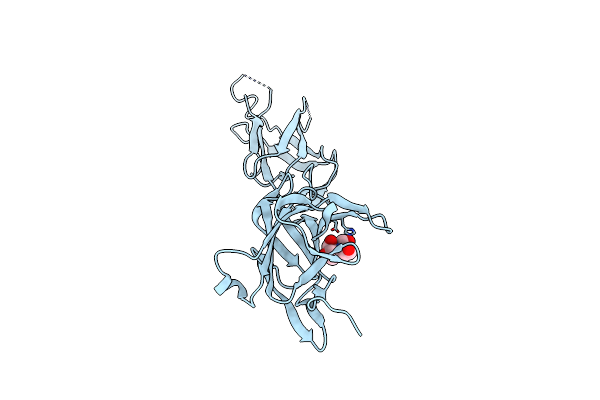 |
Crystal Structure Of Srtb-Anchored Collagen-Binding Adhesin Fragment (Residues 206-565) From Clostridioides Difficile Strain 630
Organism: Clostridioides difficile
Method: X-RAY DIFFRACTION Resolution:2.40 Å Release Date: 2021-10-20 Classification: CELL ADHESION Ligands: BTB |
Organism: Clostridioides difficile
Method: X-RAY DIFFRACTION
Release Date: 2021-10-20
Ligands: BTB
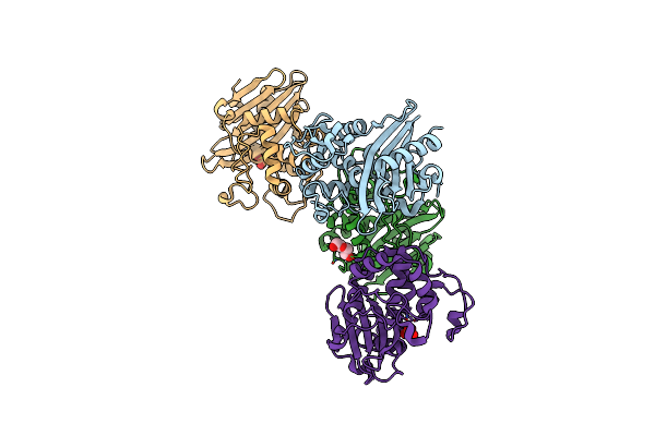 |
Crystal Structure Of C79A Mutant Of Class D Beta-Lactamase From Clostridium Difficile 630
Organism: Clostridioides difficile
Method: X-RAY DIFFRACTION Resolution:1.95 Å Release Date: 2021-08-11 Classification: HYDROLASE Ligands: PEG, SO4 |
Organism: Clostridioides difficile
Method: X-RAY DIFFRACTION
Release Date: 2021-08-11
Ligands: PEG, SO4
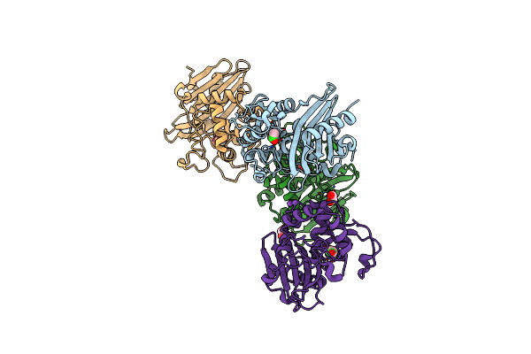 |
Crystal Structure Of K83A Mutant Of Class D Beta-Lactamase From Clostridium Difficile 630
Organism: Clostridioides difficile
Method: X-RAY DIFFRACTION Resolution:1.88 Å Release Date: 2021-08-11 Classification: HYDROLASE Ligands: CL, ACT, EDO, NA |
Organism: Clostridioides difficile
Method: X-RAY DIFFRACTION
Release Date: 2021-08-11
Ligands: CL, ACT, EDO, NA
 |
Crystal Structure Of Wild Type Class D Beta-Lactamase From Clostridium Difficile 630
Organism: Clostridioides difficile (strain 630)
Method: X-RAY DIFFRACTION Resolution:1.80 Å Release Date: 2020-05-27 Classification: HYDROLASE Ligands: PEG, NA |
Organism: Clostridioides difficile (strain 630)
Method: X-RAY DIFFRACTION
Release Date: 2020-05-27
Ligands: PEG, NA
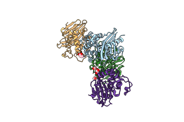 |
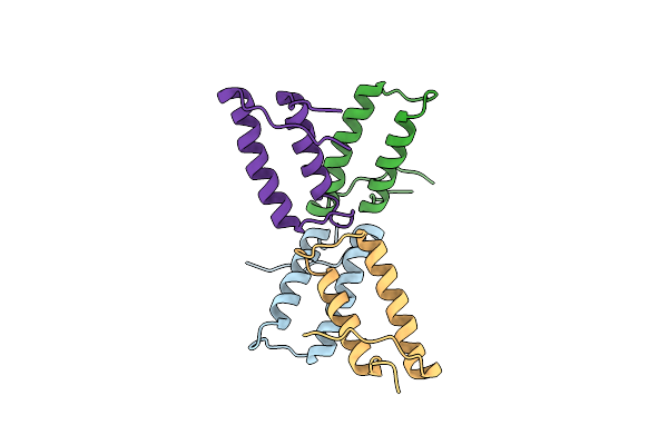
1.85 Angstrom Resolution Crystal Structure Of Class D Beta-Lactamase From Clostridium Difficile 630
Organism: Peptoclostridium difficile (strain 630)
Method: X-RAY DIFFRACTION Resolution:1.85 Å Release Date: 2019-12-25 Classification: HYDROLASE Ligands: PGE, PEG, PPI, GOL |
Organism: Peptoclostridium difficile (strain 630)
Method: X-RAY DIFFRACTION
Release Date: 2019-12-25
Ligands: PGE, PEG, PPI, GOL
 |
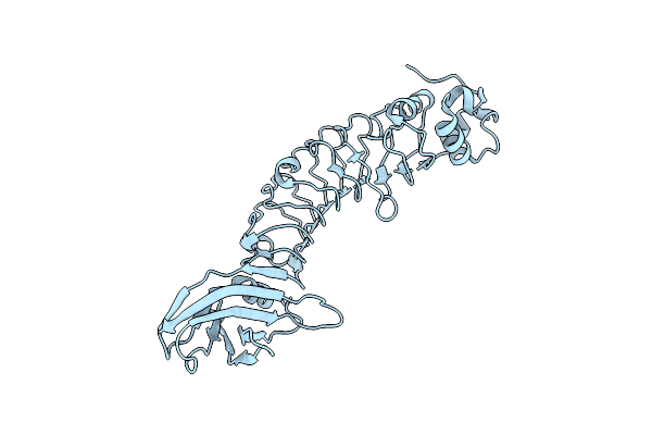
1.78 Angstrom Resolution Crystal Structure Of Hypothetical Protein Cd630_05490 From Clostridioides Difficile 630.
Organism: Peptoclostridium difficile (strain 630)
Method: X-RAY DIFFRACTION Resolution:1.78 Å Release Date: 2018-12-12 Classification: UNKNOWN FUNCTION |
Organism: Peptoclostridium difficile (strain 630)
Method: X-RAY DIFFRACTION
Release Date: 2018-12-12
 |
Listeria Monocytogenes Internalin-Like Protein Lmo2027
Organism: Listeria monocytogenes egd-e
Method: X-RAY DIFFRACTION Resolution:2.30 Å Release Date: 2017-07-26 Classification: UNKNOWN FUNCTION |
Organism: Listeria monocytogenes egd-e
Method: X-RAY DIFFRACTION
Release Date: 2017-07-26
 |
2.15 Angstrom Crystal Structure Of N-Acetylmuramoyl-L-Alanine Amidase From Staphylococcus Aureus.
Organism: Staphylococcus aureus (strain nctc 8325)
Method: X-RAY DIFFRACTION Resolution:2.15 Å Release Date: 2017-06-07 Classification: HYDROLASE |
Organism: Staphylococcus aureus (strain nctc 8325)
Method: X-RAY DIFFRACTION
Release Date: 2017-06-07
 |
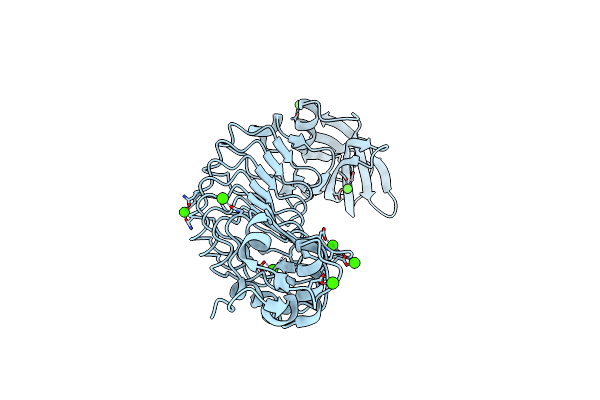
A 1.85A X-Ray Structure From Peptoclostridium Difficile 630 Of A Hypothetical Protein
Organism: Peptoclostridium difficile
Method: X-RAY DIFFRACTION Resolution:1.85 Å Release Date: 2017-02-01 Classification: STRUCTURAL GENOMICS, UNKNOWN FUNCTION |
Organism: Peptoclostridium difficile
Method: X-RAY DIFFRACTION
Release Date: 2017-02-01
 |
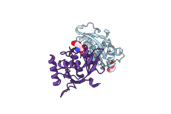
Crystal Structure Of Listeria Monocytogenes Inlp
Organism: Listeria monocytogenes egd-e
Method: X-RAY DIFFRACTION Resolution:1.40 Å Release Date: 2017-01-18 Classification: UNKNOWN FUNCTION Ligands: CA, CL |
Organism: Listeria monocytogenes egd-e
Method: X-RAY DIFFRACTION
Release Date: 2017-01-18
Ligands: CA, CL
 |
2.05 Angstrom Resolution Crystal Structure Of Peptidoglycan-Binding Protein From Clostridioides Difficile In Complex With Glutamine Hydroxamate.
Organism: Peptoclostridium difficile (strain 630)
Method: X-RAY DIFFRACTION Resolution:2.05 Å Release Date: 2016-12-14 Classification: HYDROLASE Ligands: HGA |
Organism: Peptoclostridium difficile (strain 630)
Method: X-RAY DIFFRACTION
Release Date: 2016-12-14
Ligands: HGA
 |
Crystal Structure Of The Zika Virus Ns3 Helicase.
Organism: Zika virus (strain mr 766)
Method: X-RAY DIFFRACTION Resolution:2.05 Å Release Date: 2016-12-14 Classification: HYDROLASE Ligands: K, TRS |
Organism: Zika virus (strain mr 766)
Method: X-RAY DIFFRACTION
Release Date: 2016-12-14
Ligands: K, TRS
 |
1.95 Angstrom Resolution Crystal Structure Of Stage Ii Sporulation Protein D (Spoiid) From Clostridium Difficile In Apo Conformation
Organism: Peptoclostridium difficile (strain 630)
Method: X-RAY DIFFRACTION Resolution:1.95 Å Release Date: 2016-12-14 Classification: HYDROLASE Ligands: ZN, NA, CL, FMT, DMS |
Organism: Peptoclostridium difficile (strain 630)
Method: X-RAY DIFFRACTION
Release Date: 2016-12-14
Ligands: ZN, NA, CL, FMT, DMS
 |
Crystal Structure Of Zika Virus Ns2B-Ns3 Protease In Apo-Form.
Organism: Zika virus
Method: X-RAY DIFFRACTION Resolution:3.10 Å Release Date: 2016-09-07 Classification: HYDROLASE |
Organism: Zika virus
Method: X-RAY DIFFRACTION
Release Date: 2016-09-07
 |
2.6 Angstrom Resolution Crystal Structure Of Stage Ii Sporulation Protein D (Spoiid) From Clostridium Difficile In Complex With Triacetylchitotriose
Organism: Peptoclostridium difficile (strain 630)
Method: X-RAY DIFFRACTION Resolution:2.60 Å Release Date: 2016-05-25 Classification: HYDROLASE Ligands: ZN, NA, CL, GOL |
Organism: Peptoclostridium difficile (strain 630)
Method: X-RAY DIFFRACTION
Release Date: 2016-05-25
Ligands: ZN, NA, CL, GOL
 |
First Crystal Structure Of E.Coli Based Recombinant Diphtheria Toxin Mutant Crm197
Organism: Corynebacterium diphtheriae
Method: X-RAY DIFFRACTION Resolution:2.35 Å Release Date: 2016-03-02 Classification: TRANSFERASE Ligands: SO4, GOL |
Organism: Corynebacterium diphtheriae
Method: X-RAY DIFFRACTION
Release Date: 2016-03-02
Ligands: SO4, GOL
 |
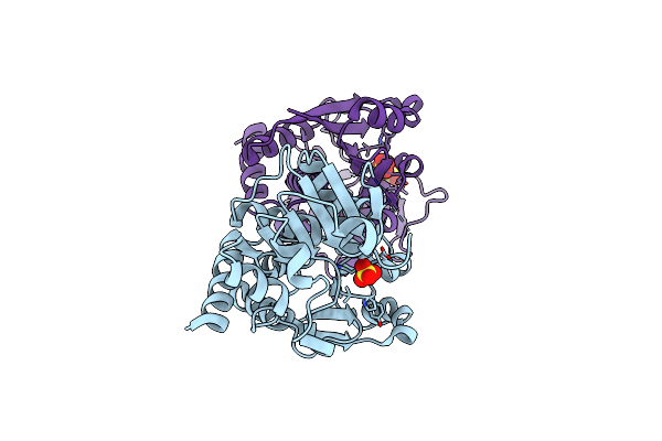
1.75 Angstrom Crystal Structure Of Superantigen-Like Protein, Exotoxin From Staphylococcus Aureus, In Complex With Sialyl-Lewisx.
Organism: Staphylococcus aureus
Method: X-RAY DIFFRACTION Resolution:1.75 Å Release Date: 2016-02-24 Classification: IMMUNE SYSTEM Ligands: PGE, PG4, NAG |
Organism: Staphylococcus aureus
Method: X-RAY DIFFRACTION
Release Date: 2016-02-24
Ligands: PGE, PG4, NAG
 |
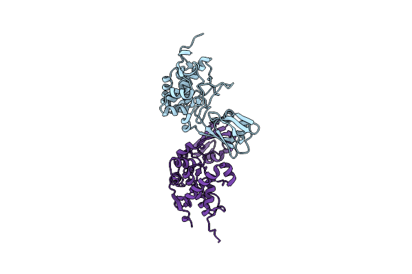
1.5 Angstrom Crystal Structure Of Shikimate Dehydrogenase 1 From Peptoclostridium Difficile.
Organism: Peptoclostridium difficile (strain 630)
Method: X-RAY DIFFRACTION Resolution:1.50 Å Release Date: 2015-10-07 Classification: OXIDOREDUCTASE Ligands: SO4 |
Organism: Peptoclostridium difficile (strain 630)
Method: X-RAY DIFFRACTION
Release Date: 2015-10-07
Ligands: SO4
 |
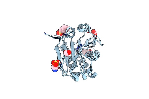
2.1 Angstrom Crystal Structure Of Stage Ii Sporulation Protein D From Bacillus Anthracis
Organism: Bacillus anthracis
Method: X-RAY DIFFRACTION Resolution:2.10 Å Release Date: 2014-12-17 Classification: VIRAL PROTEIN |
Organism: Bacillus anthracis
Method: X-RAY DIFFRACTION
Release Date: 2014-12-17
 |
The Crystal Structure Of N-Acetylmuramoyl-L-Alanine Amidase From Clostridium Difficile 630
Organism: Peptoclostridium difficile
Method: X-RAY DIFFRACTION Resolution:1.72 Å Release Date: 2014-11-05 Classification: HYDROLASE Ligands: ZN, GOL, EPE, FMT |
Organism: Peptoclostridium difficile
Method: X-RAY DIFFRACTION
Release Date: 2014-11-05
Ligands: ZN, GOL, EPE, FMT