

Search Count: 20
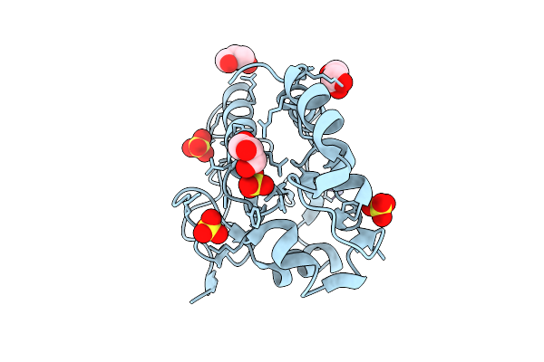 |
X-Ray Structure Of The Catalytic Domain Of Cdc25B C473S At 1.34 Angstrom Resolution
Organism: Homo sapiens
Method: X-RAY DIFFRACTION Resolution:1.34 Å Release Date: 2026-03-11 Classification: HYDROLASE Ligands: SO4, CL, ZN, GOL |
Organism: Homo sapiens
Method: X-RAY DIFFRACTION
Release Date: 2026-03-11
Ligands: SO4, CL, ZN, GOL
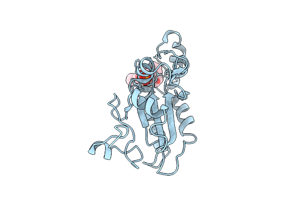 |
X-Ray Structure Of The Complex Between The Catalytic Domain Of Cdc25B C473S And 3-O-Methylfluorescein Phosphate (3-Omfp)
Organism: Homo sapiens
Method: X-RAY DIFFRACTION Resolution:2.04 Å Release Date: 2026-03-11 Classification: HYDROLASE Ligands: A1JSQ, A1JSP, CL |
Organism: Homo sapiens
Method: X-RAY DIFFRACTION
Release Date: 2026-03-11
Ligands: A1JSQ, A1JSP, CL
 |
Crystal Structure Of Hsp82-Md In Complex With The Cs Domain Of Sgt1
Organism: Saccharomyces cerevisiae
Method: X-RAY DIFFRACTION Resolution:2.10 Å Release Date: 2026-01-21 Classification: CHAPERONE |
Organism: Saccharomyces cerevisiae
Method: X-RAY DIFFRACTION
Release Date: 2026-01-21
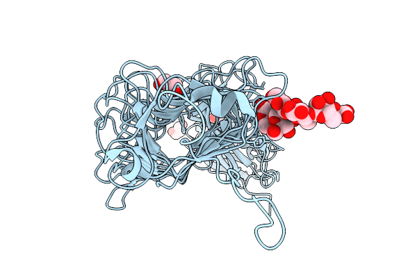 |
X-Ray Structure Of Kp32Gp38 In Complex With K21 Pyr5
Organism: Klebsiella pneumoniae
Method: X-RAY DIFFRACTION Resolution:1.80 Å Release Date: 2025-09-03 Classification: VIRAL PROTEIN Ligands: GOL, PEG |
Organism: Klebsiella pneumoniae
Method: X-RAY DIFFRACTION
Release Date: 2025-09-03
Ligands: GOL, PEG
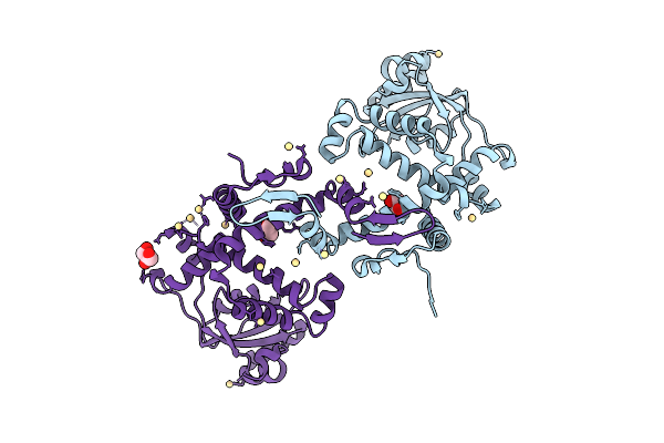 |
Crystal Structure Of The Peptidyl-Prolyl Isomerase (Ppiase) From E. Faecium
Organism: Enterococcus faecium
Method: X-RAY DIFFRACTION Resolution:2.48 Å Release Date: 2025-07-16 Classification: ISOMERASE Ligands: GOL, CD |
Organism: Enterococcus faecium
Method: X-RAY DIFFRACTION
Release Date: 2025-07-16
Ligands: GOL, CD
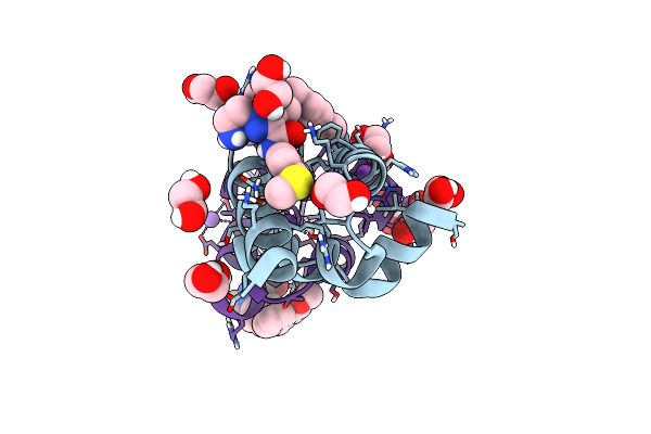 |
N-Terminal Domain Of Trypanosoma Brucei Pex14 In Complex With A Pyrazolo-Pyrazolo[4,3-C]Pyridin-3-Yl Compound Showing A Novel Binding Pose
Organism: Trypanosoma brucei brucei
Method: X-RAY DIFFRACTION Resolution:1.13 Å Release Date: 2025-01-29 Classification: SIGNALING PROTEIN Ligands: OJN, EDO, MLI, NA, CL |
Organism: Trypanosoma brucei brucei
Method: X-RAY DIFFRACTION
Release Date: 2025-01-29
Ligands: OJN, EDO, MLI, NA, CL
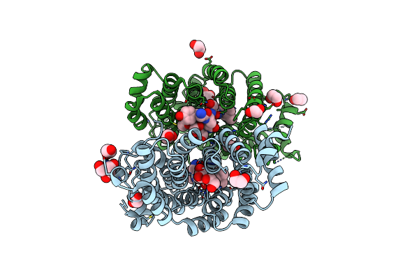 |
X-Ray Structure Of The Peroxisomal Targeting Signal 1 (Pts1) Of Trypanosoma Cruzi Pex5 In Complex With The Pts1 Peptide
Organism: Trypanosoma cruzi
Method: X-RAY DIFFRACTION Resolution:2.10 Å Release Date: 2024-10-02 Classification: TRANSPORT PROTEIN Ligands: EDO, PEG, MG, TRS |
Organism: Trypanosoma cruzi
Method: X-RAY DIFFRACTION
Release Date: 2024-10-02
Ligands: EDO, PEG, MG, TRS
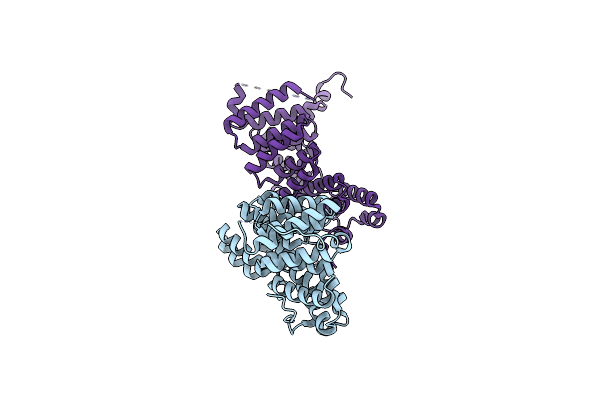 |
Crystal Structure Of The Apo Pex5 Peroxisomal Cargo Receptor From Trypanosoma Brucei
Organism: Trypanosoma brucei
Method: X-RAY DIFFRACTION Resolution:2.35 Å Release Date: 2024-10-02 Classification: PEPTIDE BINDING PROTEIN |
Organism: Trypanosoma brucei
Method: X-RAY DIFFRACTION
Release Date: 2024-10-02
 |
Crystal Structure Of Human Gpx4-R152H
Organism: Homo sapiens
Method: X-RAY DIFFRACTION Resolution:2.00 Å Release Date: 2024-08-28 Classification: OXIDOREDUCTASE |
Organism: Homo sapiens
Method: X-RAY DIFFRACTION
Release Date: 2024-08-28
 |
Crystal Structure Of Human Gpx4-U46C-I129S-L130S
Organism: Homo sapiens
Method: X-RAY DIFFRACTION Resolution:1.90 Å Release Date: 2024-08-28 Classification: OXIDOREDUCTASE |
Organism: Homo sapiens
Method: X-RAY DIFFRACTION
Release Date: 2024-08-28
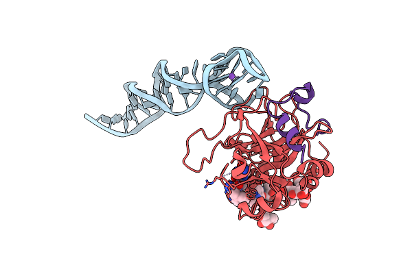 |
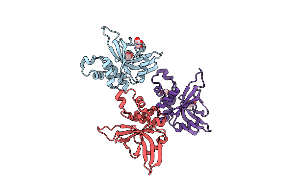
X-Ray Structure Of The Complex Between Human Alpha Thrombin And The Duplex/Quadruplex Aptamer M08S-1_41Mer
Organism: Synthetic construct, Homo sapiens
Method: X-RAY DIFFRACTION Resolution:2.80 Å Release Date: 2023-07-19 Classification: HYDROLASE Ligands: 0G6, NA |
Organism: Synthetic construct, Homo sapiens
Method: X-RAY DIFFRACTION
Release Date: 2023-07-19
Ligands: 0G6, NA
 |
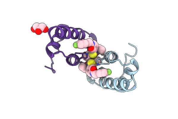
Crystal Structure Of Staphopain C From Staphylococcus Aureus
Organism: Staphylococcus aureus
Method: X-RAY DIFFRACTION Resolution:1.58 Å Release Date: 2023-06-21 Classification: HYDROLASE Ligands: GOL, CL, EDO |
Organism: Staphylococcus aureus
Method: X-RAY DIFFRACTION
Release Date: 2023-06-21
Ligands: GOL, CL, EDO
 |
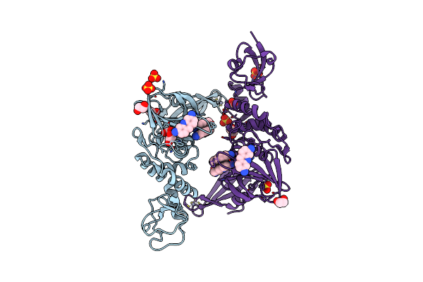
X-Ray Structure Of Trypanosoma Cruzi Pex14 In Complex With A Pex5-Pex14 Ppi Inhibitor
Organism: Trypanosoma cruzi
Method: X-RAY DIFFRACTION Resolution:2.18 Å Release Date: 2022-11-23 Classification: SIGNALING PROTEIN Ligands: GOL, ET7 |
Organism: Trypanosoma cruzi
Method: X-RAY DIFFRACTION
Release Date: 2022-11-23
Ligands: GOL, ET7
 |
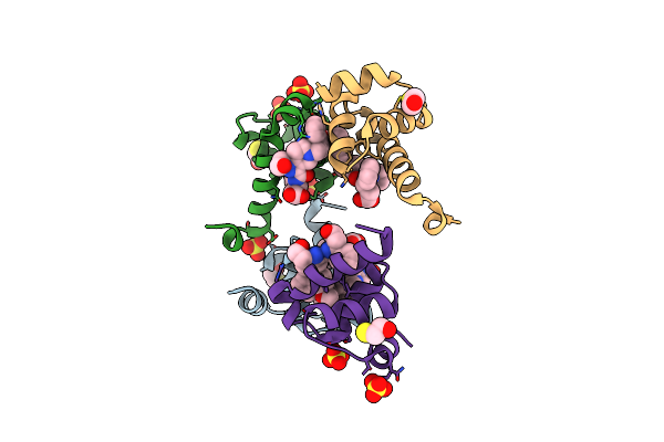
X-Ray Structure Of Scov2-Plpro In Complex With Small Molecule Inhibitor
Organism: Severe acute respiratory syndrome coronavirus 2
Method: X-RAY DIFFRACTION Resolution:2.68 Å Release Date: 2022-02-02 Classification: HYDROLASE Ligands: PRL, EDO, ZN, SO4 |
Organism: Severe acute respiratory syndrome coronavirus 2
Method: X-RAY DIFFRACTION
Release Date: 2022-02-02
Ligands: PRL, EDO, ZN, SO4
 |
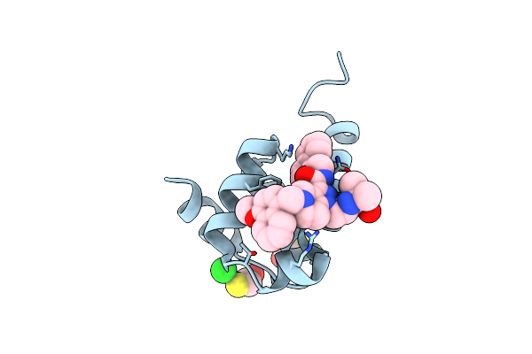
Crystal Structure Of Trypanosoma Brucei Pex14 N-Terminal Domain In Complex With Small Molecules Designed To Investigate The Water Envelope
Organism: Trypanosoma brucei brucei
Method: X-RAY DIFFRACTION Resolution:1.30 Å Release Date: 2020-04-08 Classification: SIGNALING PROTEIN Ligands: CV5, SO4, BME |
Organism: Trypanosoma brucei brucei
Method: X-RAY DIFFRACTION
Release Date: 2020-04-08
Ligands: CV5, SO4, BME
 |
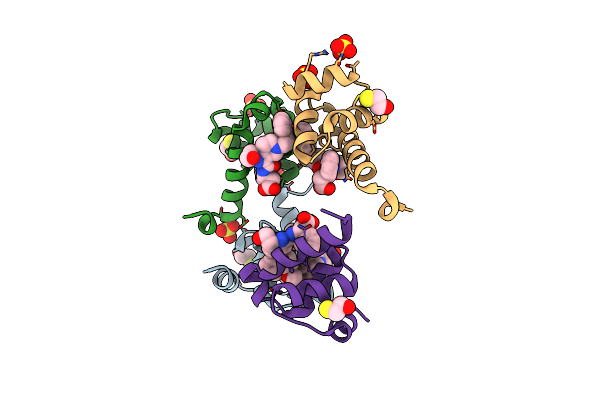
High Resolution Crystal Structure Of N-Terminal Domain Of Pex14 From Trypanosoma Brucei In Complex With The Fist Compound With Sub-Micromolar Trypanocidal Activity
Organism: Trypanosoma brucei brucei
Method: X-RAY DIFFRACTION Resolution:1.20 Å Release Date: 2020-01-01 Classification: SIGNALING PROTEIN Ligands: FTW, BME, CL |
Organism: Trypanosoma brucei brucei
Method: X-RAY DIFFRACTION
Release Date: 2020-01-01
Ligands: FTW, BME, CL
 |
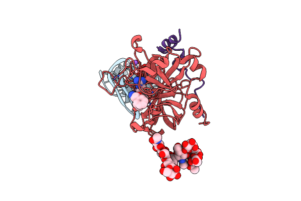
Crystal Structure Of Trypanosoma Brucei Pex14 N-Terminal Domain In Complex With Small Molecules To Investigate The Water Envelope
Organism: Trypanosoma brucei brucei
Method: X-RAY DIFFRACTION Resolution:1.50 Å Release Date: 2019-03-06 Classification: SIGNALING PROTEIN Ligands: 9YB, SO4, BME |
Organism: Trypanosoma brucei brucei
Method: X-RAY DIFFRACTION
Release Date: 2019-03-06
Ligands: 9YB, SO4, BME
 |
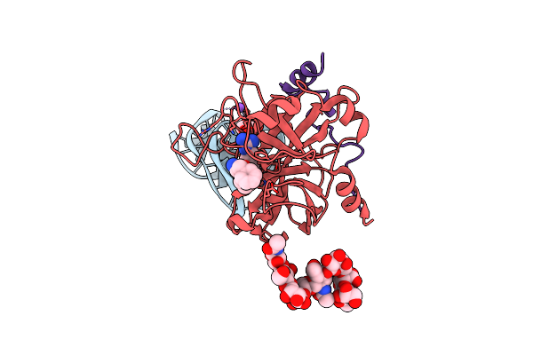
X-Ray Structure Of The Complex Between Human Alpha Thrombin And Nu172, A Duplex/Quadruplex 26-Mer Dna Aptamer, In The Presence Of Potassium Ions.
Organism: Synthetic construct, Homo sapiens
Method: X-RAY DIFFRACTION Resolution:2.50 Å Release Date: 2018-10-24 Classification: HYDROLASE Ligands: 0G6, NA, K, GOL |
Organism: Synthetic construct, Homo sapiens
Method: X-RAY DIFFRACTION
Release Date: 2018-10-24
Ligands: 0G6, NA, K, GOL
 |
X-Ray Structure Of The Complex Between Human Alpha Thrombin And Nu172, A Duplex/Quadruplex 26-Mer Dna Aptamer, In The Presence Of Sodium Ions.
Organism: Synthetic construct, Homo sapiens
Method: X-RAY DIFFRACTION Resolution:2.80 Å Release Date: 2018-10-17 Classification: HYDROLASE Ligands: 0G6, NA, GOL |
Organism: Synthetic construct, Homo sapiens
Method: X-RAY DIFFRACTION
Release Date: 2018-10-17
Ligands: 0G6, NA, GOL
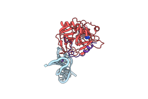 |
X-Ray Structure Of The Complex Between Human Alpha Thrombin And A Duplex/Quadruplex 31-Mer Dna Aptamer
Organism: Synthetic construct, Homo sapiens
Method: X-RAY DIFFRACTION Resolution:2.98 Å Release Date: 2016-01-13 Classification: HYDROLASE Ligands: 0G6, K |
Organism: Synthetic construct, Homo sapiens
Method: X-RAY DIFFRACTION
Release Date: 2016-01-13
Ligands: 0G6, K