

Search Count: 116
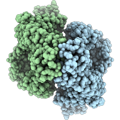 |
T. Cruzi Topoisomerase Ii Alpha Bound To Dsdna And The Covalent Inhibitor Iid432
Organism: Trypanosoma cruzi, Escherichia coli
Method: ELECTRON MICROSCOPY Resolution:2.77 Å Release Date: 2026-09-16 Classification: ISOMERASE Ligands: MG, A1KFP |
Organism: Trypanosoma cruzi, Escherichia coli
Method: ELECTRON MICROSCOPY
Release Date: 2026-09-16
Ligands: MG, A1KFP
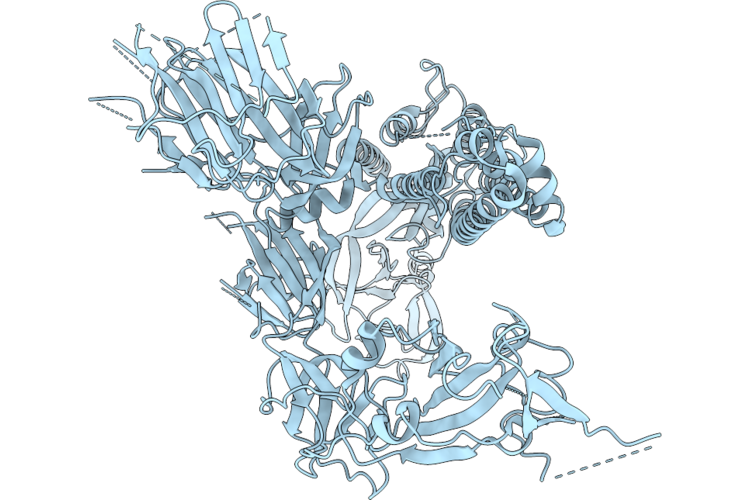 |
Cryo-Em Structure Of Sars Cov2 S Protein With Stabilising Mutations
Organism: Severe acute respiratory syndrome coronavirus 2
Method: ELECTRON MICROSCOPY Resolution:4.55 Å Release Date: 2026-04-29 Classification: VIRAL PROTEIN |
Organism: Severe acute respiratory syndrome coronavirus 2
Method: ELECTRON MICROSCOPY
Release Date: 2026-04-29
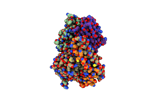 |
Structure Of Active State Mc4R In Complex With A Potent Ligand Mimicking Nanobody
Organism: Homo sapiens, Lama glama, Escherichia coli, Discosoma sp.
Method: ELECTRON MICROSCOPY Release Date: 2024-09-25 Classification: MEMBRANE PROTEIN |
Organism: Homo sapiens, Lama glama, Escherichia coli, Discosoma sp.
Method: ELECTRON MICROSCOPY
Release Date: 2024-09-25
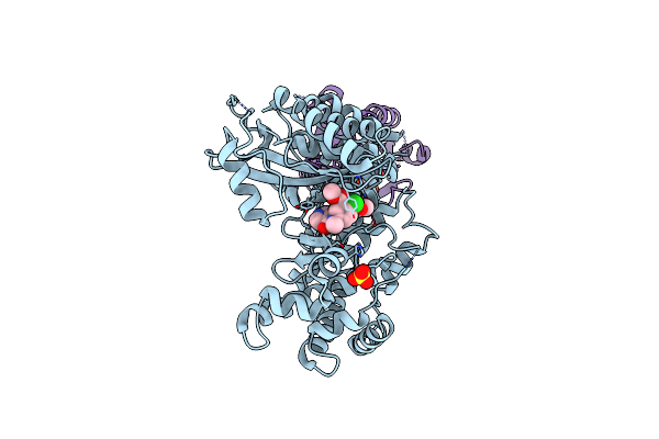 |
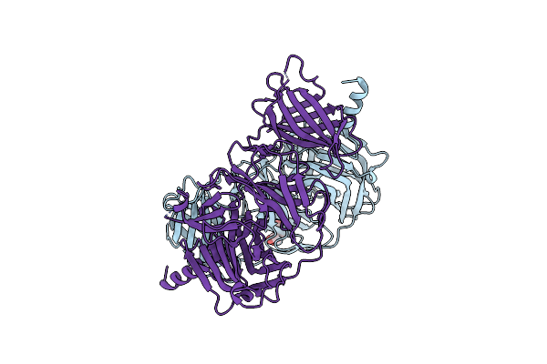
Gi Targeted Cppi4K Inhibitor
Organism: Homo sapiens
Method: X-RAY DIFFRACTION Resolution:3.00 Å Release Date: 2024-09-04 Classification: TRANSFERASE Ligands: A1ADE, SO4, GDP |
Organism: Homo sapiens
Method: X-RAY DIFFRACTION
Release Date: 2024-09-04
Ligands: A1ADE, SO4, GDP
 |
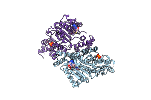
Crystal Structure Of The Tegument Protein Ul82 (Pp71) From Human Cytomegalovirus
Organism: Human betaherpesvirus 5
Method: X-RAY DIFFRACTION Resolution:2.70 Å Release Date: 2024-02-28 Classification: VIRAL PROTEIN Ligands: PG4 |
Organism: Human betaherpesvirus 5
Method: X-RAY DIFFRACTION
Release Date: 2024-02-28
Ligands: PG4
 |
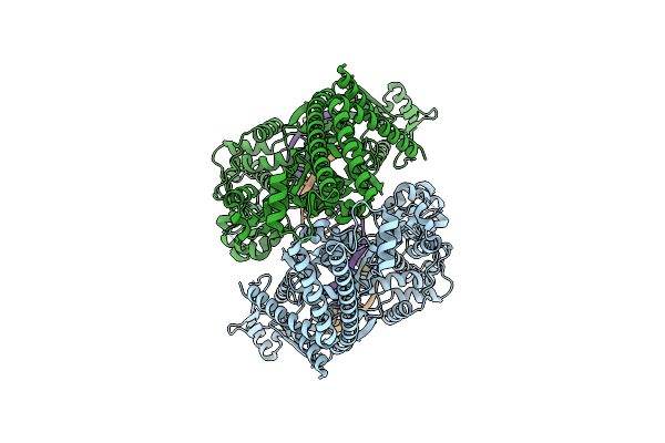
Crystal Structure Of Histone H3 Lysine 79 (H3K79) Methyltransferase Rv2067C From Mycobacterium Tuberculosis
Organism: Mycobacterium tuberculosis h37rv
Method: X-RAY DIFFRACTION Resolution:2.40 Å Release Date: 2023-10-18 Classification: TRANSFERASE Ligands: PO4, SAH |
Organism: Mycobacterium tuberculosis h37rv
Method: X-RAY DIFFRACTION
Release Date: 2023-10-18
Ligands: PO4, SAH
 |
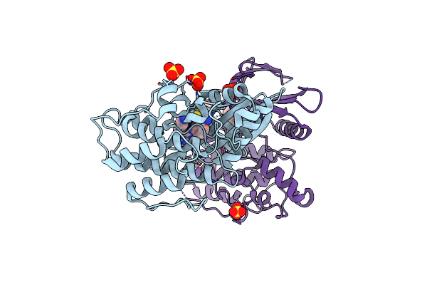
T. Cruzi Topoisomerase Ii Alpha Bound To Dsdna And The Covalent Inhibitor Ct1
Organism: Trypanosoma cruzi strain cl brener, Escherichia coli
Method: ELECTRON MICROSCOPY Release Date: 2023-07-12 Classification: ISOMERASE Ligands: YWX |
Organism: Trypanosoma cruzi strain cl brener, Escherichia coli
Method: ELECTRON MICROSCOPY
Release Date: 2023-07-12
Ligands: YWX
 |
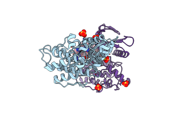
Crystal Structure Of Ap2 Associated Kinase 1 Complexed With 5-[(4-Aminopiperidin-1-Yl)Methyl]-N-{3-[5-(Propan-2-Yl)-1,3,4-Thiadiazol-2-Yl]Phenyl}Pyrrolo[2,1-F][1,2,4]Triazin-4-Amine
Organism: Homo sapiens
Method: X-RAY DIFFRACTION Resolution:2.50 Å Release Date: 2023-07-12 Classification: TRANSFERASE Ligands: YFV, SO4 |
Organism: Homo sapiens
Method: X-RAY DIFFRACTION
Release Date: 2023-07-12
Ligands: YFV, SO4
 |
Crystal Structure Of Ap2 Associated Kinase 1 Complexed With (5P)-3-({(8R)-5-[(4-Aminopiperidin-1-Yl)Methyl]Pyrrolo[2,1-F][1,2,4]Triazin-4-Yl}Amino)-5-[2-(Propan-2-Yl)-2H-Tetrazol-5-Yl]Phenol
Organism: Homo sapiens
Method: X-RAY DIFFRACTION Resolution:2.20 Å Release Date: 2023-07-12 Classification: TRANSFERASE Ligands: ZRR, SO4 |
Organism: Homo sapiens
Method: X-RAY DIFFRACTION
Release Date: 2023-07-12
Ligands: ZRR, SO4
 |
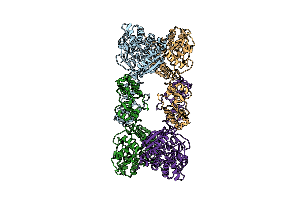
Native Cystathionine Beta-Synthase Of Mycobacterium Tuberculosis.
Organism: Mycobacterium tuberculosis h37rv
Method: ELECTRON MICROSCOPY Release Date: 2022-05-25 Classification: LYASE Ligands: PLP |
Organism: Mycobacterium tuberculosis h37rv
Method: ELECTRON MICROSCOPY
Release Date: 2022-05-25
Ligands: PLP
 |
Cystathionine Beta-Synthase Of Mycobacterium Tuberculosis In The Presence Of S-Adenosylmethionine.
Organism: Mycobacterium tuberculosis h37rv
Method: ELECTRON MICROSCOPY Release Date: 2022-05-25 Classification: LYASE |
Organism: Mycobacterium tuberculosis h37rv
Method: ELECTRON MICROSCOPY
Release Date: 2022-05-25
 |
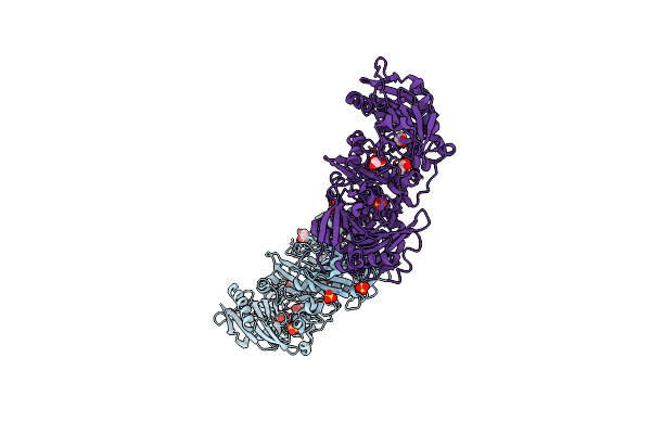
Cystathionine Beta-Synthase Of Mycobacterium Tuberculosis In The Presence Of S-Adenosylmethionine And Serine.
Organism: Mycobacterium tuberculosis h37rv
Method: ELECTRON MICROSCOPY Release Date: 2022-05-25 Classification: LYASE Ligands: PLS |
Organism: Mycobacterium tuberculosis h37rv
Method: ELECTRON MICROSCOPY
Release Date: 2022-05-25
Ligands: PLS
 |
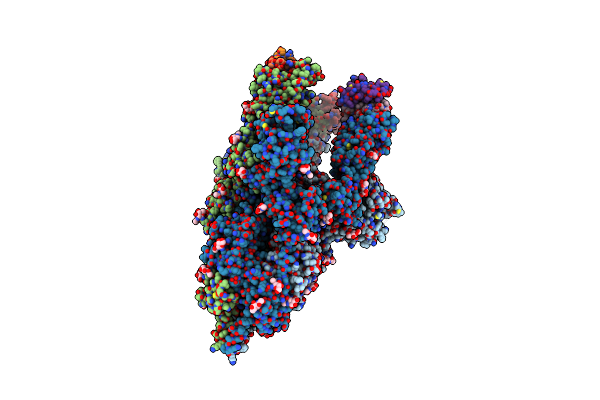
Crystal Structure Of Human Gelsolin Amyloid Mutant A551P
Organism: Homo sapiens
Method: X-RAY DIFFRACTION Resolution:3.00 Å Release Date: 2022-05-18 Classification: STRUCTURAL PROTEIN Ligands: SO4, GOL, CL |
Organism: Homo sapiens
Method: X-RAY DIFFRACTION
Release Date: 2022-05-18
Ligands: SO4, GOL, CL
 |
3D Model Of The 3-Rbd Up Single Trimeric Spike Protein Of Sars-Cov2 In The Presence Of Synthetic Peptide Sih-5.
Organism: Severe acute respiratory syndrome coronavirus 2, Synthetic construct
Method: ELECTRON MICROSCOPY Release Date: 2022-04-27 Classification: VIRAL PROTEIN Ligands: NAG |
Organism: Severe acute respiratory syndrome coronavirus 2, Synthetic construct
Method: ELECTRON MICROSCOPY
Release Date: 2022-04-27
Ligands: NAG
 |
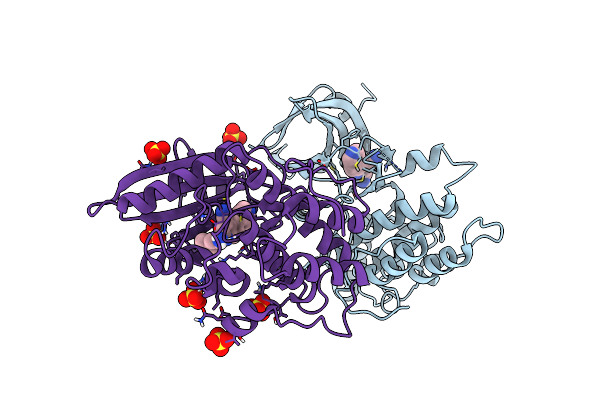
Crystal Structure Of Ap2 Associated Kinase 1 Isoform 1 Complexed With Ligand (2R)-2-Amino-N-[3-(Difluorom Ethoxy)-4-(1,3-Oxazol-5-Yl)Phenyl]-4-Methylpentanamide
Organism: Mus musculus
Method: X-RAY DIFFRACTION Resolution:2.13 Å Release Date: 2022-02-23 Classification: TRANSFERASE/TRANSFERASE INHIBITOR Ligands: YFV, SO4, 5OI |
Organism: Mus musculus
Method: X-RAY DIFFRACTION
Release Date: 2022-02-23
Ligands: YFV, SO4, 5OI
 |
Crystal Structure Of Ap2 Associated Kinase 1 Isoform 1 Complexed With Ligand 2-(1-Amino-3-Methylbutyl)-6- (Pyridin-4-Yl)Quinoline-4-Carbonitrile
Organism: Mus musculus
Method: X-RAY DIFFRACTION Resolution:2.12 Å Release Date: 2022-02-23 Classification: TRANSFERASE/TRANSFERASE INHIBITOR Ligands: 5P6, SO4 |
Organism: Mus musculus
Method: X-RAY DIFFRACTION
Release Date: 2022-02-23
Ligands: 5P6, SO4
 |
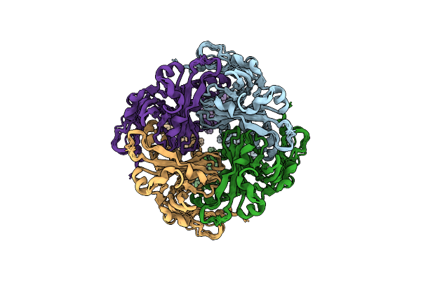
Crystal Structure Of Ap2 Associated Kinase 1 Isoform 1 Complexed With Ligand (2R)-2-Amino-N-[3-(Difluorom Ethoxy)-4-(1,3-Oxazol-5-Yl)Phenyl]-4-Methylpentanamide
Organism: Mus musculus
Method: X-RAY DIFFRACTION Resolution:2.59 Å Release Date: 2022-02-23 Classification: TRANSFERASE/TRANSFERASE INHIBITOR Ligands: 5QI, PG6, SO4, YFV |
Organism: Mus musculus
Method: X-RAY DIFFRACTION
Release Date: 2022-02-23
Ligands: 5QI, PG6, SO4, YFV
 |
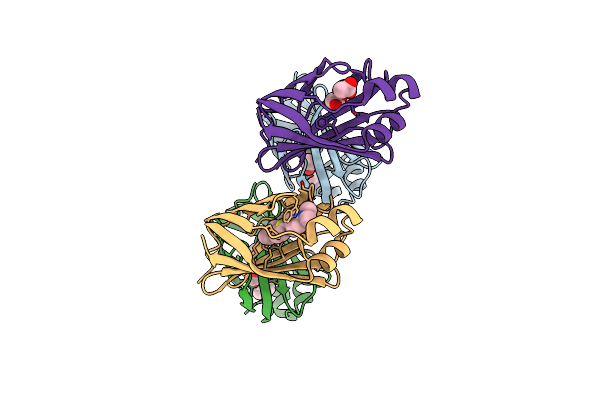
Kirbac1.1 Mutant - I131C
Organism: Burkholderia pseudomallei
Method: SOLID-STATE NMR Release Date: 2022-02-02 Classification: MEMBRANE PROTEIN |
Organism: Burkholderia pseudomallei
Method: SOLID-STATE NMR
Release Date: 2022-02-02
 |
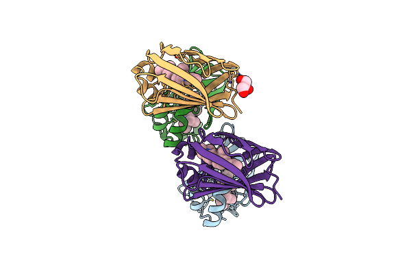
The Crystal Structure Of Q108K:K40E:T53A:R58W:Q38F:Q4F:Y19W Mutant Of Hcrbpii Bound With Lizfluor Chromophore Showing Excited State Intermolecular Proton Transfer
Organism: Homo sapiens
Method: X-RAY DIFFRACTION Resolution:1.67 Å Release Date: 2022-01-12 Classification: LIPID BINDING PROTEIN Ligands: ACT, ZFG |
Organism: Homo sapiens
Method: X-RAY DIFFRACTION
Release Date: 2022-01-12
Ligands: ACT, ZFG
 |
The Crystal Structure Of Q108K:K40H:T53A:R58L:Q38F:Q4F Mutant Of Hcrbpii Bound With Fr1 Chromophore Showing Excited State Intermolecular Proton Transfer
Organism: Homo sapiens
Method: X-RAY DIFFRACTION Resolution:1.59 Å Release Date: 2022-01-12 Classification: LIPID BINDING PROTEIN Ligands: ZFJ, GOL |
Organism: Homo sapiens
Method: X-RAY DIFFRACTION
Release Date: 2022-01-12
Ligands: ZFJ, GOL