

Search Count: 571
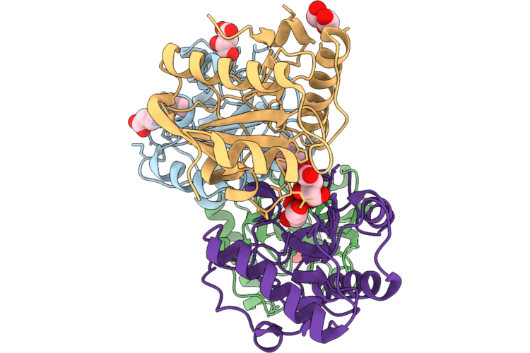 |
Crystal Structure Of The Complex Of Short Peptidoglycan Recognition Protein From Camelus Dromedarius With Nonanoic Acid At 1.83 A Resolution
Organism: Camelus dromedarius
Method: X-RAY DIFFRACTION Resolution:1.83 Å Release Date: 2026-06-17 Classification: IMMUNE SYSTEM Ligands: TLA, KNA, EDO, GOL |
Organism: Camelus dromedarius
Method: X-RAY DIFFRACTION
Release Date: 2026-06-17
Ligands: TLA, KNA, EDO, GOL
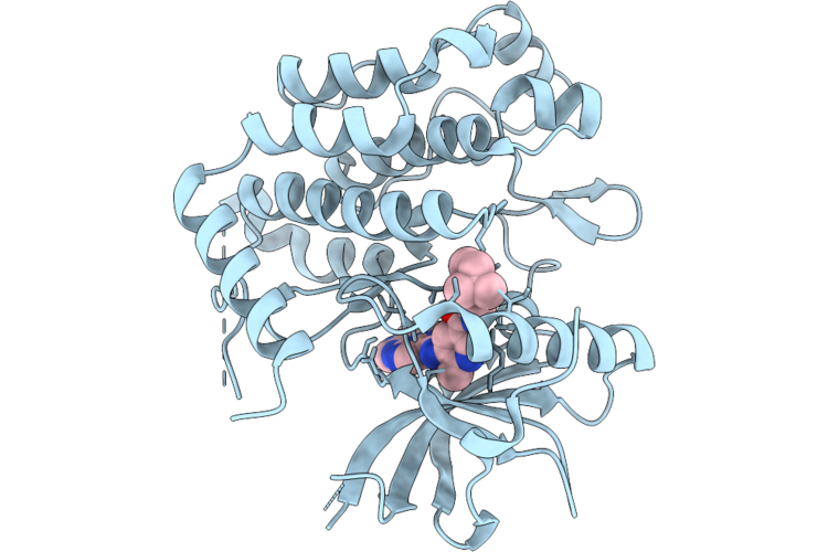 |
Structure Of Pdgfrb Kinase Domain Bound To Jnj-Pdgfrbi-1
Organism: Homo sapiens
Method: X-RAY DIFFRACTION Resolution:2.32 Å Release Date: 2026-05-20 Classification: TRANSFERASE Ligands: A1DA1 |
Organism: Homo sapiens
Method: X-RAY DIFFRACTION
Release Date: 2026-05-20
Ligands: A1DA1
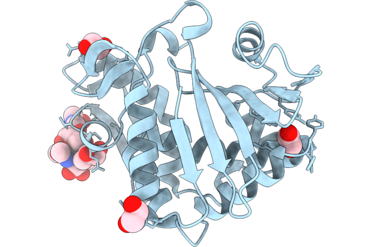 |
Crystal Structure Of A New Isoform Of Ribosome Inactivating Protein From Momordica Balsamina At 1.41 A Resolution
Organism: Momordica balsamina
Method: X-RAY DIFFRACTION Resolution:1.41 Å Release Date: 2026-05-20 Classification: HYDROLASE Ligands: NAG, EDO, GOL |
Organism: Momordica balsamina
Method: X-RAY DIFFRACTION
Release Date: 2026-05-20
Ligands: NAG, EDO, GOL
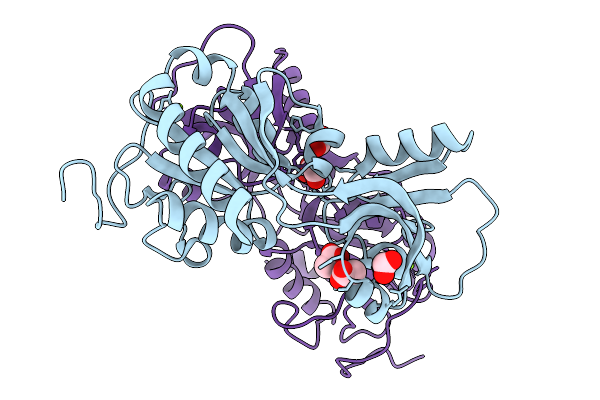 |
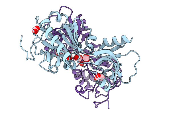
Crystal Structure Of Short-Form Adenosine Triphosphate Phosphoribosyltransferase From Acinetobacter Baumannii At 2.18 Angstrom Resolution.
Organism: Acinetobacter baumannii
Method: X-RAY DIFFRACTION Resolution:2.18 Å Release Date: 2026-02-18 Classification: TRANSFERASE Ligands: ACT, FMT, MG |
Organism: Acinetobacter baumannii
Method: X-RAY DIFFRACTION
Release Date: 2026-02-18
Ligands: ACT, FMT, MG
 |
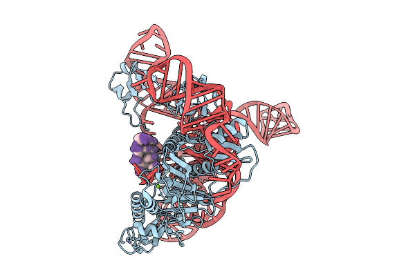
Crystal Structure Of Short-Form Adenosine Triphosphate Phosphoribosyltransferase From Acinetobacter Baumannii At 1.94 Angstrom Resolution
Organism: Acinetobacter baumannii
Method: X-RAY DIFFRACTION Resolution:1.94 Å Release Date: 2026-02-18 Classification: TRANSFERASE Ligands: FMT, ACT, MG |
Organism: Acinetobacter baumannii
Method: X-RAY DIFFRACTION
Release Date: 2026-02-18
Ligands: FMT, ACT, MG
 |
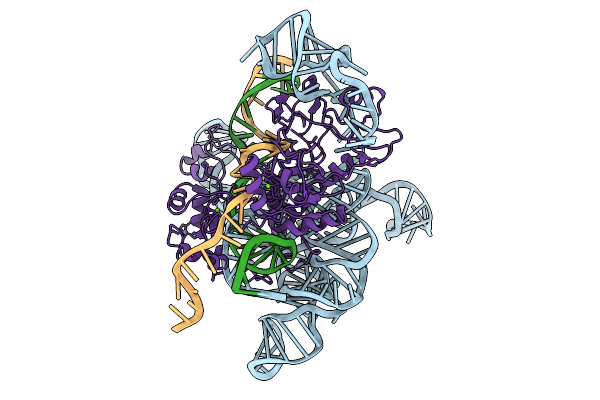
Cryoem Structures Uncover The Unexpected Hinges Of Iscb For Enhanced Gene Editing
Organism: Synthetic construct
Method: ELECTRON MICROSCOPY Resolution:3.04 Å Release Date: 2026-01-21 Classification: RNA BINDING PROTEIN Ligands: ZN, MG |
Organism: Synthetic construct
Method: ELECTRON MICROSCOPY
Release Date: 2026-01-21
Ligands: ZN, MG
 |
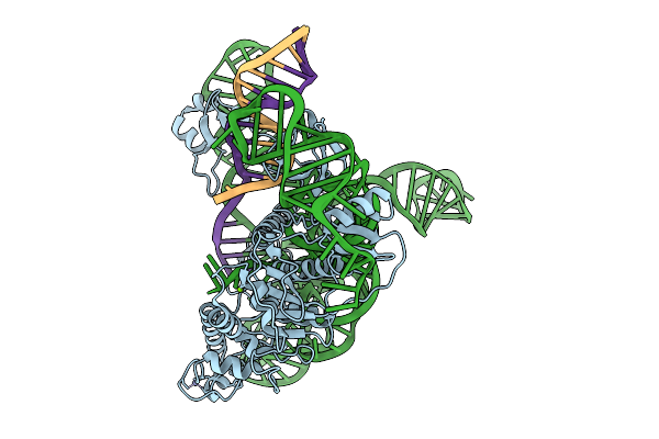
Cryoem Structures Uncover The Unexpected Hinges Of Iscb For Enhanced Gene Editing
Organism: Synthetic construct
Method: ELECTRON MICROSCOPY Resolution:3.10 Å Release Date: 2026-01-21 Classification: DNA BINDING PROTEIN Ligands: MG, ZN |
Organism: Synthetic construct
Method: ELECTRON MICROSCOPY
Release Date: 2026-01-21
Ligands: MG, ZN
 |
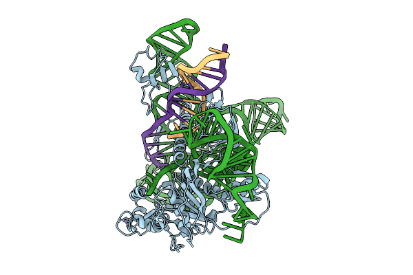
Cryoem Structures Uncover The Unexpected Hinges Of Iscb For Enhanced Gene Editing
Organism: Synthetic construct
Method: ELECTRON MICROSCOPY Resolution:3.39 Å Release Date: 2026-01-21 Classification: RNA BINDING PROTEIN Ligands: ZN, MG |
Organism: Synthetic construct
Method: ELECTRON MICROSCOPY
Release Date: 2026-01-21
Ligands: ZN, MG
 |
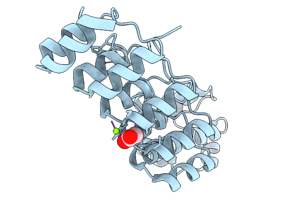
Cryoem Structures Uncover The Unexpected Hinges Of Iscb For Enhanced Gene Editing
Organism: Synthetic construct
Method: ELECTRON MICROSCOPY Resolution:3.09 Å Release Date: 2026-01-21 Classification: DNA BINDING PROTEIN Ligands: ZN, MG |
Organism: Synthetic construct
Method: ELECTRON MICROSCOPY
Release Date: 2026-01-21
Ligands: ZN, MG
 |
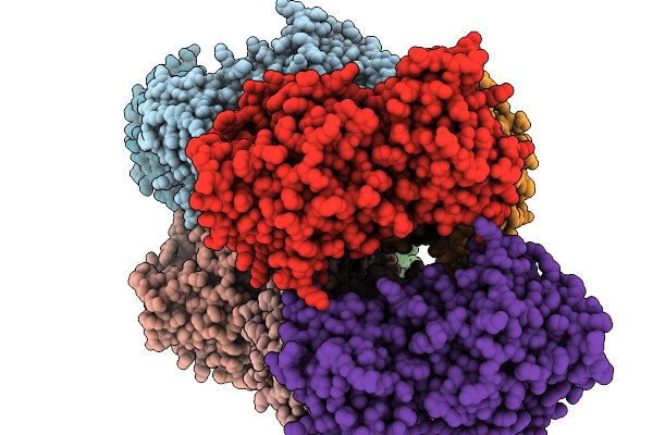
Crystal Structure Of A Darpin Fused To The 1Tel Crystallization Chaperone Via A Direct Helical Fusion In A 3-Fold Crystal Form
Organism: Homo sapiens, Synthetic construct
Method: X-RAY DIFFRACTION Resolution:1.77 Å Release Date: 2026-01-14 Classification: PROTEIN BINDING Ligands: ACY, CL, MG |
Organism: Homo sapiens, Synthetic construct
Method: X-RAY DIFFRACTION
Release Date: 2026-01-14
Ligands: ACY, CL, MG
 |
Crystal Structure Of Pseudopedobacter Saltans Gh43 Beta-Xylosidase In Complex With Xylose.
Organism: Pseudopedobacter saltans dsm 12145
Method: X-RAY DIFFRACTION Resolution:2.80 Å Release Date: 2026-01-07 Classification: HYDROLASE Ligands: CA, XLS |
Organism: Pseudopedobacter saltans dsm 12145
Method: X-RAY DIFFRACTION
Release Date: 2026-01-07
Ligands: CA, XLS
 |
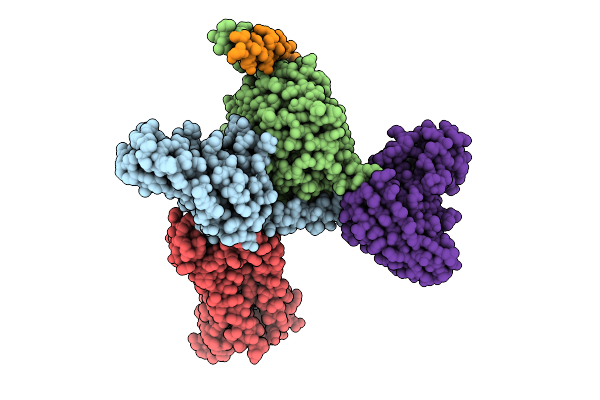
Crystal Structure Of The Chymotrypsin-Cleaved Iron-Free C-Lobe Of Bovine Lactoferrin At 2.82 Angstrom Resolution
Organism: Bos taurus
Method: X-RAY DIFFRACTION Resolution:2.82 Å Release Date: 2025-12-31 Classification: METAL BINDING PROTEIN Ligands: EDO, GOL, ACT, NAG, SO4 |
Organism: Bos taurus
Method: X-RAY DIFFRACTION
Release Date: 2025-12-31
Ligands: EDO, GOL, ACT, NAG, SO4
 |
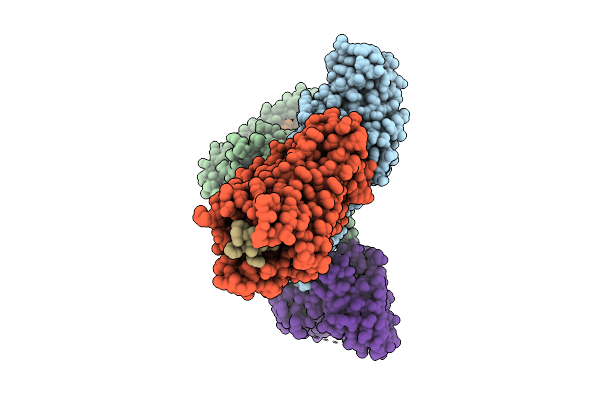
Structure Of Jr14A Bound To Human C3Ar In Complex With Go
Organism: Homo sapiens, Mus musculus
Method: ELECTRON MICROSCOPY Release Date: 2025-11-26 Classification: SIGNALING PROTEIN Ligands: A1D9A |
Organism: Homo sapiens, Mus musculus
Method: ELECTRON MICROSCOPY
Release Date: 2025-11-26
Ligands: A1D9A
 |
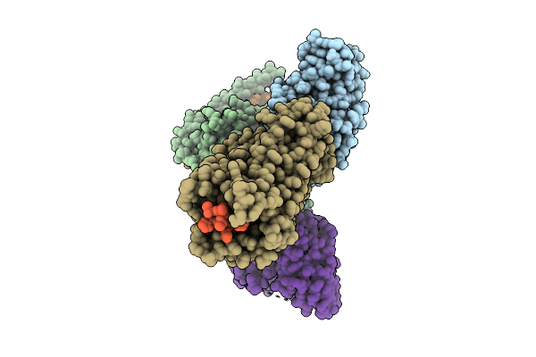
Structure Of Mouse Tlqp21 Bound To Mouse C3Ar In Complex With Go
Organism: Homo sapiens, Mus musculus
Method: ELECTRON MICROSCOPY Release Date: 2025-11-26 Classification: SIGNALING PROTEIN |
Organism: Homo sapiens, Mus musculus
Method: ELECTRON MICROSCOPY
Release Date: 2025-11-26
 |
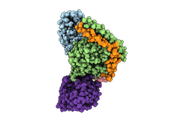
Structure Of Human Tlqp21 Bound To Mouse C3Ar In Complex With Go
Organism: Homo sapiens, Mus musculus
Method: ELECTRON MICROSCOPY Release Date: 2025-11-26 Classification: SIGNALING PROTEIN |
Organism: Homo sapiens, Mus musculus
Method: ELECTRON MICROSCOPY
Release Date: 2025-11-26
 |
Structure Of Sb290157 Bound To Human C3Ar In Complex With Go (Full Map)
Organism: Homo sapiens, Mus musculus
Method: ELECTRON MICROSCOPY Release Date: 2025-11-26 Classification: SIGNALING PROTEIN Ligands: A1L6Y |
Organism: Homo sapiens, Mus musculus
Method: ELECTRON MICROSCOPY
Release Date: 2025-11-26
Ligands: A1L6Y
 |
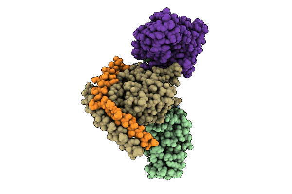
Structure Of Mouse C5A Bound Mouse C5Ar1 In Complex With Go
Organism: Homo sapiens, Mus musculus
Method: ELECTRON MICROSCOPY Release Date: 2025-11-26 Classification: SIGNALING PROTEIN |
Organism: Homo sapiens, Mus musculus
Method: ELECTRON MICROSCOPY
Release Date: 2025-11-26
 |
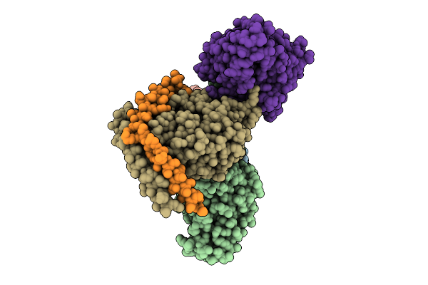
Structure Of Mouse C5A-Desarg Bound Mouse C5Ar1 In Complex With Go
Organism: Homo sapiens, Mus musculus
Method: ELECTRON MICROSCOPY Release Date: 2025-11-26 Classification: SIGNALING PROTEIN |
Organism: Homo sapiens, Mus musculus
Method: ELECTRON MICROSCOPY
Release Date: 2025-11-26
 |
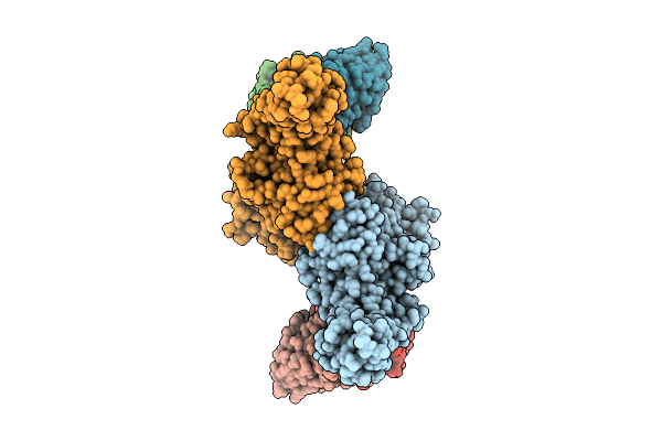
Structure Of Human C5A-Desarg Bound Mouse C5Ar1 In Complex With Go
Organism: Homo sapiens, Mus musculus
Method: ELECTRON MICROSCOPY Release Date: 2025-11-26 Classification: SIGNALING PROTEIN |
Organism: Homo sapiens, Mus musculus
Method: ELECTRON MICROSCOPY
Release Date: 2025-11-26
 |
Structure Of Beta-Arrestin2 In Complex With Mouse C5Ar1Pp
Organism: Bos taurus, Mus musculus
Method: ELECTRON MICROSCOPY Release Date: 2025-11-26 Classification: SIGNALING PROTEIN |
Organism: Bos taurus, Mus musculus
Method: ELECTRON MICROSCOPY
Release Date: 2025-11-26