

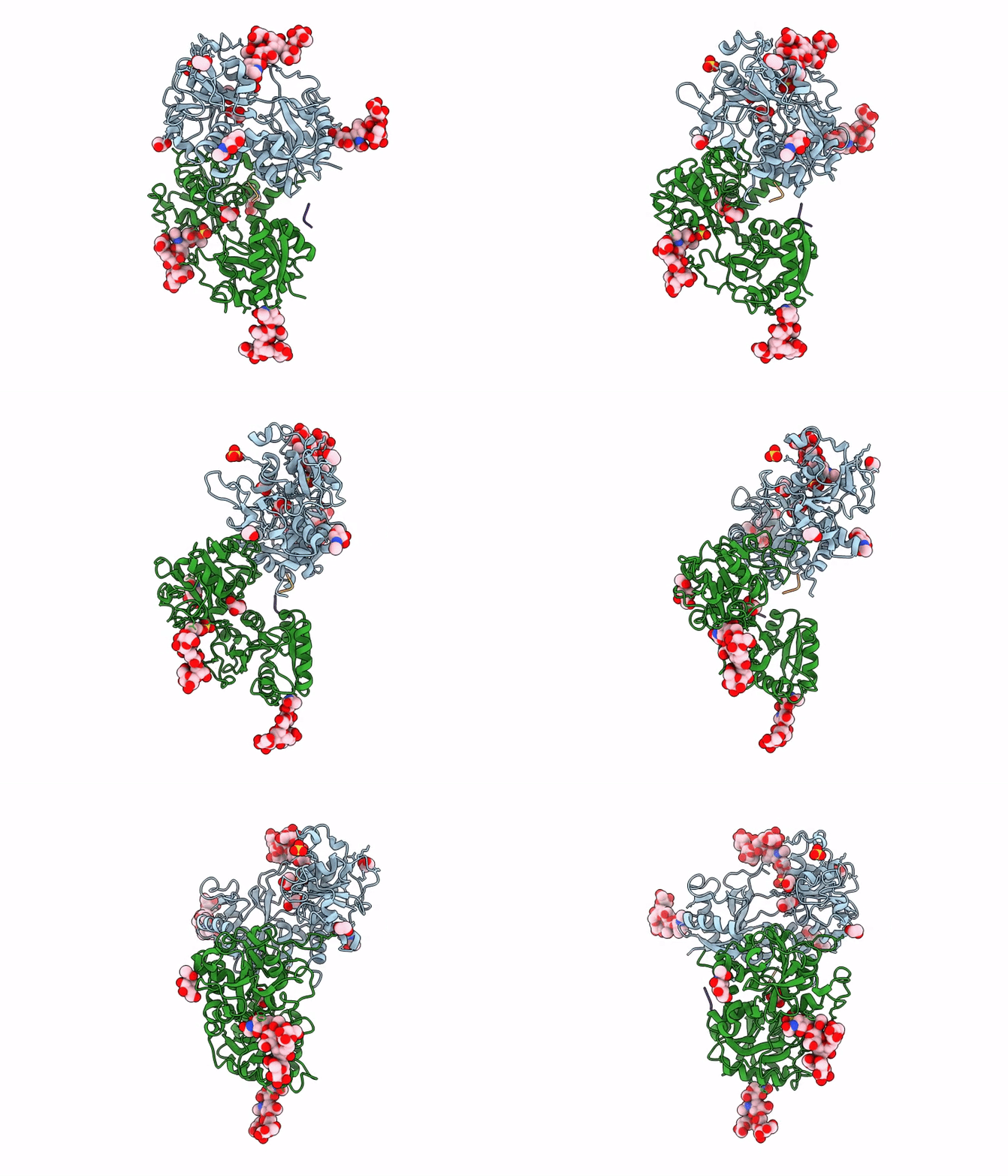
9XXU image
Deposition Date 2025-12-01
Release Date 2025-12-31
Last Version Date 2026-09-16
Entry Detail
PDB ID:
9XXU
Title:
Crystal structure of the chymotrypsin-cleaved iron-free C-lobe of bovine lactoferrin at 2.82 Angstrom resolution
Biological Source:
Source Organism(s):
Bos taurus (Taxon ID: 9913)
Method Details:
Experimental Method:
Resolution:
2.82 Å
R-Value Free:
0.26
R-Value Work:
0.21
Space Group:
C 2 2 21
Macromolecular Entities
Polymer Type:polypeptide(L)
Molecule:Lactotransferrin
Gene (Uniprot):LTF
Chain IDs:A, C (auth: B)
Chain Length:0
Number of Molecules:2
Biological Source:Bos taurus
Polymer Type:polypeptide(L)
Molecule:C-terminal fragment of Lactot
Gene (Uniprot):LTF
Chain IDs:B (auth: C), D
Chain Length:0
Number of Molecules:2
Biological Source:Bos taurus
Primary Citation
Structural Basis for Single-Site Cleavage of Lactoferrin by Diverse Proteases for Prolonged Antibacterial Action: Structure of the Chymotrypsin-Cleaved Lactoferrin C-Lobe.
Proteins 94 1658 1670 (2026)
PMID: 42230792 DOI: 10.1002/prot.70148

Abstact

The stable lactoferrin C-lobe offers strong potential for therapeutic applications as an antibacterial agent. Lactoferrin is a 78 kDa (Ala1Arg689) iron-binding glycoprotein which is composed of two homologous N- and C-lobes, connected by an 11-residue alpha-helical linker (Thr334Arg344). The limited proteolysis of lactoferrin, carried out using chymotrypsin, generated a 40 kDa, fully functional C-lobe. The structure determination revealed that the protein chain consisted of residues from Thr343 to Leu680 together with a disulfide-linked tripeptide, Ala683Cys684Ala685. It showed that the cleavage occurred specifically at the Tyr342Thr343 peptide bond within the inter-lobe 11-residue-long peptide. Remarkably, previous studies using proteinase K, trypsin, and pepsin also produced an identical C-lobe. Thus, the inter-lobe region seems to be stereochemically designed by nature for the single-site cleavage by multiple digestive enzymes. The proteolytically generated C-lobe, with three observed glycosylation sites, remains stable for 3 days in the presence of digestive enzymes. The stable C-lobe continues to sequester iron, thus showing a prolonged antibacterial property. This is a unique example of evolutionary convergence whereby multiple digestive enzymes cleave a native protein into a stable half molecule with full antibacterial action.

Legend

Protein

Chemical

Disease

Primary Citation of related structures
Feedback Form
Name
Email
Institute
Feedback