
Search Count: 567
All
Selected
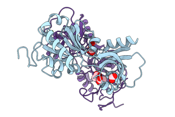 |
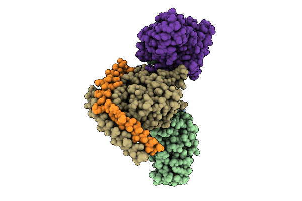
Crystal Structure Of Short-Form Adenosine Triphosphate Phosphoribosyltransferase From Acinetobacter Baumannii At 2.18 Angstrom Resolution.
Organism: Acinetobacter baumannii
Method: X-RAY DIFFRACTION Resolution:2.18 Å Release Date: 2026-02-18 Classification: TRANSFERASE Ligands: ACT, FMT, MG |
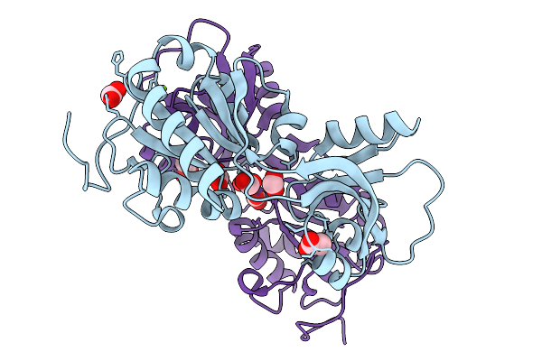 |
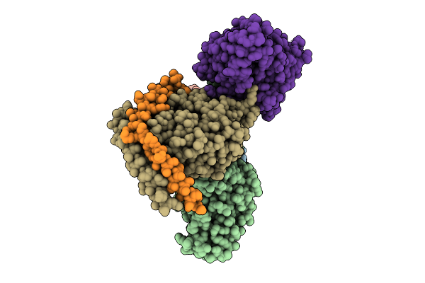
Crystal Structure Of Short-Form Adenosine Triphosphate Phosphoribosyltransferase From Acinetobacter Baumannii At 1.94 Angstrom Resolution
Organism: Acinetobacter baumannii
Method: X-RAY DIFFRACTION Resolution:1.94 Å Release Date: 2026-02-18 Classification: TRANSFERASE Ligands: FMT, ACT, MG |
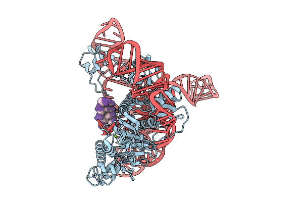 |
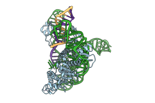
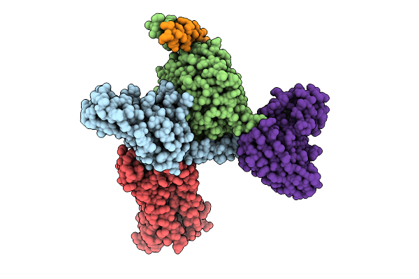
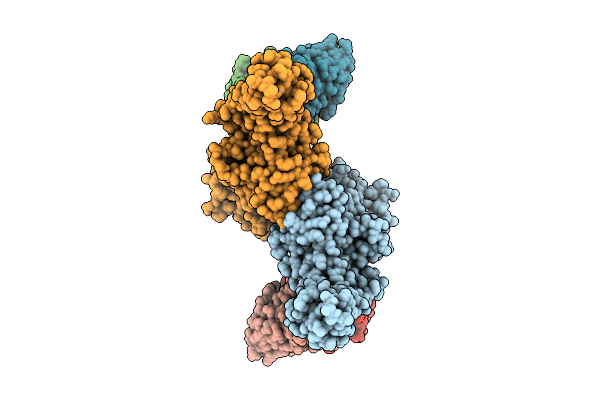
Cryoem Structures Uncover The Unexpected Hinges Of Iscb For Enhanced Gene Editing
Organism: Synthetic construct
Method: ELECTRON MICROSCOPY Resolution:3.04 Å Release Date: 2026-01-21 Classification: RNA BINDING PROTEIN Ligands: ZN, MG |
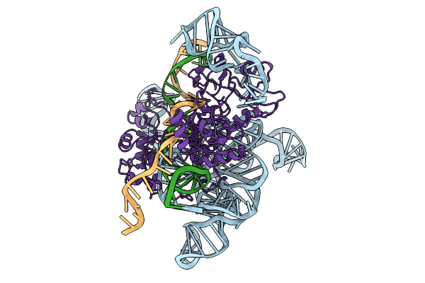 |
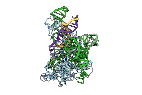
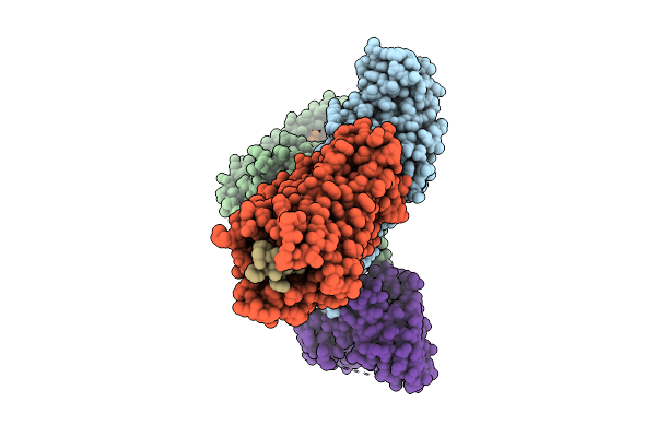
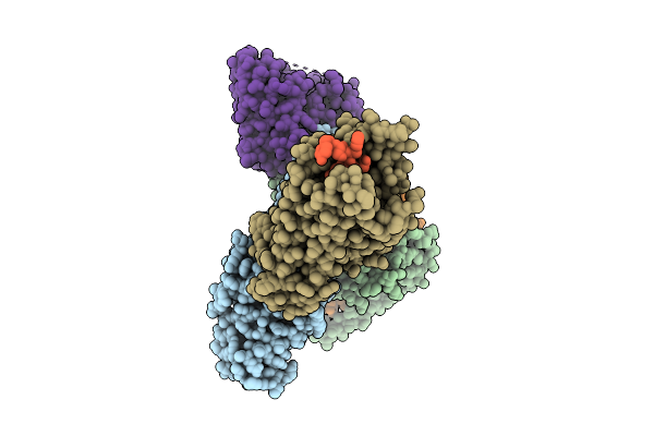
Cryoem Structures Uncover The Unexpected Hinges Of Iscb For Enhanced Gene Editing
Organism: Synthetic construct
Method: ELECTRON MICROSCOPY Resolution:3.10 Å Release Date: 2026-01-21 Classification: DNA BINDING PROTEIN Ligands: MG, ZN |
 |
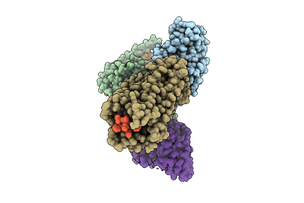
Cryoem Structures Uncover The Unexpected Hinges Of Iscb For Enhanced Gene Editing
Organism: Synthetic construct
Method: ELECTRON MICROSCOPY Resolution:3.39 Å Release Date: 2026-01-21 Classification: RNA BINDING PROTEIN Ligands: ZN, MG |
 |
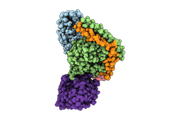
Cryoem Structures Uncover The Unexpected Hinges Of Iscb For Enhanced Gene Editing
Organism: Synthetic construct
Method: ELECTRON MICROSCOPY Resolution:3.09 Å Release Date: 2026-01-21 Classification: DNA BINDING PROTEIN Ligands: ZN, MG |
 |
Crystal Structure Of Pseudopedobacter Saltans Gh43 Beta-Xylosidase In Complex With Xylose.
Organism: Pseudopedobacter saltans dsm 12145
Method: X-RAY DIFFRACTION Resolution:2.80 Å Release Date: 2026-01-07 Classification: HYDROLASE Ligands: CA, XLS |
 |
Crystal Structure Of The Chymotrypsin-Cleaved Iron-Free C-Lobe Of Bovine Lactoferrin At 2.82 Angstrom Resolution
Organism: Bos taurus
Method: X-RAY DIFFRACTION Resolution:2.82 Å Release Date: 2025-12-31 Classification: METAL BINDING PROTEIN Ligands: EDO, GOL, ACT, NAG, SO4 |
 |
Organism: Homo sapiens, Mus musculus
Method: ELECTRON MICROSCOPY Release Date: 2025-11-26 Classification: SIGNALING PROTEIN |
 |
Organism: Homo sapiens, Mus musculus
Method: ELECTRON MICROSCOPY Release Date: 2025-11-26 Classification: SIGNALING PROTEIN Ligands: A1D9A |
 |
Organism: Homo sapiens, Mus musculus
Method: ELECTRON MICROSCOPY Release Date: 2025-11-26 Classification: SIGNALING PROTEIN |
 |
Organism: Homo sapiens, Mus musculus
Method: ELECTRON MICROSCOPY Release Date: 2025-11-26 Classification: SIGNALING PROTEIN |
 |
Organism: Homo sapiens, Mus musculus
Method: ELECTRON MICROSCOPY Release Date: 2025-11-26 Classification: SIGNALING PROTEIN Ligands: A1L6Y |
 |
Organism: Homo sapiens, Mus musculus
Method: ELECTRON MICROSCOPY Release Date: 2025-11-26 Classification: SIGNALING PROTEIN |
 |
Organism: Homo sapiens, Mus musculus
Method: ELECTRON MICROSCOPY Release Date: 2025-11-26 Classification: SIGNALING PROTEIN |
 |
Organism: Homo sapiens, Mus musculus
Method: ELECTRON MICROSCOPY Release Date: 2025-11-26 Classification: SIGNALING PROTEIN |
 |
Organism: Homo sapiens, Mus musculus
Method: ELECTRON MICROSCOPY Release Date: 2025-11-26 Classification: SIGNALING PROTEIN |
 |
Organism: Bos taurus, Mus musculus
Method: ELECTRON MICROSCOPY Release Date: 2025-11-26 Classification: SIGNALING PROTEIN |
 |
Organism: Rattus norvegicus, Mus musculus
Method: ELECTRON MICROSCOPY Release Date: 2025-11-26 Classification: SIGNALING PROTEIN |
 |
Organism: Homo sapiens, Mus musculus
Method: ELECTRON MICROSCOPY Release Date: 2025-11-26 Classification: SIGNALING PROTEIN |