
Search Count: 2,688
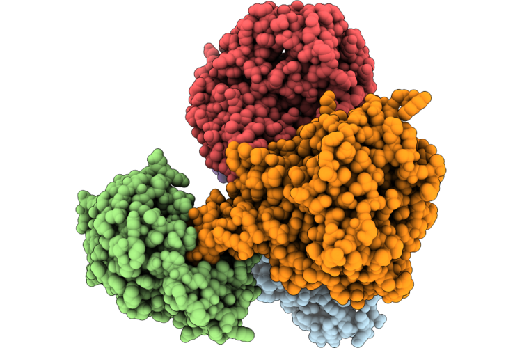 |
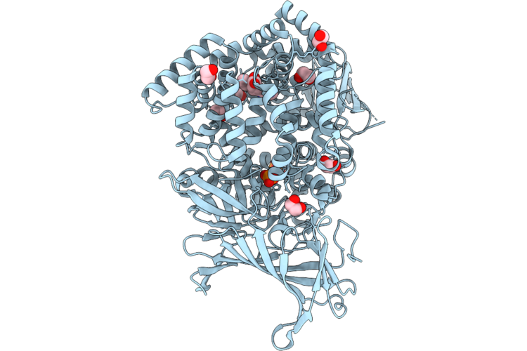
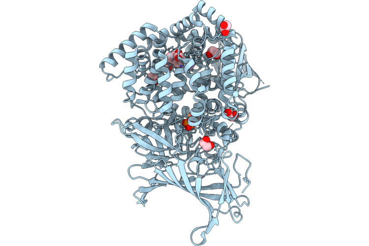
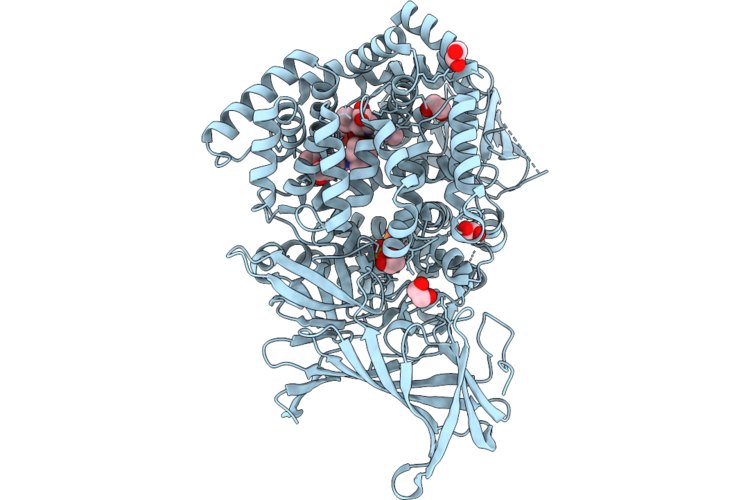
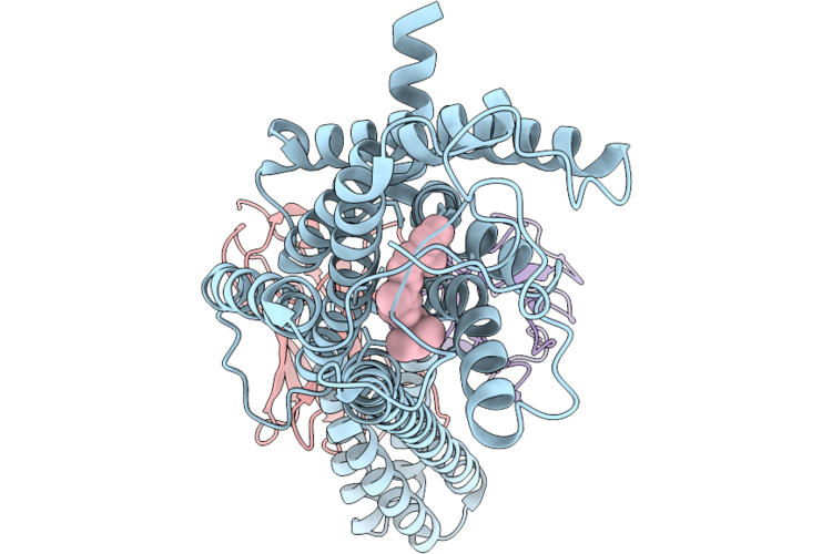
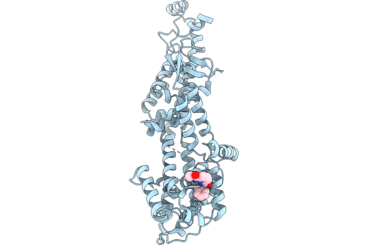
E. Coli Sufe Bound To Sufbc2D
Organism: Escherichia coli
Method: ELECTRON MICROSCOPY Release Date: 2026-07-15 Classification: METAL BINDING PROTEIN |
Organism: Escherichia coli
Method: ELECTRON MICROSCOPY
Release Date: 2026-07-15
 |
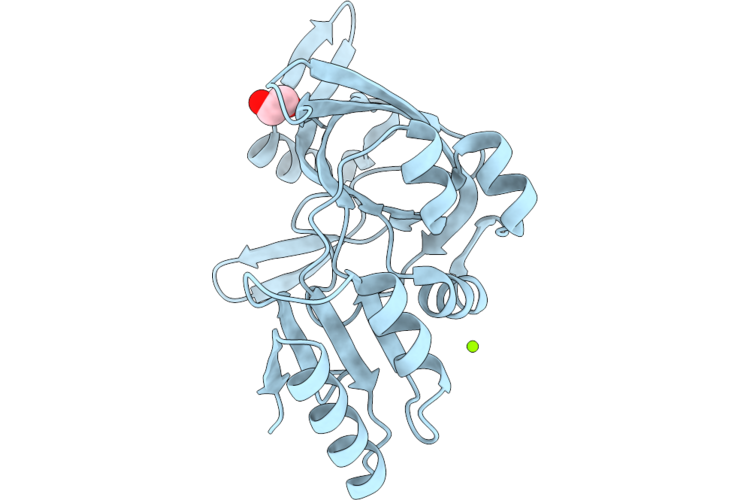
Crystal Structure Of The Peptide-Binding Protein Nika From Streptococcus Agalactiae In Complex With Zinc And L-Histidine.
Organism: Streptococcus agalactiae 2603v/r
Method: X-RAY DIFFRACTION Resolution:1.95 Å Release Date: 2026-07-15 Classification: TRANSPORT PROTEIN Ligands: NI, HIS |
Organism: Streptococcus agalactiae 2603v/r
Method: X-RAY DIFFRACTION
Release Date: 2026-07-15
Ligands: NI, HIS
 |
Crystal Structure Of The Peptide-Binding Protein Nika From Streptococcus Agalactiae In Complex With Zinc, L-Histidine And Tris Buffer.
Organism: Streptococcus agalactiae 2603v/r
Method: X-RAY DIFFRACTION Resolution:1.80 Å Release Date: 2026-07-15 Classification: TRANSPORT PROTEIN Ligands: NI, HIS, TRS |
Organism: Streptococcus agalactiae 2603v/r
Method: X-RAY DIFFRACTION
Release Date: 2026-07-15
Ligands: NI, HIS, TRS
 |
Crystal Structure Of The Peptide-Binding Protein Nika From Streptococcus Agalactiae In Complex With Zinc, L-Histidine, Imidazole And Ethylene Glycol.
Organism: Streptococcus agalactiae 2603v/r
Method: X-RAY DIFFRACTION Resolution:1.95 Å Release Date: 2026-07-15 Classification: TRANSPORT PROTEIN Ligands: NI, HIS, IMD, EDO |
Organism: Streptococcus agalactiae 2603v/r
Method: X-RAY DIFFRACTION
Release Date: 2026-07-15
Ligands: NI, HIS, IMD, EDO
 |
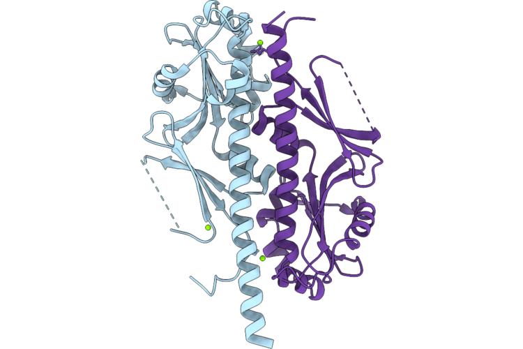
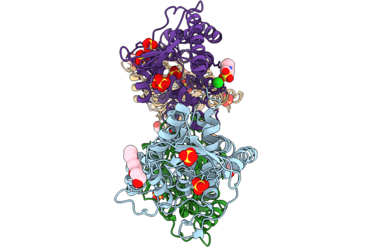
Crystal Structure Of Braf:Mek1 Complex With Symmetric Dimer Interface Bound To Amppnp
Organism: Homo sapiens
Method: X-RAY DIFFRACTION Resolution:2.91 Å Release Date: 2026-07-15 Classification: SIGNALING PROTEIN Ligands: ANP, CA, GOL, CL, PEG |
Organism: Homo sapiens
Method: X-RAY DIFFRACTION
Release Date: 2026-07-15
Ligands: ANP, CA, GOL, CL, PEG
 |
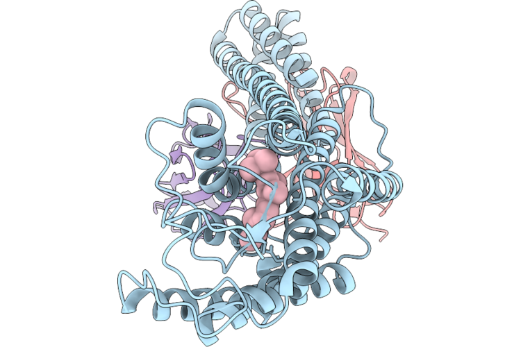
Crystal Structure Of Braf:Mek1 Complex With Asymmetric Dimer Interface Bound To Amppnp
Organism: Homo sapiens
Method: X-RAY DIFFRACTION Resolution:2.60 Å Release Date: 2026-07-15 Classification: SIGNALING PROTEIN Ligands: ANP, MG |
Organism: Homo sapiens
Method: X-RAY DIFFRACTION
Release Date: 2026-07-15
Ligands: ANP, MG
 |
Crystal Structure Of Braf:Mek1(Ps222) Complex With Asymmetric Dimer Interface Bound To Adp
Organism: Homo sapiens
Method: X-RAY DIFFRACTION Resolution:2.30 Å Release Date: 2026-07-15 Classification: SIGNALING PROTEIN Ligands: ADP, CA, GOL, NA |
Organism: Homo sapiens
Method: X-RAY DIFFRACTION
Release Date: 2026-07-15
Ligands: ADP, CA, GOL, NA
 |
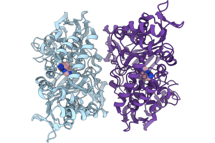
Erap1 In Complex With (3S,4S)-1-(3-Cyano-6-Methylpyridin-2-Yl)-4-(Propan-2-Yl)Pyrrolidine-3-Carboxylic Acid
Organism: Homo sapiens
Method: X-RAY DIFFRACTION Resolution:1.87 Å Release Date: 2026-07-08 Classification: PEPTIDE BINDING PROTEIN Ligands: ZN, PO4, EDO, A1JVC |
Organism: Homo sapiens
Method: X-RAY DIFFRACTION
Release Date: 2026-07-08
Ligands: ZN, PO4, EDO, A1JVC
 |
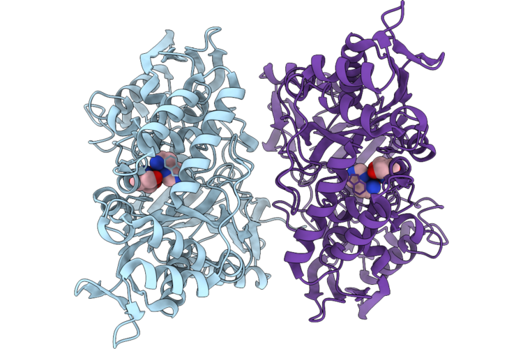
Erap1 In Complex With (3R,4R)-1-(3-Cyano-6-Methylpyridin-2-Yl)-4-(Propan-2-Yl)Pyrrolidine-3-Carboxylic Acid
Organism: Homo sapiens
Method: X-RAY DIFFRACTION Resolution:1.63 Å Release Date: 2026-07-08 Classification: PEPTIDE BINDING PROTEIN Ligands: ZN, PO4, EDO, A1JVB |
Organism: Homo sapiens
Method: X-RAY DIFFRACTION
Release Date: 2026-07-08
Ligands: ZN, PO4, EDO, A1JVB
 |
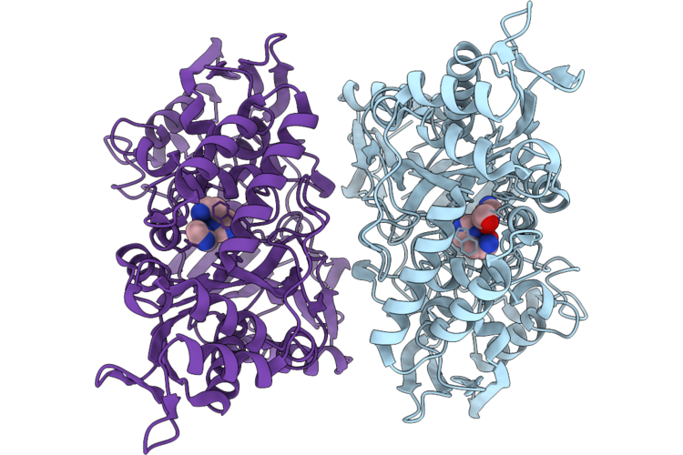
Erap1 In Complex With (3R,4R)-1-[3-Cyano-6-(Cyclopropylamino)Pyridin-2-Yl]-4-(Propan-2-Yl)Pyrrolidine-3-Carboxylic Acid
Organism: Homo sapiens
Method: X-RAY DIFFRACTION Resolution:1.73 Å Release Date: 2026-07-08 Classification: PEPTIDE BINDING PROTEIN Ligands: ZN, PO4, EDO, A1JVL |
Organism: Homo sapiens
Method: X-RAY DIFFRACTION
Release Date: 2026-07-08
Ligands: ZN, PO4, EDO, A1JVL
 |
Erap1 In Complex With (3R,4R)-1-{3-Cyano-4-Methyl-6-[(4-Methyloxan-4-Yl)Amino]Pyridin-2-Yl}-4-(Propan-2-Yl)Pyrrolidine-3-Carboxylic Acid
Organism: Homo sapiens
Method: X-RAY DIFFRACTION Resolution:1.74 Å Release Date: 2026-07-08 Classification: PEPTIDE BINDING PROTEIN Ligands: ZN, PO4, EDO, A1JV2 |
Organism: Homo sapiens
Method: X-RAY DIFFRACTION
Release Date: 2026-07-08
Ligands: ZN, PO4, EDO, A1JV2
 |
Dark-State Structure Of Human Medium-Wave-Sensitive Cone Opsin (Opn1Mw)
Organism: Homo sapiens, Escherichia coli, Synthetic construct
Method: ELECTRON MICROSCOPY Resolution:3.70 Å Release Date: 2026-06-24 Classification: MEMBRANE PROTEIN Ligands: RET |
Organism: Homo sapiens, Escherichia coli, Synthetic construct
Method: ELECTRON MICROSCOPY
Release Date: 2026-06-24
Ligands: RET
 |
Dark-State Structure Of Human Short-Wave-Sensitive Opsin (Opn1Sw)
Organism: Homo sapiens, Escherichia coli, Synthetic construct
Method: ELECTRON MICROSCOPY Resolution:3.22 Å Release Date: 2026-06-24 Classification: MEMBRANE PROTEIN Ligands: RET |
Organism: Homo sapiens, Escherichia coli, Synthetic construct
Method: ELECTRON MICROSCOPY
Release Date: 2026-06-24
Ligands: RET
 |
Nucleotide Binding Domain (Residues 475-720) Of Abc3 Transporter Permease From Clostridioides Difficile Strain 630
Organism: Clostridioides difficile 630
Method: X-RAY DIFFRACTION Resolution:2.00 Å Release Date: 2026-06-24 Classification: TRANSPORT PROTEIN Ligands: MG, CL, EDO |
Organism: Clostridioides difficile 630
Method: X-RAY DIFFRACTION
Release Date: 2026-06-24
Ligands: MG, CL, EDO
 |
Crystal Structure Of The Ggdef Domain (Residues 31-260) Of Diguanylate Cyclase From Vibrio Cholerae Serotype O1
Organism: Vibrio cholerae o1 biovar el tor str. n16961
Method: X-RAY DIFFRACTION Resolution:2.00 Å Release Date: 2026-06-24 Classification: OXIDOREDUCTASE Ligands: MG, CL |
Organism: Vibrio cholerae o1 biovar el tor str. n16961
Method: X-RAY DIFFRACTION
Release Date: 2026-06-24
Ligands: MG, CL
 |
Pre-Active Dark-State Structure Of Human Short-Wave-Sensitive Opsin (Opn1Sw)
Organism: Homo sapiens, Escherichia coli, Synthetic construct
Method: ELECTRON MICROSCOPY Resolution:3.06 Å Release Date: 2026-06-24 Classification: MEMBRANE PROTEIN Ligands: RET |
Organism: Homo sapiens, Escherichia coli, Synthetic construct
Method: ELECTRON MICROSCOPY
Release Date: 2026-06-24
Ligands: RET
 |
Sos1 In Complex With Compound 3
Organism: Homo sapiens
Method: X-RAY DIFFRACTION Resolution:1.92 Å Release Date: 2026-06-24 Classification: SIGNALING PROTEIN Ligands: A1JWK |
Organism: Homo sapiens
Method: X-RAY DIFFRACTION
Release Date: 2026-06-24
Ligands: A1JWK
 |
Crystal Structure Of Conserved Hypothetical Protein From Stenotrophomonas Maltophilia (Strain K279A)
Organism: Stenotrophomonas maltophilia k279a
Method: X-RAY DIFFRACTION Resolution:1.85 Å Release Date: 2026-06-17 Classification: UNKNOWN FUNCTION Ligands: SO4, PGE, CL, EDO, EPE |
Organism: Stenotrophomonas maltophilia k279a
Method: X-RAY DIFFRACTION
Release Date: 2026-06-17
Ligands: SO4, PGE, CL, EDO, EPE
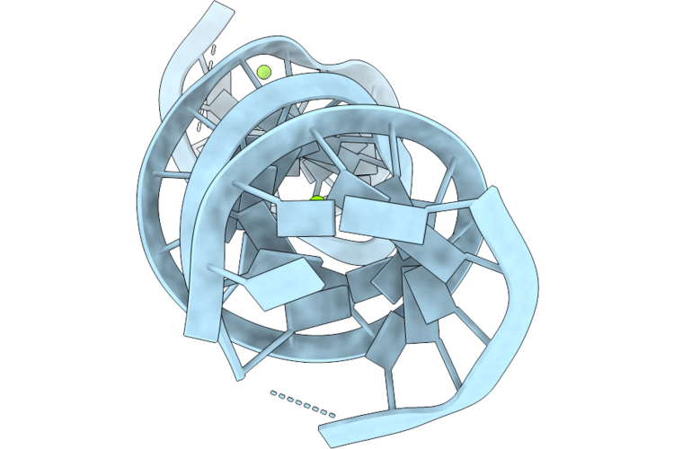 |
The Structure Of Egalitarian In Complex With The K10 Mrna Localization Signal Reveals A Modular Binding Surface Required For Function
Organism: Drosophila melanogaster
Method: X-RAY DIFFRACTION Resolution:3.60 Å Release Date: 2026-06-10 Classification: RNA BINDING PROTEIN Ligands: MG |
Organism: Drosophila melanogaster
Method: X-RAY DIFFRACTION
Release Date: 2026-06-10
Ligands: MG
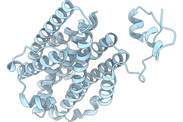 |
Crystal Structure Of Class Ie Ribonucleotide Reductase R2 Subunit From Mesoplasma Florum With A D212N Mutation
Organism: Mesoplasma florum l1
Method: X-RAY DIFFRACTION Resolution:1.70 Å Release Date: 2026-05-20 Classification: OXIDOREDUCTASE |
Organism: Mesoplasma florum l1
Method: X-RAY DIFFRACTION
Release Date: 2026-05-20