
Search Count: 78,081
All
Selected
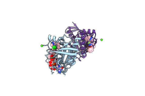 |
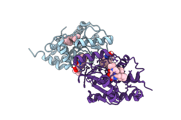
Organism: Homo sapiens
Method: X-RAY DIFFRACTION Resolution:1.50 Å Release Date: 2026-04-15 Classification: ONCOPROTEIN Ligands: CA, A1C6Y, GOL, GDP |
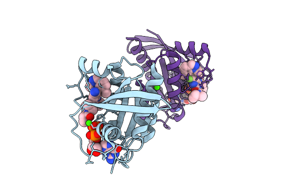 |
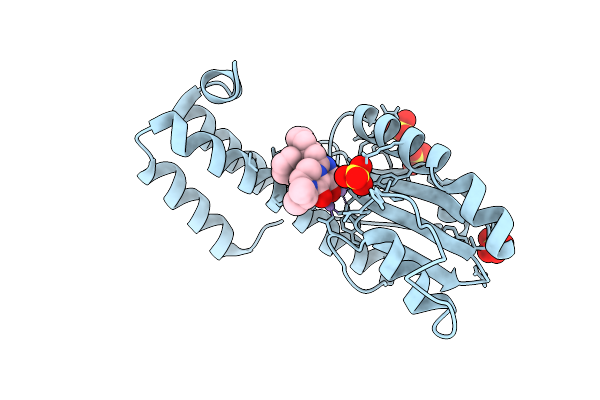
Organism: Homo sapiens
Method: X-RAY DIFFRACTION Resolution:1.52 Å Release Date: 2026-04-15 Classification: ONCOPROTEIN Ligands: GDP, A1C60, CA |
 |
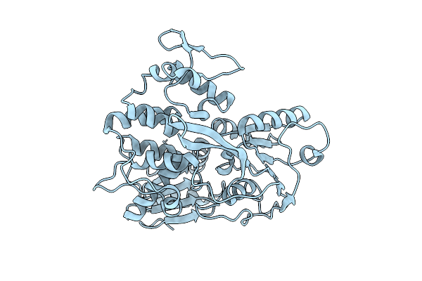
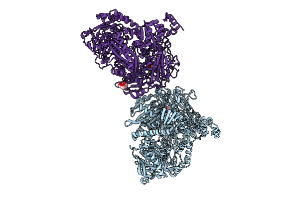
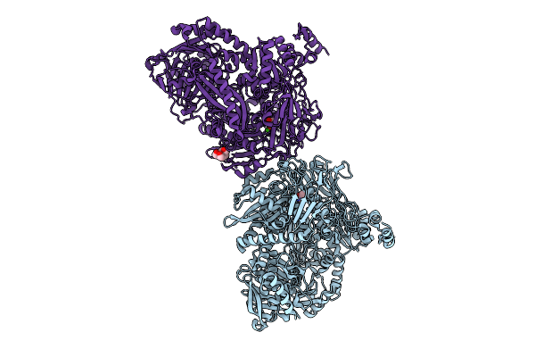
Organism: Homo sapiens
Method: ELECTRON MICROSCOPY Resolution:3.40 Å Release Date: 2026-04-15 Classification: SIGNALING PROTEIN |
 |
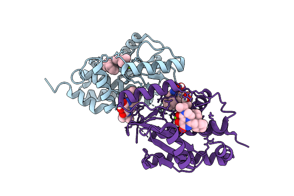
Organism: Cacipacore virus
Method: X-RAY DIFFRACTION Resolution:2.07 Å Release Date: 2026-04-15 Classification: HYDROLASE |
 |
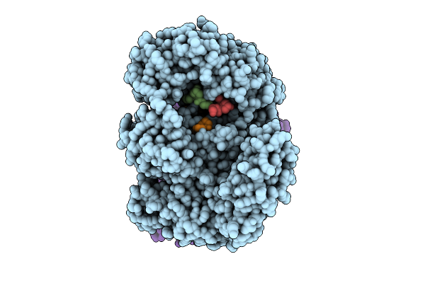
Three Viral Endonucleases Bound To The Same Inhibitor (9-Hydroxy-3,4-Dihydro-2H-Pyrazino[1,2-C]Pyrimidine-1,8-Dione Derivative). (1A) Influenza Virus A/Ph1N1 Pa Subunit Endonuclease With Magnesium Ions.
Organism: Influenza a virus
Method: X-RAY DIFFRACTION Resolution:1.86 Å Release Date: 2026-04-15 Classification: RNA BINDING PROTEIN Ligands: MG, A1J4F, GOL |
 |
Three Viral Endonucleases Bound To The Same Inhibitor (9-Hydroxy-3,4-Dihydro-2H-Pyrazino[1,2-C]Pyrimidine-1,8-Dione Derivative). (1B) Influenza Virus A/Ph1N1 Polymerase Pa Subunit Endonuclease With Manganese Ions.
Organism: Influenza a virus
Method: X-RAY DIFFRACTION Resolution:2.00 Å Release Date: 2026-04-15 Classification: RNA BINDING PROTEIN Ligands: MN, A1J4F |
 |
Three Viral Endonucleases Bound To The Same Inhibitor (9-Hydroxy-3,4-Dihydro-2H-Pyrazino[1,2-C]Pyrimidine-1,8-Dione Derivative). (2) La Crosse Virus L Protein Endonuclease With Manganese Ions.
Organism: La crosse virus
Method: X-RAY DIFFRACTION Resolution:2.80 Å Release Date: 2026-04-15 Classification: RNA BINDING PROTEIN Ligands: MN, A1J4F, SO4 |
 |
Three Viral Endonucleases Bound To The Same Inhibitor (9-Hydroxy-3,4-Dihydro-2H-Pyrazino[1,2-C]Pyrimidine-1,8-Dione Derivative). (3) Lassa Virus L Protein Endonuclease With Manganese Ions.
Organism: Mammarenavirus lassaense
Method: X-RAY DIFFRACTION Resolution:2.70 Å Release Date: 2026-04-15 Classification: RNA BINDING PROTEIN Ligands: MN, A1J4F |
 |
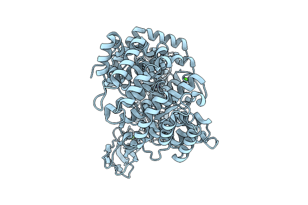
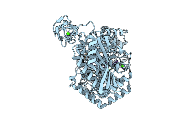
Crystal Structure Of N-Methylhydantoinase In Complex With 1-Methylimidazolidine-2,4-Dione
Organism: Glutamicibacter protophormiae
Method: X-RAY DIFFRACTION Resolution:2.07 Å Release Date: 2026-04-15 Classification: HYDROLASE Ligands: CA, A1BC1, NH4, BTB |
 |
Crystal Structure Of N-Methylhydantoinase In Complex With 1-Methylimidazolidine-2,4-Dione, Iodide Soak
Organism: Glutamicibacter protophormiae
Method: X-RAY DIFFRACTION Resolution:2.62 Å Release Date: 2026-04-15 Classification: HYDROLASE Ligands: CA, A1BC1, NH4, IOD, BTB |
 |
Organism: Glutamicibacter protophormiae
Method: X-RAY DIFFRACTION Resolution:3.13 Å Release Date: 2026-04-15 Classification: HYDROLASE Ligands: ANP, MG, NH4 |
 |
Crystal Structure Of N-Methylhydantoinase In Complex With 1-Methylimidazolidine-2,4-Dione, C2221 Form
Organism: Glutamicibacter protophormiae
Method: X-RAY DIFFRACTION Resolution:2.80 Å Release Date: 2026-04-15 Classification: HYDROLASE Ligands: CA, A1BC1, NH4, MES, SO4 |
 |
Organism: Glutamicibacter protophormiae
Method: X-RAY DIFFRACTION Resolution:2.58 Å Release Date: 2026-04-15 Classification: HYDROLASE Ligands: ANP, ZN, NH4, ACT |
 |
Crystal Structure Of N-Methylhydantoinase In Complex With 1-Methylimidazolidine-2,4-Dione, C-Terminal Residues Visible
Organism: Glutamicibacter protophormiae
Method: X-RAY DIFFRACTION Resolution:2.07 Å Release Date: 2026-04-15 Classification: HYDROLASE Ligands: CA, A1BC1, BTB, NH4, SO4 |
 |
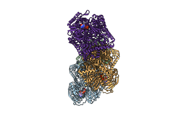
Cryo-Em Structure Of Collagenase H (E416Q Mutant) From Hathewaya Histolytica Bound To C-Terminal Region Of Collagen Model Peptide (Pro-Hyp-Gly)10
Organism: Hathewaya histolytica, Homo sapiens
Method: ELECTRON MICROSCOPY Release Date: 2026-04-15 Classification: METAL BINDING PROTEIN Ligands: CA, ZN |
 |
Organism: Hathewaya histolytica
Method: ELECTRON MICROSCOPY Release Date: 2026-04-15 Classification: METAL BINDING PROTEIN Ligands: ZN, CA |
 |
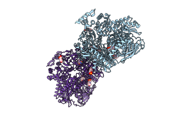
Cryo-Em Structure Of Collagenase H (E416Q Mutant) From Hathewaya Histolytica In Complex With Collagen Model Peptide (Pro-Hyp-Gly)10
Organism: Hathewaya histolytica, Homo sapiens
Method: ELECTRON MICROSCOPY Release Date: 2026-04-15 Classification: METAL BINDING PROTEIN Ligands: CA, ZN |
 |
Cryo-Em Structure Of Collagenase H (E416Q Mutant) From Hathewaya Histolytica In Complex With Collagen Model Peptide (Pro-Hyp-Gly)12
Organism: Hathewaya histolytica, Homo sapiens
Method: ELECTRON MICROSCOPY Release Date: 2026-04-15 Classification: METAL BINDING PROTEIN Ligands: CA, ZN |
 |
Organism: Homo sapiens
Method: X-RAY DIFFRACTION Resolution:1.85 Å Release Date: 2026-04-15 Classification: HYDROLASE, LYASE/DNA Ligands: SO4 |
 |
Organism: Homo sapiens, Synthetic construct
Method: X-RAY DIFFRACTION Resolution:2.00 Å Release Date: 2026-04-15 Classification: HYDROLASE, LYASE/DNA Ligands: EDO |