

Search Count: 16
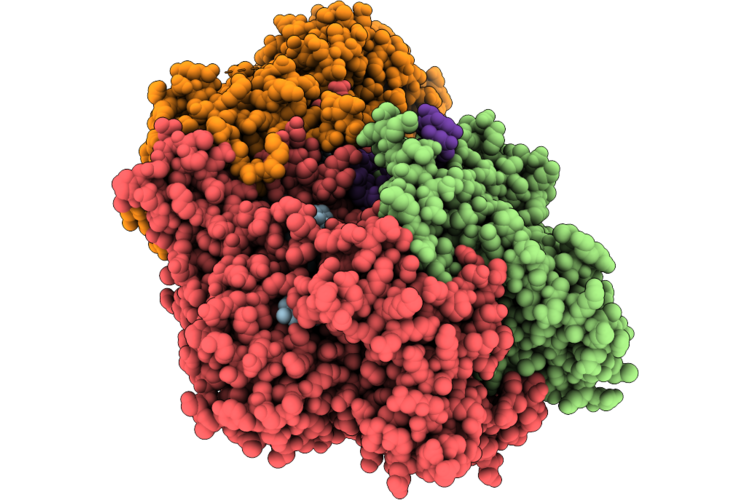 |
Cryo-Em Structure Of The Type Iii-D2 Crispr-Cas Effector Complex Bound To A Cognate Target Rna In The Pre-Cleavage State
Organism: Gammaproteobacteria bacterium
Method: ELECTRON MICROSCOPY Release Date: 2026-06-24 Classification: IMMUNE SYSTEM Ligands: MG, ANP, SAM, ZN |
Organism: Gammaproteobacteria bacterium
Method: ELECTRON MICROSCOPY
Release Date: 2026-06-24
Ligands: MG, ANP, SAM, ZN
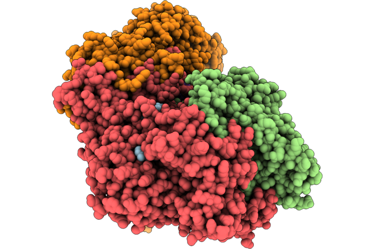 |
Cryo-Em Structure Of The Type Iii-D2 Crispr-Cas Effector Complex Bound To A Cognate Target Rna In The Post-Cleavage State
Organism: Gammaproteobacteria bacterium
Method: ELECTRON MICROSCOPY Release Date: 2026-06-24 Classification: IMMUNE SYSTEM Ligands: MG, ANP, SAM, ZN |
Organism: Gammaproteobacteria bacterium
Method: ELECTRON MICROSCOPY
Release Date: 2026-06-24
Ligands: MG, ANP, SAM, ZN
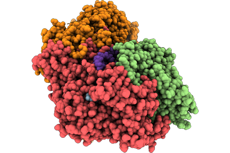 |
Cryo-Em Structure Of The Type Iii-D2 Crispr-Cas Effector Complex Bound To A Non-Cognate Target Rna In The Pre-Cleavage State
Organism: Gammaproteobacteria bacterium
Method: ELECTRON MICROSCOPY Release Date: 2026-06-24 Classification: IMMUNE SYSTEM Ligands: MG, ANP, SAM, ZN |
Organism: Gammaproteobacteria bacterium
Method: ELECTRON MICROSCOPY
Release Date: 2026-06-24
Ligands: MG, ANP, SAM, ZN
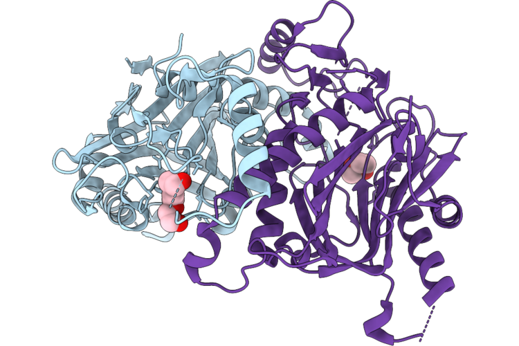 |
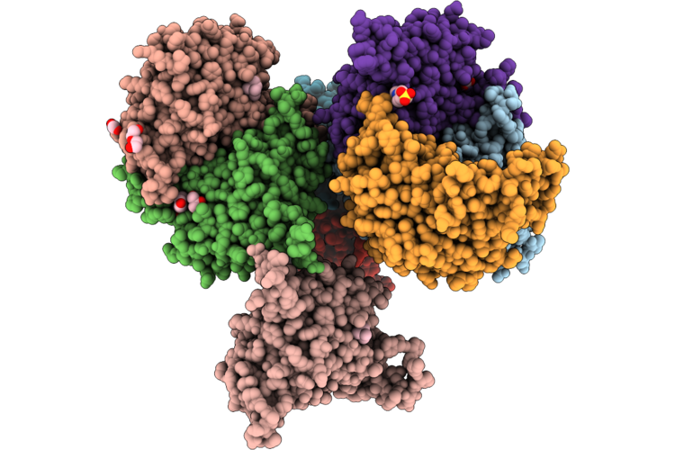
Crystal Structure Of The Polycaprolactam (Nylon6) And Poly(Hexamethylene Adipamide) (Nylon66) Hydrolase Nyl12 At Room Temperature
Organism: Gammaproteobacteria bacterium
Method: X-RAY DIFFRACTION Resolution:1.90 Å Release Date: 2026-06-03 Classification: HYDROLASE Ligands: PGE, PEG |
Organism: Gammaproteobacteria bacterium
Method: X-RAY DIFFRACTION
Release Date: 2026-06-03
Ligands: PGE, PEG
 |
Crystal Structure Of The Polycaprolactam (Nylon6) And Poly(Hexamethylene Adipamide) (Nylon66) Hydrolase Nyl12 At Cryo Temperature
Organism: Gammaproteobacteria bacterium
Method: X-RAY DIFFRACTION Resolution:1.75 Å Release Date: 2026-06-03 Classification: HYDROLASE Ligands: CHT, ACT, EPE, GOL, EDO, PEG |
Organism: Gammaproteobacteria bacterium
Method: X-RAY DIFFRACTION
Release Date: 2026-06-03
Ligands: CHT, ACT, EPE, GOL, EDO, PEG
 |
Structure Of G2L4 Rt Apoenzyme
Organism: Gammaproteobacteria
Method: X-RAY DIFFRACTION Resolution:2.61 Å Release Date: 2025-08-06 Classification: DNA BINDING PROTEIN Ligands: URE |
Organism: Gammaproteobacteria
Method: X-RAY DIFFRACTION
Release Date: 2025-08-06
Ligands: URE
 |
Structure Of G2L4 Rt In Complex With 15 Nucleotide Snapback Substrate
Organism: Gammaproteobacteria, Synthetic construct
Method: X-RAY DIFFRACTION Resolution:2.77 Å Release Date: 2025-07-30 Classification: DNA BINDING PROTEIN Ligands: MN, DCP |
Organism: Gammaproteobacteria, Synthetic construct
Method: X-RAY DIFFRACTION
Release Date: 2025-07-30
Ligands: MN, DCP
 |
Cryo-Em Structure Of Retinal-Free Proteoopsin Bound To Decanoate
Organism: Uncultured gammaproteobacteria bacterium
Method: ELECTRON MICROSCOPY Release Date: 2024-07-03 Classification: PROTON TRANSPORT Ligands: DKA |
Organism: Uncultured gammaproteobacteria bacterium
Method: ELECTRON MICROSCOPY
Release Date: 2024-07-03
Ligands: DKA
 |
Cryo-Em Structure Of Pentameric Proteorhodopsin A18L Mutant
Organism: Uncultured gammaproteobacteria bacterium
Method: ELECTRON MICROSCOPY Release Date: 2024-07-03 Classification: PROTON TRANSPORT Ligands: RET |
Organism: Uncultured gammaproteobacteria bacterium
Method: ELECTRON MICROSCOPY
Release Date: 2024-07-03
Ligands: RET
 |
Cryo-Em Structure Of Hexameric Proteorhodopsin A18L Mutant
Organism: Uncultured gammaproteobacteria bacterium
Method: ELECTRON MICROSCOPY Release Date: 2024-07-03 Classification: PROTON TRANSPORT Ligands: RET |
Organism: Uncultured gammaproteobacteria bacterium
Method: ELECTRON MICROSCOPY
Release Date: 2024-07-03
Ligands: RET
 |
Wilavidin Biotin Complex
Organism: Gammaproteobacteria bacterium
Method: X-RAY DIFFRACTION Resolution:2.63 Å Release Date: 2021-11-17 Classification: UNKNOWN FUNCTION Ligands: BTN, GOL |
Organism: Gammaproteobacteria bacterium
Method: X-RAY DIFFRACTION
Release Date: 2021-11-17
Ligands: BTN, GOL
 |
Wilavidin Apo Form (P1 Form)
Organism: Gammaproteobacteria bacterium
Method: X-RAY DIFFRACTION Resolution:1.95 Å Release Date: 2021-11-17 Classification: UNKNOWN FUNCTION |
Organism: Gammaproteobacteria bacterium
Method: X-RAY DIFFRACTION
Release Date: 2021-11-17
 |
Short Wilavidin Apo Form
Organism: Gammaproteobacteria bacterium
Method: X-RAY DIFFRACTION Resolution:2.18 Å Release Date: 2021-11-17 Classification: UNKNOWN FUNCTION Ligands: 1PE, P6G, PEG, PGE |
Organism: Gammaproteobacteria bacterium
Method: X-RAY DIFFRACTION
Release Date: 2021-11-17
Ligands: 1PE, P6G, PEG, PGE
 |
Short Wilavidin Biotin Complex
Organism: Gammaproteobacteria bacterium
Method: X-RAY DIFFRACTION Resolution:1.67 Å Release Date: 2021-11-17 Classification: UNKNOWN FUNCTION Ligands: BTN, PG4, PGE, EDO, PEG, P33, NA |
Organism: Gammaproteobacteria bacterium
Method: X-RAY DIFFRACTION
Release Date: 2021-11-17
Ligands: BTN, PG4, PGE, EDO, PEG, P33, NA
 |
Cryo-Em Structure Of The Green-Light Absorbing Proteorhodopsin
Organism: Uncultured gammaproteobacteria bacterium
Method: ELECTRON MICROSCOPY Release Date: 2021-06-16 Classification: PROTON TRANSPORT Ligands: RET |
Organism: Uncultured gammaproteobacteria bacterium
Method: ELECTRON MICROSCOPY
Release Date: 2021-06-16
Ligands: RET
 |
Crystal Structure Of N2, A Member Of 4-Oxalocrotonate Tautomerase (4-Ot) Family
Organism: Gammaproteobacteria bacterium sg8_31
Method: X-RAY DIFFRACTION Resolution:1.65 Å Release Date: 2021-06-02 Classification: ISOMERASE |
Organism: Gammaproteobacteria bacterium sg8_31
Method: X-RAY DIFFRACTION
Release Date: 2021-06-02